Review
. 2010 Aug 5;119(10):407-21.
doi: 10.1042/CS20100094.

The vascular contribution to Alzheimer's disease

Affiliations
Review

The vascular contribution to Alzheimer's disease

Robin Altman et al. Clin Sci (Lond). .

Abstract

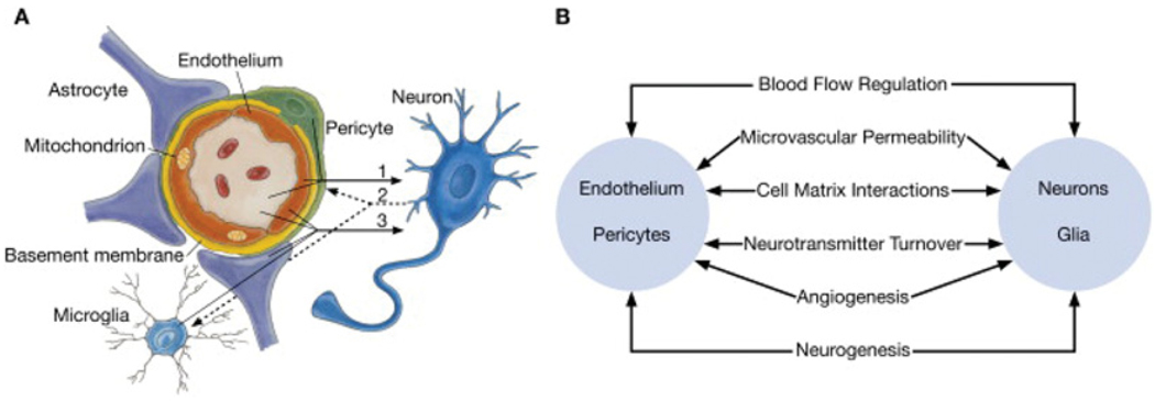
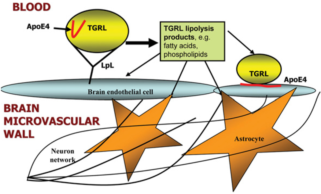
AD (Alzheimer's disease) is a progressive neurodegenerative disease of unknown origin. Despite questions as to the underlying cause(s) of this disease, shared risk factors for both AD and atherosclerotic cardiovascular disease indicate that vascular mechanisms may critically contribute to the development and progression of both AD and atherosclerosis. An increased risk of developing AD is linked to the presence of the apoE4 (apolipoprotein E4) allele, which is also strongly associated with increased risk of developing atherosclerotic cardiovascular disease. Recent studies also indicate that cardiovascular risk factors, including elevated blood cholesterol and triacylglycerol (triglyceride), increase the likelihood of AD and vascular dementia. Lipids and lipoproteins in the circulation interact intimately with the cerebrovasculature, and may have important effects on its constituent brain microvascular endothelial cells and the adjoining astrocytes, which are components of the neurovascular unit. The present review will examine the potential mechanisms for understanding the contributions of vascular factors, including lipids, lipoproteins and cerebrovascular Abeta (amyloid beta), to AD, and suggest therapeutic strategies for the attenuation of this devastating disease process. Specifically, we will focus on the actions of apoE, TGRLs (triacylglycerol-rich lipoproteins) and TGRL lipolysis products on injury of the neurovascular unit and increases in blood-brain barrier permeability.


Figures

Figure 1
Figure 1. Schematic representation of the neurovascular unit
(A) Endothelial cells and pericytes are separated by the basement membrane. Pericyte processes sheathe most of the outer side of the basement membrane. At points of contact, pericytes communicate directly with endothelial cells through the synapse-like peg-socket contacts. Astrocytic endfoot processes unsheathe the microvessel wall, which is made up of endothelial cells and pericytes. Resting microglia have a ‘ramified’ shape. In cases of neuronal disorders that have a primary vascular origin, circulating neurotoxins may cross the BBB to reach their neuronal targets, or pro-inflammatory signals from the vascular cells or reduced capillary blood flow may disrupt normal synaptic transmission and trigger neuronal injury (arrow 1). Microglia recruited from the blood or within the brain and the vessel wall can sense signals from neurons (arrow 2). Activated endothelium, microglia and astrocytes signal back to neurons, which in most cases aggravates the neuronal injury (arrow 3). In the case of a primary neuronal disorder, signals from neurons are sent to the vascular cells and microglia (arrow 2), which activate the vasculo–glial unit and contributes to the progression of the disease (arrow 3). (B) Co-ordinated regulation of normal neurovascular functions depends on vascular cells (endothelium and pericytes), neurons and astrocytes. Reprinted from Neuron volume 57, Zlokovic, B.V., The blood–brain barrier in health and chronic neurodegenerative disorders, pp. 178–201, Copyright (2008), with permission from Elsevier (http://www.sciencedirect.com/science/journal/08966273).
Figure 2
Figure 2. Schematic representation of apoE4 before and after a moderately high-fat meal
An apoE4 mutant was employed to study the domain interaction of the apoE4 mutant using EPR (electron paramagnetic resonance) spectroscopy. The dotted line represents a salt bridge between Arg61 (R61) and Glu255 (E255) of apoE4 showing a domain interaction. In this mutant, the serine residue at position 76 and the alanine residue at position 241 were mutated to cysteine and subsequently labelled with a nitroxyl spin label. The left-hand panel represents the structural conformation of apoE4 during the fasting state, and the right-hand panel represents the conformation in the postprandial state. ApoE4 had a reduced domain interaction during the postprandial state, implying a more linear protein [82]. The left-hand Figure was reproduced with permission from [82]. Copyright (2006) American Society for Biochemistry and Molecular Biology. The right-hand Figure was reproduced with permission from [222]. Copyright (2010) American Society for Biochemistry and Molecular Biology.
Figure 3
Figure 3. Overview of LDL metabolism in humans
Dietary cholesterol and triacylglycerols are packaged with apolipoproteins in the enterocytes of the small intestine and secreted into the lymphatic system as chylomicrons (CM). As chylomicrons circulate, the core triacylglycerols are hydrolysed by LpL, resulting in the formation of chylomicron remnants (CM Rem), which are rapidly removed by the liver. Dietary cholesterol has four possible fates once it reaches the liver: it can be (i) esterified and stored as cholesteryl esters in hepatocytes; (ii) packaged into VLDL particles and secreted into the plasma; (iii) secreted directly into the bile; or (iv) converted into bile acids and secreted into the bile. VLDL particles secreted into the plasma undergo lipolysis to form VLDL remnants (VLDL Rem). Approx. 50 % of VLDL remnants are removed by the liver via the LDLR and the remainder mature into LDL, the major cholesterol transport particle in the blood. An estimated 70 % of circulating LDL is cleared by LDLR in the liver. ABCG5 and ABCG8 (ABC family G, members 5 and 8 respectively) are located predominantly in the enterocytes of the duodenum and jejunum, the sites of uptake of dietary sterols, and in hepatocytes, where they participate in sterol trafficking into bile. ApoB, apolipoprotein B; ARH, autosomal recessive hypercholesterolaemia protein; FFA, non-esterified (‘free’) fatty acid Journal of Clinical Investigation. Online by Rader, D.J., Cohen, J. and Hobbs, H.H. Copyright 2003 by American Society for Clinical Investigation. Reproduced with permission of American Society for Clinical Investigation in the format Journal via Copyright Clearance Center.
Figure 4
Figure 4. Vascular disease model illustrating how TGRL lipolysis products and apoE4 may interact with brain microvascular endothelial cells and astrocytes
Hydrolysis of TGRLs by LpL present in the circulation results in the release of lipolysis products, including TGRL remnant particles, mono-, di- and tri-acylglycerols, phospholipids and NEFAs. Lipolysis products may influence the inflammatory environment of the brain through two pathways: (i) direct injury to brain microvascular endothelial cells or astrocytes, or (ii) indirect injury to glial cells and neurons through cascades that begin with damaged endothelial cells. In addition, apoE4 associated with TGRL undergoes a conformational change to a more linear species in the presence of TGRL lipolysis products. This conformational change may influence the binding of apoE4 to brain microvascular endothelial cells. Negative effects to the endothelial cells due to the altered apoE4 conformation may disrupt the barrier function of the cerebrovasculature and allow TGRL lipolysis products to access the brain parenchyma, causing damage to neurons and glial cells.
Figure 5
Figure 5. ZO-1 immunofluorescence before and after treatment with TGRL lipolysis products
Images of human aortic endothelial cell monolayers showing (A) continuous ZO-1 staining with treatment with TGRL (150 mg/dl triacylglycerol), and (B) discontinuity and radial rearrangement of ZO-1 (arrow) during exposure to lipolysis products generated from co-incubation of TGRL (150 mg/dl) + LpL (2 units/ml), causing increased endothelial layer permeability. This Figure was reproduced from [166] and is used with permission. Copyright (2007) American Physiological Society.

References

    1. Aggarwal NT, Decarli C. Vascular dementia: emerging trends. Semin. Neurol. 2007;27:66–77. - PubMed
    1. Chui HC, Zarow C, Mack WJ, Ellis WG, Zheng L, Jagust WJ, Mungas D, Reed BR, Kramer JH, Decarli CC, et al. Cognitive impact of subcortical vascular and Alzheimer’s disease pathology. Ann. Neurol. 2006;60:677–687. - PMC - PubMed
    1. DeCarli C. The role of cerebrovascular disease in dementia. Neurologist. 2003;9:123–136. - PubMed
    1. DeCarli CS. When two are worse than one: stroke and Alzheimer disease. Neurology. 2006;67:1326–1327. - PMC - PubMed
    1. Berglund L, Wiklund O, Eggertsen G, Olofsson SO, Eriksson M, Linden T, Bondjers G, Angelin B. Apolipoprotein E phenotypes in familial hypercholesterolaemia: importance for expression of disease and response to therapy. J. Intern. Med. 1993;233:173–178. - PubMed

Publication types