

Extramammary Paget's disease with the appearance of a nodule: a case report
- PMID: 20684770
- PMCID: PMC2921398
- DOI: 10.1186/1471-2407-10-405
Extramammary Paget's disease with the appearance of a nodule: a case report
Abstract
Background: Extramammary Paget's disease (EMPD) remains a rare condition with only a limited number of cases reported in the literature. EMPD is mainly composed of intraepidermal Paget cells, and possesses variable clinical behaviors and histological appearances, leading to difficulty in the diagnosis of this disease.
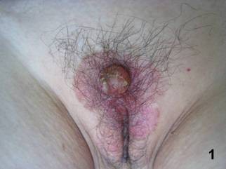
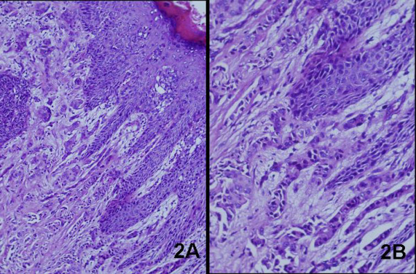
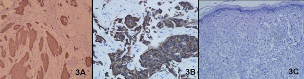
Case presentation: We here report a case of primary EMPD with the appearance of a nodule on the background of erythema. Histological assessment showed Paget cell infiltration throughout the epidermis with dermal spread. Using immunohistochemistry, the expressions of CK7, CK19, CK20, GCDFP-15, CEA, S-100 protein and bcl-2 were examined to elucidate the cellular differentiation of the carcinoma.
Conclusion: According to the histological assessment, this case was diagnosed as primary EMPD with carcinoma cells invading into the dermis, but without lymph node infiltration.
Figures
Similar articles
-
Intraepidermal cytokeratin 7 expression is not restricted to Paget cells but is also seen in Toker cells and Merkel cells.Am J Surg Pathol. 1999 Feb;23(2):212-9. doi: 10.1097/00000478-199902000-00011. Am J Surg Pathol. 1999. PMID: 9989849
-
Primary Extra Mammary Paget's Disease of Vulva, With Apocrine Adenocarcinoma, Signet Ring Cell Differentiation and Distant Metastasis.J Family Reprod Health. 2020 Dec;14(4):276-280. doi: 10.18502/jfrh.v14i4.5213. J Family Reprod Health. 2020. PMID: 34055001 Free PMC article.
-
Clinical and pathological characteristics of extramammary Paget's disease: report of 246 Chinese male patients.Int J Clin Exp Pathol. 2015 Oct 1;8(10):13233-40. eCollection 2015. Int J Clin Exp Pathol. 2015. PMID: 26722523 Free PMC article.
-
Ectopic extramammary Paget's disease: case report and literature review.Acta Derm Venereol. 2010 Sep;90(5):502-5. doi: 10.2340/00015555-0892. Acta Derm Venereol. 2010. PMID: 20814627 Review.
-
Perianal Paget's disease: presentation of six cases and literature review.Int J Colorectal Dis. 2010 Jan;25(1):1-7. doi: 10.1007/s00384-009-0797-9. Epub 2009 Aug 26. Int J Colorectal Dis. 2010. PMID: 19707774 Review.
Cited by
-
Extramammary Paget's disease of vulva: A rare entity.Indian J Sex Transm Dis AIDS. 2017 Jan-Jun;38(1):76-77. doi: 10.4103/0253-7184.196891. Indian J Sex Transm Dis AIDS. 2017. PMID: 28442808 Free PMC article.
-
Pemetrexed induced a durable response in heavily pretreated metastatic extramammary Paget's disease.Int J Clin Exp Med. 2015 Jun 15;8(6):9595-8. eCollection 2015. Int J Clin Exp Med. 2015. PMID: 26309631 Free PMC article.
-
Perianal Extramammary Paget's Disease: More Than Meets the Eye.Dig Dis Sci. 2018 Nov;63(11):2853-2857. doi: 10.1007/s10620-018-5089-1. Dig Dis Sci. 2018. PMID: 29696480 No abstract available.
-
Perianal extramammary Paget's disease with adenocarcinoma of perianal skin area, a case report.J Surg Case Rep. 2023 Jun 16;2023(6):rjad291. doi: 10.1093/jscr/rjad291. eCollection 2023 Jun. J Surg Case Rep. 2023. PMID: 37337540 Free PMC article.
-
A true epidermotropic apocrine neoplasm in the form of perianal Paget's disease: a case report.J Med Case Rep. 2013 Jun 20;7:162. doi: 10.1186/1752-1947-7-162. J Med Case Rep. 2013. PMID: 23786719 Free PMC article.
References
-
- Besa P, Rich TA, Delclos L, Edwards CL, Ota DM, Wharton JT. Extramammary Paget's disease of the perineal skin: role of radiotherapy. Int J Radiat Oncol Biol Phys. 1992;24(1):73–8. - PubMed
-
- de Blois GG, Patterson JW, Hunter SB. Extramammary Paget's disease: Arising in knee region in association with sweat gland carcinoma. Arch Pathol Lab Med. 1984;108(9):713–6. - PubMed
Publication types
MeSH terms
Substances
LinkOut - more resources
Full Text Sources
Medical
Research Materials
