. 2010 Sep;225(1):104-13.
doi: 10.1016/j.expneurol.2010.05.020. Epub 2010 Jun 2.

Widespread cortical expression of MANF by AAV serotype 7: localization and protection against ischemic brain injury

Affiliations

Widespread cortical expression of MANF by AAV serotype 7: localization and protection against ischemic brain injury

Mikko Airavaara et al. Exp Neurol. 2010 Sep.

Abstract

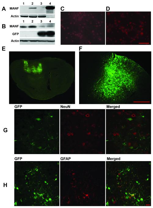
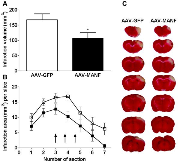
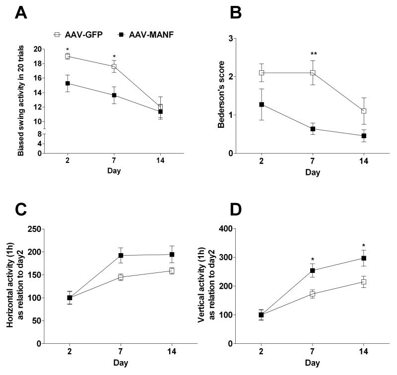
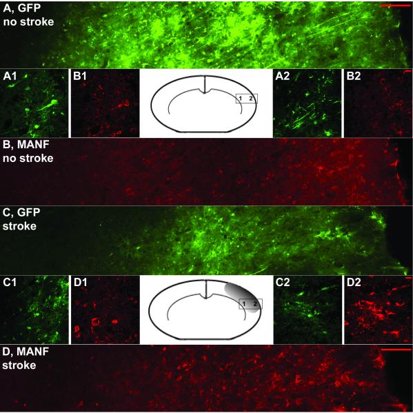
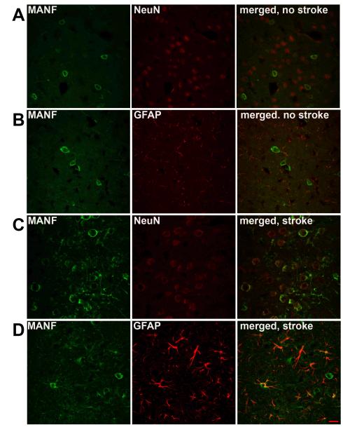
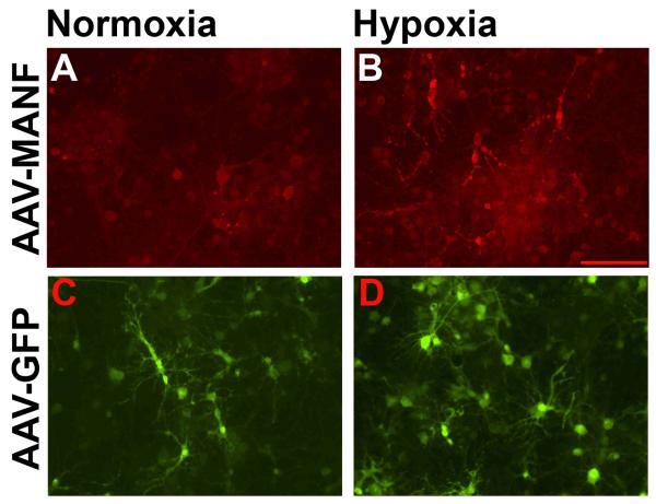
Mesencephalic astrocyte-derived neurotrophic factor (MANF) is a secreted protein which reduces endoplasmic reticulum (ER) stress and has neurotrophic effects on dopaminergic neurons. Intracortical delivery of recombinant MANF protein protects tissue from ischemic brain injury in vivo. In this study, we examined the protective effect of adeno-associated virus serotype 7 encoding MANF in a rodent model of stroke. An AAV vector containing human MANF cDNA (AAV-MANF) was constructed and verified for expression of MANF protein. AAV-MANF or an AAV control vector was administered into three sites in the cerebral cortex of adult rats. One week after the vector injections, the right middle cerebral artery (MCA) was ligated for 60min. Behavioral monitoring was conducted using body asymmetry analysis, neurological testing, and locomotor activity. Standard immunohistochemical and western blotting procedures were conducted to study MANF expression. Our data showed that AAV-induced MANF expression is redistributed in neurons and glia in cerebral cortex after ischemia. Pretreatment with AAV-MANF reduced the volume of cerebral infarction and facilitated behavioral recovery in stroke rats. In conclusion, our data suggest that intracortical delivery of AAV-MANF increases MANF protein production and reduces ischemic brain injury. Ischemia also caused redistribution of AAV-mediated MANF protein suggesting an injury-induced release.


Figures

Figure 1
Figure 1. Characterization of AAV-MANF and AAV-GFP serotype 7 transduction in rat cortex
(A) HEK293 cells were transfected with the AAV-MANF packaging plasmid for 24 hours and then protein extracts were analyzed by western blot (A1-untreated cells, 2-AAV-MANF, 3-AAV-GFP, 4-recombinant MANF protein, 2 ug). Rat primary cortical cultures were transduced with AAV-GFP (C) or AAV-MANF (D) virus for 48 hours then fixed and immunostained for MANF (red). (B) AAV-MANF and AAV-GFP transduction in rat cortex was verified with western blot analysis (B1 and B2 – AAV-MANF; B3 and B4 – AAV-GFP). Delivery of AAV-GFP (serotype 7) into three cortical sites produced wide-spread GFP expression at 1 week post-injection as shown in sagittal (E) and coronal (F) brain sections. AAV-induced GFP expression is detected in NeuN+(G, neuronal marker) and GFAP+ (H, glial marker) cells. Scale bars: C,D = 100μm, F = 500μm, G,H = 10μm.
Figure 2
Figure 2. AAV-MANF decreases infarction volume in stroke rats
Rats received three intracortical injections of AAV-MANF or AAV-GFP one week prior to a 60 minute MCAo. Tissue was sectioned (2 mm) and stained with TTC at 48 hours post-stroke. (A) AAV-MANF significantly reduced infarction volume compared to AAV-GFP (*p=0.0447, Student’s t-test). (B) Area of infarction per 2-mm section (7 sections/brain; anterior to posterior) was significantly reduced for all sections in the AAV-MANF group with the most notable difference in section 4 (two-way ANOVA, p<0.0001). Three cortical injections of AAV-MANF or AAV-GFP were made between sections 3 to 5 (arrows). C) Representative TTC stained sections.
Figure 3
Figure 3. AAV-MANF promotes recovery after cerebral ischemia
Rats received three intracortical injections of AAV-MANF or AAV-GFP one week prior to a 60 minute MCAo. Behavioral tests were carried out at 2, 7 and 14 days after the MCAo and analyzed with 2-way ANOVA. AAV-MANF injection significantly reduced body asymmetry in an elevated body swing test (A) and neurological abnormality scores in Bederson’s test (B). AAV-MANF increased horizontal activity (C) and vertical activity (D). Results of Bonferroni post-hoc tests are indicated by an asterisk. X-axis shows days after the MCAo.
Figure 4
Figure 4. MCAo alters MANF-IR in AAV-MANF injected animals
Rats were injected with mixture of AAV-MANF and AAV-GFP (1:1) and MANF (red) and GFP (green) expression were analyzed before and after MCAo. Figure 4A and 4B shows the expression patterns induced by the AAV vector before MCAo. After 1h MCAo and 6h reperfusion, there was broad change in the pattern of MANF expression in the ischemic cortex (Fig. 4D) which was not seen for GFP expression. Left and right sides of the figure are proximal to the corpus callosum and infarction core, respectively. Panels A/B and C/D are from the same brain section of non-stroke and stroke animals, respectively. Inset pictures (1 and 2) are high magnification images taken from indicated regions on the inset diagram of the coronal brain section. Note the change in MANF-IR is more prominent in the ischemic core (D2 versus D1). Scale bars: A,B, C, D = 100μm, A1-2, B1-2, C1-2, D1-2 = 10μm.
Figure 5
Figure 5. MANF-IR colocalizes to both neurons and astrocytes after MCAo
Before MCAo, MANF-IR in AAV-MANF injected cortical tissue colocalizes to NeuN+ (A, neuronal marker) cells and to a lesser extent GFAP+ (B, glial marker) cells. Six hours after reperfusion MANF is co-localized with both NeuN+ (C) and GFAP+ (D) cells ,with more prominent labeling of the cellular processes. A punctate pattern not associated with cell bodies is observed following stroke suggesting an extracellular location. Scale bar = 10μm.
Figure 6
Figure 6. Hypoxia causes redistribution of MANF-IR in primary cortical neurons transduced with AAV-MANF
Primary cortical neurons were transduced with AAV-MANF (A,B) or AAV-GFP (C,D) on DIV6 and given 8 hours hypoxia and 24 hours normoxia on DIV12. MANF-IR in AAV-MANF transduced cultures associates with cellular processes following hypoxia (A vs. B). No gross changes in GFP fluorescence is observed in AAV-GFP transduced cultures after hypoxia (C vs. D). Scale bars = 100μm.

Similar articles

Cited by

References

    1. Airavaara M, Shen H, Kuo CC, Peranen J, Saarma M, Hoffer B, Wang Y. Mesencephalic astrocyte-derived neurotrophic factor reduces ischemic brain injury and promotes behavioral recovery in rats. J Comp Neurol. 2009;515:116–124. - PMC - PubMed
    1. Apostolou A, Shen Y, Liang Y, Luo J, Fang S. Armet, a UPR-upregulated protein, inhibits cell proliferation and ER stress-induced cell death. Exp Cell Res. 2008;314:2454–2467. - PMC - PubMed
    1. Bederson JB, Pitts LH, Tsuji M, Nishimura MC, Davis RL, Bartkowski H. Rat middle cerebral artery occlusion: evaluation of the model and development of a neurologic examination. Stroke. 1986;17:472–476. - PubMed
    1. Borlongan CV, Tajima Y, Trojanowski JQ, Lee VM, Sanberg PR. Cerebral ischemia and CNS transplantation: differential effects of grafted fetal rat striatal cells and human neurons derived from a clonal cell line. Neuroreport. 1998;9:3703–3709. - PubMed
    1. Chang CF, Lin SZ, Chiang YH, Morales M, Chou J, Lein P, Chen HL, Hoffer BJ, Wang Y. Intravenous administration of bone morphogenetic protein-7 after ischemia improves motor function in stroke rats. Stroke. 2003;34:558–564. - PubMed

Publication types

MeSH terms