Review
. 2010 Nov;92(11):1587-96.
doi: 10.1016/j.biochi.2010.05.011. Epub 2010 Jun 2.

Molecular mechanisms of antithrombin-heparin regulation of blood clotting proteinases. A paradigm for understanding proteinase regulation by serpin family protein proteinase inhibitors

Affiliations
Review

Molecular mechanisms of antithrombin-heparin regulation of blood clotting proteinases. A paradigm for understanding proteinase regulation by serpin family protein proteinase inhibitors

Steven T Olson et al. Biochimie. 2010 Nov.

Abstract

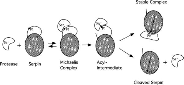
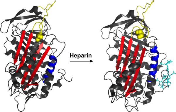
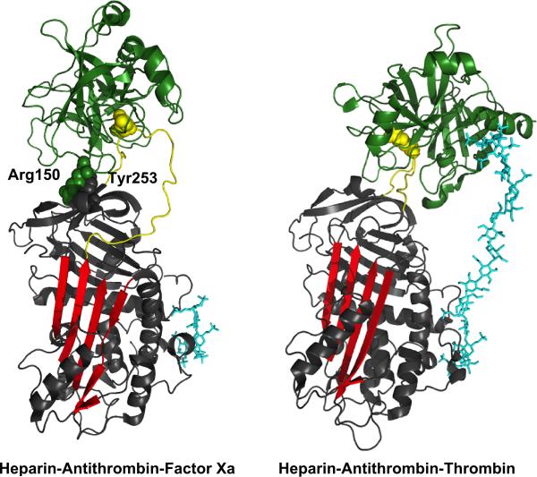
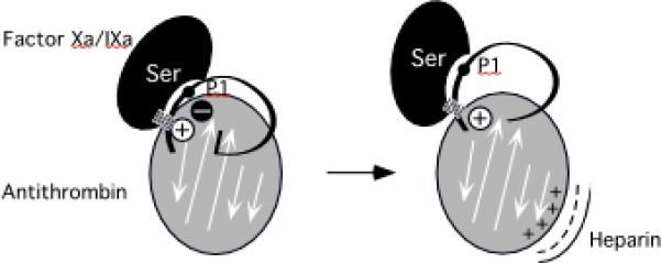
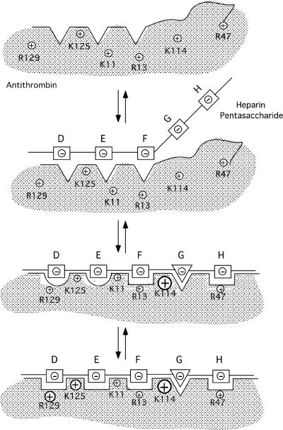
Serpin family protein proteinase inhibitors regulate the activity of serine and cysteine proteinases by a novel conformational trapping mechanism that may itself be regulated by cofactors to provide a finely-tuned time and location-dependent control of proteinase activity. The serpin, antithrombin, together with its cofactors, heparin and heparan sulfate, perform a critical anticoagulant function by preventing the activation of blood clotting proteinases except when needed at the site of a vascular injury. Here, we review the detailed molecular understanding of this regulatory mechanism that has emerged from numerous X-ray crystal structures of antithrombin and its complexes with heparin and target proteinases together with mutagenesis and functional studies of heparin-antithrombin-proteinase interactions in solution. Like other serpins, antithrombin achieves specificity for its target blood clotting proteinases by presenting recognition determinants in an exposed reactive center loop as well as in exosites outside the loop. Antithrombin reactivity is repressed in the absence of its activator because of unfavorable interactions that diminish the favorable RCL and exosite interactions with proteinases. Binding of a specific heparin or heparan sulfate pentasaccharide to antithrombin induces allosteric activating changes that mitigate the unfavorable interactions and promote template bridging of the serpin and proteinase. Antithrombin has thus evolved a sophisticated means of regulating the activity of blood clotting proteinases in a time and location-dependent manner that exploits the multiple conformational states of the serpin and their differential stabilization by glycosaminoglycan cofactors.


Figures

Fig. 1
Fig. 1. Branched pathway suicide substrate mechanism by which serpins inhibit serine and cysteine proteinases
The pathway begins with the proteinase active-site recognizing and binding to a substrate sequence contained within an exposed reactive center loop (RCL) of the serpin (gray-shaded) to form a Michaelis complex. The active-site serine or cysteine of the proteinase proceeds to attack a reactive P1-P1' bond in the serpin RCL sequence to form an acyl-intermediate complex in which the bond is cleaved and the P1 residue has become covalently linked to the attacking serine or cysteine. RCL cleavage triggers the suicide mechanism by inducing the metastable serpin to undergo a large conformational change in which the RCL inserts into the center of β-sheet A (shown as arrows), causing the tethered proteinase to be dragged to the opposite end of the protein and inactivated by conformational deformation. The trapped acyl-intermediate complex is kinetically stable and deacylates extremely slowly. A normal deacylation of the acyl-intermediate competes with conformational trapping to release active proteinase and cleaved serpin.
Fig. 2
Fig. 2. X-ray structures of the intermediate serpin-proteinase Michaelis complex and the final trapped acyl-intermediate complex
Ribbon representations of the Michaelis complex of a P1Arg variant of the serpin, α1-proteinase inhibitor, with catalytically inactivated S195A trypsin on the left (pdb 1OPH) and the trapped acyl-intermediate complex of wild-type α1-proteinase inhibitor (P1Met) with porcine pancreatic elastase on the right (pdb 1EZX). The proteinase is depicted in green and the serpin in gray with the A-sheet highlighted in red, the RCL in blue and the serpin P1 residue and proteinase catalytic serine shown in space-filling representation.
Fig. 3
Fig. 3. Mechanisms of heparin activation of antithrombin
The scheme depicts two mechanisms by which heparin or heparan sulfate glycosaminoglycans activate antithrombin to rapidly inhibit blood clotting proteinases. In both mechanisms a positively charged site on antithrombin (AT) binds to a sequence-specific negatively charged pentasaccharide (shaded) on the heparin or heparan sulfate polysaccharide (H). This binding induces an activating conformational change in the heparin binding site of the serpin that is allosterically transmitted to the proteinase binding region. The allosteric changes specifically activate antithrombin reactivity with factors Xa and IXa by relieving unfavorable interactions and strengthening favorable interactions of these proteinases with the serpin RCL and exosites. The allosteric mechanism minimally affects antithrombin reactivity with thrombin since thrombin does not interact with the factor Xa and IXa-specific exosites. Longer chain heparin or heparan sulfate glycosaminoglycans activate antithrombin reactivity with thrombin and further augment antithrombin reactivity with factors Xa and IXa through a second bridging mechanism in which positively charged sites on the proteinase bind to the negatively charged polysaccharide at an exosite adjacent to bound antithrombin so as to position the proteinase for engagement of the serpin RCL.
Fig. 4
Fig. 4. Structure of the antithrombin binding pentasaccharide sequence of heparin and heparan sulfate
The O-methyl glycoside of the pentasaccharide sequence is shown from the nonreducing end to the reducing end with sulfates and carboxylates that are important determinants of pentasaccharide binding to antithrombin labeled with an asterisk. The unique 3-O-sulfate marker of the pentasaccharide sequence is indicated by a double asterisk. The saccharides are designated DEFGH based on the historical record of discovery of the sequence [15]. The dominant structure of heparin and of certain highly sulfated domains of heparan sulfate is a polymer of repeating GH disaccharide units.
Fig. 5
Fig. 5. X-ray structures of free and heparin pentasaccharide-complexed antithrombin
Ribbon representations of free antithrombin on the left (pdb 1E05) and heparin pentasaccharide complexed antithrombin on the right (1E03) reveal the activating conformational changes induced in antithrombin by the binding of the pentasaccharide (a mimetic shown in cyan stick representation). These include an extension of helix D and formation of a new P helix in the heparin binding site and an expulsion of the P14 serine residue (space-filling) of the serpin RCL (yellow), initially buried in β-sheet A (red) in free antithrombin, from the A sheet and closing of the gap in the A sheet.
Fig. 6
Fig. 6. Closeup of the heparin binding site of free and heparin pentasaccharide-complexed antithrombin structures
Highlighted in ribbon representation are the three regions of antithrombin that comprise the heparin binding site: helix D and the loops that extend from its C- and N-terminal ends (blue), helix A (red) and the N-terminal region (yellow). Residues involved in binding the pentasaccharide are shown in stick representation and include Lys11, Arg13, Arg46, Arg47, Lys114, Lys125 and Arg129. The pentasaccharide is represented in stick (cyan). Conformational changes induced in antithrombin by pentasaccharide binding include an extension of helix D at the C-terminal end, formation of a new P helix in the loop preceding the N-terminal end of helix D and rotations of the three heparin binding regions to position basic residues for binding the critical sulfates and carboxylates of the pentasaccharide.
Fig. 7
Fig. 7. X-ray structures of heparin-antithrombin-proteinase Michaelis complexes
Shown in ribbon representation are the structures of ternary Michaelis complexes of heparin pentasaccharide-antithrombin with S195A factor Xa on the left (pdb 2GD4) and heparin hexadecasaccharide-antithrombin with S195A thrombin on the right (1TB6). The hexadecasaccharide contains a pentasaccharide mimetic, an uncharged saccharide linker and five terminal sulfated glucose saccharides. Heparin molecules are depicted in cyan (stick), proteinases in green and antithrombin in gray. The A-sheet is highlighted in red and the RCL in yellow. The critical Tyr253 exosite residue in strand 3C of antithrombin and the complementary Arg150 exosite residue of factor Xa as well as the P1 Arg are shown in space-filling representation. The structures reveal distinct orientations of the proteinase bound to the serpin RCL, with factor Xa bending downward toward the serpin body to form the critical exosite-exosite interaction and thrombin extending away from the serpin surface and bending in the opposite direction to interact with a heparin exosite on the extended polysaccharide chain.
Fig. 8
Fig. 8. Model for allosteric activation of antithrombin reactivity with factor Xa and IXa
Antithrombin is proposed to exist in a repressed reactivity state as a result of unfavorable interactions (symbolized by the black circled minus sign) that mitigate favorable interactions of an exosite (symbolized by the white circled plus sign) and the RCL with factors Xa and IXa (hatched rectangle denotes the favorable exosite interaction). This mitigation is exacerbated by the burial of the RCL hinge in sheet A (shown as arrows) which causes the bound proteinase to closely approach the serpin surface. Heparin releases antithrombin from its repressed reactivity state by inducing allosteric activating changes that diminish the negative interactions but retain the positive exosite interactions. The unfavorable interactions with proteinase are reduced both by changes in the serpin surface electrostatics and by extension of the RCL away from the serpin surface.
Fig. 9
Fig. 9. Induced-fit model for heparin pentasaccharide binding to and allosteric activation of antithrombin
The pentasaccharide is proposed to bind and allosterically activate antithrombin in 3-steps. ln step one, the conformationally rigid nonreducing end DEF trisaccharide recognizes and binds to the D helix and N-terminal segments of the heparin binding site of antithrombin primarily through electrostatic interactions involving Lys125 and Lys11. The conformationally flexible GH disaccharide, the consequence of an equilibrium between chair and skew boat forms of iduronate residue G, minimally interacts in this step [16]. DEF binding triggers a first set of induced-fit conformational changes in the heparin binding site in step two that include formation of the P helix, rotation of the D helix and bending of the A helix. These changes allow the critical Lys114 interaction with the pentasaccharide 3-O-sulfate to be made and serve to position Lys114 together with Arg13, Arg46, and Arg47 for binding the reducing end GH disaccharide in the skew boat form (triangle). The changes additionally improve the DEF trisaccharide interactions with Lys11, Lys125 and Arg129. A second set of induced-fit conformational changes follows in step three in which the D helix extends at its C-terminal end, causing the buried Tyr131 to become exposed and the hydrophobic core to compact. Such changes increase pentasaccharide complementarity with the heparin binding site by repositioning Lys125 and Arg129 for optimal interactions with the DEF trisaccharide. The relative importance of each basic residue in binding the pentasaccharide in each step is reflected by the size of the positive charge symbols for each residue.
Fig. 10
Fig. 10. Comparison of the structures of the antithrombin-heparin pentasaccharide complex in native, latent and intermediate forms
A closeup of the D- and P-helices and parts of the N-terminal region and the A-helix of the heparin binding site of native antithrombin (A), latent antithrombin (B) and an intermediate native form (C) in complex with a heparin pentasaccharide mimetic (large images, pdb codes 1E03 and 1NQ9) and in the unbound state (inset, pdb code 1E05) are shown in ribbon representation (gray). Antithrombin basic residues that participate in binding the heparin pentasaccharide as well as Tyr131, Arg132 and Lys133 in the C-terminal extension of helix D are shown in ball-and-stick representation. Carbon atoms are in black, nitrogen atoms are blue and oxygen atoms are red. The pentasaccharide atoms are shown in green ball-and-stick. The latent and intermediate antithrombin-pentasaccharide complex structures show similar induced-fit conformational changes including P helix formation, D helix rotation and A helix bending but have not undergone helix D extension and the flipping of Tyr131 that is evident in the native antithrombin structure.

References

    1. Gettins P. Serpin Structure, Mechanism, and Function. Chem. Rev. 2002;102:4751–4803. - PubMed
    1. Huntington JA. Shape-shifting serpins-advantages of a mobile mechanism. Trends Biochem. Sci. 2006;31:427–435. - PubMed
    1. Irving JA, Pike RN, Dai W, Bromme D, Worrall DM, Silverman GA, Coetzer THT, Dennison C, Bottomley SP, Whisstock JC. Evidence That Serpin Architecture Intrinsically Supports Papain-like Cysteine Protease Inhibition: Engineering α1-Antitrypsin To Inhibit Cathepsin Proteases. Biochemistry. 2002;41:4998–5004. - PubMed
    1. Björk I, Olson ST. Antithrombin. A Bloody Important Serpin. In: Church FC, Cunningham DD, Ginsburg D, Hoffman M, Stone SR, Tollefsen DM, editors. Chemistry and Biology of Serpins. Plenum Press; New York: 1997. pp. 17–33. - PubMed
    1. Church FC, Pike RN, Tollefsen DM, Buckle DM, Ciaccia AV, Olson ST. Regulation of hemostasis by heparin-binding serpins. In: Silverman GA, Lomas DA, editors. Molecular and Cellular Aspects of the Serpinopathies and Disorders of Serpin Activity. World Scientific Publishing; Singapore: 2007. pp. 509–554.