. 2010 Oct;139(4):1375-84.
doi: 10.1053/j.gastro.2010.05.074. Epub 2010 Jun 1.

Reduced nicotinamide adenine dinucleotide phosphate oxidase 2 plays a key role in stellate cell activation and liver fibrogenesis in vivo

Affiliations

Reduced nicotinamide adenine dinucleotide phosphate oxidase 2 plays a key role in stellate cell activation and liver fibrogenesis in vivo

Joy X Jiang et al. Gastroenterology. 2010 Oct.

Abstract

Background & aims: Hepatocyte apoptosis and activation of hepatic stellate cells (HSC) are critical events in fibrogenesis. We previously demonstrated that phagocytosis of apoptotic hepatocytes by HSC is profibrogenic. Based on this, as well as the observation that reduced nicotinamide adenine dinucleotide phosphate oxidase (NADPH) oxidase induction is central to fibrogenesis, our aim was to study the phagocytic NADPH oxidase NOX2.

Methods: An in vivo phagocytosis model was developed by injecting wild type (wt) or NOX2(-/-) mice with lentiviral-green fluorescence protein (GFP) containing a hepatocyte-specific promoter, and adeno-tumor necrosis factor-related apoptosis-inducing ligand (ad-TRAIL). Fibrosis was evaluated in bile duct ligated (BDL) wt and NOX2(-/-) mice with or without gadolinium treatment. NOX2 expression was studied in human liver samples and in HSC isolated from fibrotic livers. The fibrogenic activity of NOX2 was assessed by collagen reporter assays.

Results: In the phagocytosis model, engulfment of GFP-labeled apoptotic bodies was seen, and the expression of α-smooth muscle actin (α-SMA) and collagen I increased significantly in the wt but not in the NOX2(-/-) mice. Inhibiting apoptosis decreased the profibrogenic response. NOX2(-/-) animals exhibited significantly less fibrosis following BDL. Inactivating macrophages in wt BDL mice did not lower collagen production to the level observed in NOX2(-/-) mice, suggesting that NOX2-expressing HSC are important in fibrogenesis. NOX2 was up-regulated in HSC from fibrotic livers, and phagocytosis-induced NOX2 expression and activity were demonstrated. Based on reporter assays, production of NOX2-mediated reactive oxygen species directly induced collagen promoter activity in HSC.

Conclusions: Apoptosis and phagocytosis of hepatocytes directly induce HSC activation and initiation of fibrosis. NOX2, the phagocytic NADPH oxidase, plays a key role in this process and in liver fibrogenesis in vivo.


Figures

Figure 1
Figure 1. The expression of NOX2 is upregulated in HSC during liver fibrogenesis
(A) Myofibroblasts in patients with HCV (a, b, c,) and PBC (d, e, f) express NOX2 (green channel, a, d), α-SMA (red channel b, e), and co-localization images (c, f), by confocal microscopy. Bar=30 μm. (B) HSC were isolated from BDL and sham rats and NOX2 mRNA and protein expression were assessed. Both NOX2 mRNA and protein (*p<0.05) were significantly upregulated compared to HSC from sham-operated animals (shown in fold expression). (C) HSC were isolated from rats and exposed to AB for 24 hours. NOX2 expression was determined by real-time PCR. Phagocytosis of AB induced NOX2 upregulation in HSC, shown as fold increase (**p≤0.01). Error bars indicate 1 SED.
Figure 2
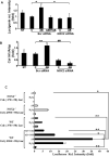
Figure 2. NOX2 activation in HSC results in superoxide production and upregulation of collagen I mRNA by inducing its transcriptional activation
(A) Primary rat HSC were treated with either scrambled siRNA or NOX2 siRNA and exposed to AB. After 6 hours superoxide production was assessed by the lucigenin assay. In the scrambled siRNA-transfected HSC, phagocytosis induced superoxide production, and this was inhibited in the NOX2 siRNA-transfected cells (NT: non-transfected, C: control, not exposed to AB, *p<0.05, average of 4 experiments, bars indicate 1 SED, shown in fold expression). (B) Collagen IA1 expression was assessed by real-time PCR. Collagen IA1 expression was upregulated in the scrambled siRNA-transfected cells after AB exposure (shown in fold expression, **p≤0.01), and it decreased significantly in the NOX2 siRNA-transfected primary HSC (##p≤0.0001). (C) Primary wild type or NOX2-/- HSC were transfected by constructs containing the truncated collagen promoter Col1A2 P1-Luc (-378/+58) or with a construct where the peroxide-responsive area is intact [Col1A2 P1-Luc (-2900/+58)], or the empty vector. The cells were exposed to AB in the presence or absence of the reducing agent GSH or catalase. Phagocytosis resulted in a significant induction of the COLIA2 promoter activity in the Col1A2 P1-Luc (-2900/+58)-transfected cells. This was abrogated in the Col1A2 P1-Luc (-378/+58)-transfected cells, or decreased after exposure to catalase, or GSH. In wt cells the collagen promoter activity thus resulted from NOX2-mediated peroxide production following phagocytosis. In the NOX2-/- cells the luciferase activity was significantly blunted after exposure to AB (C: control cells, AB: apoptotic bodies, cata:catalase, GSH: glutathione *p<0.05, **p≤0.01).
Figure 3
Figure 3. The phagocytic activity is decreased in NOX2-/- HSC
(A) Primary wt or NOX2-/- HSC were exposed to TAMRA-labeled AB, and the rate of phagocytosis was assessed after 24 hours. The engulfment was significantly decreased in HSC isolated from NOX2-/- mice compared to wt mice (#p<0.001). (B) Pull-down of active GTP-bound Rac1 from the membrane fractions of wt and NOX2-/- HSC demonstrated significantly more GTP-Rac1 in the membrane fraction of wt cells following AB exposure compared to NOX2-/- cells. Rac1 immunostaining was detectable at the phagosomal membrane forming around the TAMRA-labeled AB (red) in wt HSC (arrow), while in the NOX2-/- HSC AB attachment is seen but impaired phagosome formation (arrowhead), bar=10μm.
Figure 4
Figure 4. In vivo model of hepatocyte apoptosis and phagocytosis
To follow the fate of apoptotic hepatocytes, mice were first injected with the α1-AT-LV-GFP via the portal vein then 7 days later Ad-TRAIL was injected into the tail vein of the same mice. As control, mice were injected only with Ad-TRAIL, or Ad-GFP (images not shown) or with α1-AT-LV-GFP. To inhibit apoptosis, a separate group of mice were injected with Q-VD-OPH, pancaspase inhibitor before and after the Ad-TRAIL injection. The liver from only Ad-TRAIL, or LV-injected mice showed no significant injury or infiltration by inflammatory cells, and the ALT values remained normal (Figure 4b, c). In the LV plus Ad-TRAIL-injected animals the liver showed mild to moderate hepatocyte injury, increased ALT values, (*p<0.05), and no significant infiltration by inflammatory cells (Figure 4d). In the Q-VD-OPH treated animals the liver showed normal histology (Figure 4e) and decreased ALT values (*p<0.05) compared to LV plus Ad-TRAIL injected mice. To assess apoptosis, TUNEL assays were done on all liver samples. In the LV plus Ad-TRAIL infected livers hepatocyte apoptosis was increased (Figure 4d’) compared to only Ad-TRAIL (Figure 4b’) or only LV (Figure 4c’)-injected animals. In the Q-VD-OPH treated animals apoptosis decreased (Figure 4e’). bar=50 μm.
Figure 5
Figure 5. Phagocytosis of apoptotic hepatocytes in vivo results in a fibrogenic response
Immunohistochemistry and confocal microscopy were performed to visualize GFP-labeled hepatocytes and their AB and activated HSC (α-SMA, red). (GFP was only expressed in hepatocytes because of the hepatocyte specific α1-AT promoter). There was an increase in the number of activated HSC (α-SMA, red) in the liver of mice injected with α1-AT-LV-GFP plus Ad-TRAIL (Figure 5d, e), while the only Ad-TRAIL (Figure 5b), or only LV-GFP (Figure 5c) injected mice had the same amount of activated HSC as control, uninjected mice (Figure 5a), bar=50 μm. At higher magnification in the α-SMA-positive HSC, GFP-labeled AB could be seen, indicating phagocytosis of hepatocyte-derived AB (arrow, arrowhead depicts apoptotic hepatocytes and AB, Figure 5f), bar=20 μm.
Figure 6
Figure 6. NOX2-/- mice exhibit decreased profibrogenic activity
Wt and NOX2-/- mice were injected with Ad-TRAIL, or LV or LV plus Ad-TRAIL. Immunohistochemistry showed much less α-SMA positive HSC in NOX2-/- mice after injection of LV plus Ad-TRAIL, suggesting that in these animals less HSC activation have occurred (Figure 6b, bar=50 μm). In wt animals treated with Q-VD-OPH less α-SMA positive HSC are seen. Real-time PCR was performed from the liver tissues of wt and NOX2-/- mice in the above experiments using α-SMA, collagen IA1 and TGF-β1-specific primers. The expression of the fibrosis-related transcripts have increased in wt Ad-TRAIL plus LV-injected mice, while no increase was detected in NOX2-/- animals (*p<0.05, **p<0.01). In wt animals treated with Q-VD-OPH the increase in α-SMA expression was significantly lower than in vehicle treated wt mice (*p≤0.05), and collagen IA1 and TGF-β1 levels decreased significantly, as well (*p≤0.05). Data expressed as fold over control (control mice, not injected).
Figure 7
Figure 7. Liver fibrosis is decreased in NOX2-/- mice
(A) Wt or NOX2-/- mice were bile duct ligated (BDL) then sacrificed after 3 weeks (in the case of some NOX2-/- mice after 6 weeks) following surgery. Picrosirius staining was done to assess fibrosis stage. In NOX2-/- animals the fibrosis stage was significantly lower compared to that of wt animals following BDL (Figure 7A b, c, d). NOX2-/- mice survived longer with less fibrosis compared to wt animals (Figure 7A, b, d). ALT and bilirubin values were increased in both the wt and NOX2-/- BDL animals (*p<0.05). (B) Mice undergone BDL were injected with GdCl3, throughout the experiment, to inhibit macrophage function. The fibrosis decreased in GdCl3-injected wt animals after BDL (Figure 7B, a) compared to PBS-injected controls (Figure 7A, b), while no significant change was seen in NOX2-/- livers, bars=50μm. Real-time PCR was performed using collagen IA1 specific primers, and OH-proline incorporation assay to assess the amount of collagen in all samples. In NOX2-/- BDL mice the expression of collagen IA1 (**p≤0.01), and OH-proline incorporation significantly decreased (*p<0.05) compared to wt BDL animals. Inhibiting macrophages decreased collagen IA1 expression (*p<0.05) and OH-proline incorporation (*p<0.05) in wt BDL animals, compared to PBS-injected animals, but not to the extent seen in NOX2-/- BDL mice. In GdCl3-injected mice the ALT values were overall lower (*p<0.05) than in PBS-injected mice however, the values among GdCl3-injected wt and NOX2-/- animals were not significantly different.

Similar articles

Cited by

References

    1. Friedman SL. Mechanisms of hepatic fibrogenesis. Gastroenterology. 2008;134:1655–69. - PMC - PubMed
    1. Malhi H, Gores GJ. Cellular and molecular mechanisms of liver injury. Gastroenterology. 2008;134:1641–54. - PMC - PubMed
    1. Birge RB, Ucker DS. Innate apoptotic immunity: the calming touch of death. Cell Death Differ. 2008;15:1096–102. - PubMed
    1. Zhan SS, Jiang JX, Wu J, Halsted C, Friedman SL, Zern MA, Torok NJ. Phagocytosis of apoptotic bodies by hepatic stellate cells induces NADPH oxidase and is associated with liver fibrosis in vivo. Hepatology. 2006;43:435–43. - PubMed
    1. Jiang JX, Mikami K, Venugopal S, Li Y, Torok NJ. Apoptotic body engulfment by hepatic stellate cells promotes their survival by the JAK/STAT and Akt/NF-kappaB-dependent pathways. J Hepatol. 2009;51:139–48. - PMC - PubMed

Publication types