

Influenza virus-like particles coated onto microneedles can elicit stimulatory effects on Langerhans cells in human skin
- PMID: 20685601
- PMCID: PMC3371415
- DOI: 10.1016/j.vaccine.2010.05.055
Influenza virus-like particles coated onto microneedles can elicit stimulatory effects on Langerhans cells in human skin
Abstract
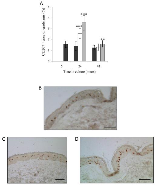
Virus-like particles (VLPs) have a number of features that make them attractive influenza vaccine candidates. Microneedle (MN) devices are being developed for the convenient and pain-free delivery of vaccines across the skin barrier layer. Whilst MN-based vaccines have demonstrated proof-of-concept in mice, it is vital to understand how MN targeting of VLPs to the skin epidermis affects activation and migration of Langerhans cells (LCs) in the real human skin environment. MNs coated with vaccine reproducibly penetrated freshly excised human skin, depositing 80% of the coating within 60 s of insertion. Human skin experiments showed that H1 (A/PR/8/34) and H5 (A/Viet Nam/1203/04) VLPs, delivered via MN, stimulated LCs resulting in changes in cell morphology and a reduction in cell number in epidermal sheets. LC response was significantly more pronounced in skin treated with H1 VLPs, compared with H5 VLPs. Our data provides strong evidence that MN-facilitated delivery of influenza VLP vaccines initiates a stimulatory response in LCs in human skin. The results support and validate animal data, suggesting that dendritic cells (DCs) targeted through deposition of the vaccine in skin generate immune response. The study also demonstrates the value of using human skin alongside animal studies for preclinical testing of intra-dermal (ID) vaccines.
Copyright 2010 Elsevier Ltd. All rights reserved.
Figures

Similar articles
-
Microneedle delivery of H5N1 influenza virus-like particles to the skin induces long-lasting B- and T-cell responses in mice.Clin Vaccine Immunol. 2010 Sep;17(9):1381-9. doi: 10.1128/CVI.00100-10. Epub 2010 Jul 14. Clin Vaccine Immunol. 2010. PMID: 20631330 Free PMC article.
-
Host responses in human skin after conventional intradermal injection or microneedle administration of virus-like-particle influenza vaccine.Adv Healthc Mater. 2013 Oct;2(10):1401-10. doi: 10.1002/adhm.201300006. Epub 2013 Apr 8. Adv Healthc Mater. 2013. PMID: 23564440 Free PMC article.
-
Microneedle vaccination with stabilized recombinant influenza virus hemagglutinin induces improved protective immunity.Clin Vaccine Immunol. 2011 Apr;18(4):647-54. doi: 10.1128/CVI.00435-10. Epub 2011 Feb 2. Clin Vaccine Immunol. 2011. PMID: 21288996 Free PMC article.
-
Plant-made virus-like particle vaccines bearing the hemagglutinin of either seasonal (H1) or avian (H5) influenza have distinct patterns of interaction with human immune cells in vitro.Vaccine. 2017 May 2;35(19):2592-2599. doi: 10.1016/j.vaccine.2017.03.058. Vaccine. 2017. PMID: 28389100
-
Microneedle-based vaccines.Curr Top Microbiol Immunol. 2009;333:369-93. doi: 10.1007/978-3-540-92165-3_18. Curr Top Microbiol Immunol. 2009. PMID: 19768415 Free PMC article. Review.
Cited by
-
Microneedles coated with peanut allergen enable desensitization of peanut sensitized mice.J Control Release. 2019 Nov 28;314:38-47. doi: 10.1016/j.jconrel.2019.09.022. Epub 2019 Oct 15. J Control Release. 2019. PMID: 31626861 Free PMC article.
-
Progress in developing virus-like particle influenza vaccines.Expert Rev Vaccines. 2016 Oct;15(10):1281-93. doi: 10.1080/14760584.2016.1175942. Epub 2016 May 5. Expert Rev Vaccines. 2016. PMID: 27058302 Free PMC article. Review.
-
Micro Electromechanical Systems (MEMS) Based Microfluidic Devices for Biomedical Applications.Int J Mol Sci. 2011;12(6):3648-704. doi: 10.3390/ijms12063648. Epub 2011 Jun 7. Int J Mol Sci. 2011. PMID: 21747700 Free PMC article. Review.
-
Polymeric microneedles for transdermal protein delivery.Adv Drug Deliv Rev. 2018 Mar 1;127:106-118. doi: 10.1016/j.addr.2018.01.015. Epub 2018 Jan 31. Adv Drug Deliv Rev. 2018. PMID: 29408182 Free PMC article. Review.
-
Transdermal delivery of PeptiCRAd cancer vaccine using microneedle patches.Bioact Mater. 2024 Nov 19;45:115-127. doi: 10.1016/j.bioactmat.2024.11.006. eCollection 2025 Mar. Bioact Mater. 2024. PMID: 39639878 Free PMC article.
References
-
- Fiore AE, Shay DK, Broder K, Iskander JK, Uyeki TM, Mootrey G, Bresee JS, Cox NS, Centers for Disease Control and Prevention (CDC) Advisory Committee on Immunization Practices (ACIP) Prevention and control of influenza: recommendations of the Advisory Committee on Immunization Practices (ACIP) MMWR Recomm Rep. 2008;57:1–60. 2008. - PubMed
-
- Capua I, Alexander DJ. Ecology, epidemiology and human health implications of avian influenza viruses: why do we need to share genetic data? Zoonoses Public Health. 2008;55(1):2–15. - PubMed
Publication types
MeSH terms
Substances
Grants and funding
LinkOut - more resources
Full Text Sources
Other Literature Sources
Medical
Research Materials
