. 2010 Oct 22;285(43):33475-33484.
doi: 10.1074/jbc.M110.156240. Epub 2010 Aug 3.

A specific docking site for DNA polymerase {alpha}-primase on the SV40 helicase is required for viral primosome activity, but helicase activity is dispensable

Affiliations

A specific docking site for DNA polymerase {alpha}-primase on the SV40 helicase is required for viral primosome activity, but helicase activity is dispensable

Hao Huang et al. J Biol Chem. .

Abstract

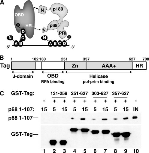
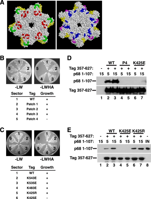
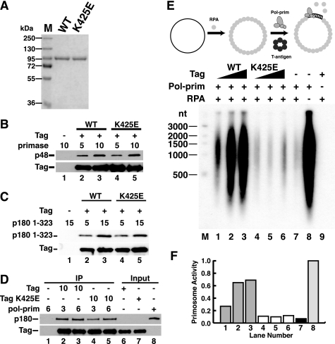
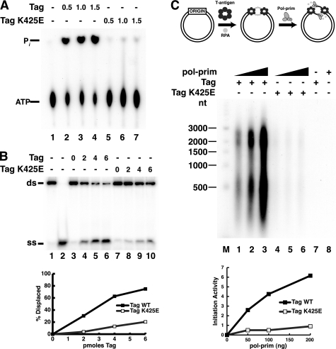
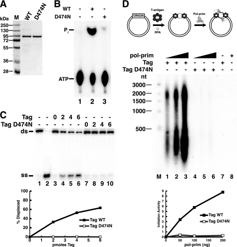
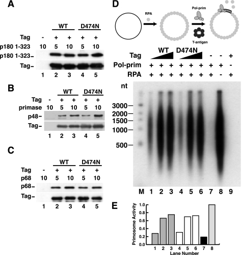
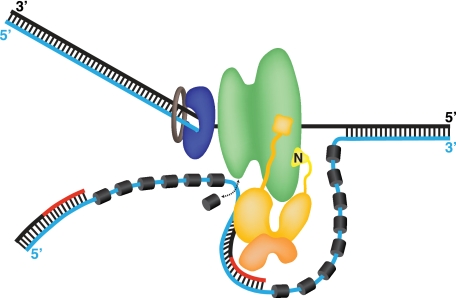
Replication of simian virus 40 (SV40) DNA, a model for eukaryotic chromosomal replication, can be reconstituted in vitro using the viral helicase (large tumor antigen, or Tag) and purified human proteins. Tag interacts physically with two cellular proteins, replication protein A and DNA polymerase α-primase (pol-prim), constituting the viral primosome. Like the well characterized primosomes of phages T7 and T4, this trio of proteins coordinates parental DNA unwinding with primer synthesis to initiate the leading strand at the viral origin and each Okazaki fragment on the lagging strand template. We recently determined the structure of a previously unrecognized pol-prim domain (p68N) that docks on Tag, identified the p68N surface that contacts Tag, and demonstrated its vital role in primosome function. Here, we identify the p68N-docking site on Tag by using structure-guided mutagenesis of the Tag helicase surface. A charge reverse substitution in Tag disrupted both p68N-binding and primosome activity but did not affect docking with other pol-prim subunits. Unexpectedly, the substitution also disrupted Tag ATPase and helicase activity, suggesting a potential link between p68N docking and ATPase activity. To assess this possibility, we examined the primosome activity of Tag with a single residue substitution in the Walker B motif. Although this substitution abolished ATPase and helicase activity as expected, it did not reduce pol-prim docking on Tag or primosome activity on single-stranded DNA, indicating that Tag ATPase is dispensable for primosome activity in vitro.


Figures

FIGURE 1.
FIGURE 1.
Tag 357–627 is sufficient to bind to pol-prim p68 1–107. A, a molecular handoff model for SV40 primosome activity on RPA-coated ssDNA. The four ssDNA-binding domains (A–D) of RPA (dark gray) occlude up to 30 nucleotides of ssDNA (straight line). Flexible linkers (wavy lines) join the N-terminal domain of RPA70 and the C-terminal domain of RPA32 to the RPA/ssDNA. Tag contacts with RPA32C and RPA70AB remodel it into a more compact, lower affinity ssDNA-binding mode and stabilize it as a ternary complex (22, 23, 35), transiently exposing the template ssDNA. pol-prim (light gray) contacts the Tag helicase domains (HEL) through p68N (36), the N terminus of p180 DNA polymerase, and unknown surfaces of primase p58/p48 (PRI) (27, 29, 30). The ensemble of these interactions is proposed to position primase on the exposed template to synthesize an RNA primer (not shown). B, domain architecture of SV40 Tag. The DnaJ chaperone domain (72), SV40 OBD (73), and helicase domain (42, 43) are depicted. The structure of the host-range (HR) domain is not known (74). C, GST-tagged Tag fragments 131–259 (lanes 2 and 3), 251–627 (lanes 4 and 5), 303–627 (lanes 6 and 7), or 357–627 (lanes 8 and 9) adsorbed to glutathione beads were incubated with increasing amounts of His-tagged p68 1–107 as indicated. Proteins bound to the beads were separated by SDS-PAGE and visualized by Western blotting with anti-His antibody (top) or anti-GST antibody (bottom). Glutathione beads lacking GST-Tag protein (lane 1) are shown as negative control. Lane 10 shows 200 ng of input p68 1–107.
FIGURE 2.
FIGURE 2.
Structure-guided mutagenesis of Tag surface residues to map the p68N-docking site. A, diagram of conserved patches of charged surface residues of the Tag hexamer (residues 266–627) (45) (reprinted with permission). In the left view, the six zinc subdomains face the reader; the right view is rotated 180° so that the AAA+ subdomains face the reader. Green, patch 1; yellow, patch 2; red, patch 3; blue, patch 4; magenta, patch 5. Patch mutants (B) and single residue substitutions of pGADT7-fused Tag (C) were screened in yeast two-hybrid assays for interaction with pGBKT7-fused p68 1–107. The numbered sectors are identified in the tables below. Left panel, control plate -Leu -Trp; right panel, selective plate -Leu -Trp -His -Ade. D, glutathione beads alone (lane 1) or adsorbed to WT (lanes 2 and 3), patch 4 (P4) mutant (lanes 4 and 5), or K425E GST-Tag 357–627 (lanes 6 and 7) were incubated with 5 or 15 μg of His-tagged p68 1–107 as indicated. Bound proteins were analyzed by Western blotting with anti-His (top) or anti-GST antibody (bottom). E, glutathione beads alone (lane 1) or adsorbed to GST-Tag 357–627 WT (lanes 2 and 3), K425E (lanes 3 and 4), or K425R (lanes 6 and 7) were incubated with 5 or 15 μg of p68 1–107 as indicated. Bound proteins were analyzed by Western blotting with anti-His (top) or anti-GST antibody (bottom). Lane 8 shows 200 ng of input p68 1–107.
FIGURE 3.
FIGURE 3.
K425E Tag binds to primase and p180 pol-prim but lacks primosome activity. A, purified WT and K425E Tag were separated by SDS-PAGE and stained with Coomassie Brilliant Blue. M, protein size markers. B and C, Pab101 beads alone (lane 1) or bound to WT (lanes 2 and 3) or K425E Tag (lanes 4 and 5) were incubated with increasing amounts of primase dimer (B) or His-p180 1–323 (C) as indicated. Bound proteins were detected by SDS-PAGE and immunoblotting with anti-p48, anti-His (catalog no. A00186, Genscript), or Pab101 against Tag. D, anti-Tag beads alone (lane 1) or adsorbed to 10 μg of WT (lanes 2 and 3) or K425E Tag (lanes 4 and 5) were incubated with 3 or 6 μg of pol-prim as indicated. Bound proteins were analyzed by Western blotting with anti-Tag and anti-p180 antibody as indicated. Input, 100 ng. E and F, primosome activity of 200, 400, or 600 ng of Tag WT (lanes 1–3) or K425E (lanes 4–6) was assayed on 100 ng of ssDNA precoated with 1 μg of RPA in the presence of 600 ng of pol-prim. Control reactions lacked Tag (lane 7), Tag and RPA (lane 8), or pol-prim (lane 9). Reaction products were analyzed by alkaline electrophoresis and visualized by autoradiography (E). DNA size markers are shown (M). F, reaction products were quantified; signal in the negative control reaction (lane 9) was subtracted from that in lanes 1–8. Incorporation in lanes 1–7 is expressed as a fraction of that in lane 8.
FIGURE 4.
FIGURE 4.
K425E Tag is defective in ATPase activity and initiation of SV40 replication. A, ATPase reactions were carried out without Tag (lane 1) or with increasing amounts of Tag WT (lanes 2–4) or K425E (lanes 5–7) as indicated. After 10 min, the reaction products were separated by ascending thin layer chromatography and visualized by autoradiography. B, to assess helicase activity, 10 fmol of DNA substrate was incubated without Tag (lanes 3 and 7) or with increasing amounts (2, 4, or 6 pmol) of WT (lanes 4–6) or K425E (lanes 8–10) Tag. Lanes 1 and 2 contain DNA substrate or boiled substrate alone. C, SV40 initiation activity of Tag WT (lanes 1–3) or K425E (lanes 4–6) (600 ng) was assayed in monopolymerase reactions with 50, 100, or 200 ng of pol-prim. Radiolabeled DNA products were visualized by alkaline agarose electrophoresis and autoradiography. Products from control reactions without pol-prim (lane 7) or Tag (lane 8) are shown as indicated (−). End-labeled DNA fragments of the indicated sizes are shown at the left (M). Reaction products were quantified; background (lanes 7 and 8) was subtracted from incorporation in lanes 1–6 (lower panel). ss, single strand; ds, double strand.
FIGURE 5.
FIGURE 5.
A single residue substitution in the Walker B motif of Tag abolishes ATPase/helicase activity and initiation of SV40 replication. A, purified WT and D474N Tag were separated by SDS-PAGE and stained with Coomassie Brilliant Blue. M, protein size markers. B, ATPase reactions were carried out without Tag (lane 1) or with 1 μg of Tag WT (lane 2) or D474N (lane 3). The reaction products were separated by ascending thin layer chromatography and visualized by autoradiography. C, to assess helicase activity, 10 fmol of DNA substrate was incubated without Tag (lanes 3 and 7) or with increasing amounts (2, 4, or 6 pmol) of WT (lanes 4–6) or D474N (lanes 8–10) Tag. Lanes 1 and 2 contain DNA substrate or boiled substrate alone. D, SV40 initiation activity of 600 ng Tag WT (lanes 1–3) or D474N (lanes 4–6) was assayed in monopolymerase reactions with 50, 100, or 200 ng of pol-prim. Radiolabeled DNA products were visualized by alkaline agarose electrophoresis and autoradiography. Products of control reactions without pol-prim (lane 7) or Tag (lane 8) are shown as indicated (−). End-labeled DNA fragments of the indicated sizes are shown at the left (M). Reaction products were quantified; background (lanes 7 and 8) was subtracted from incorporation in lanes 1–6 (lower panel). ss, single strand; ds, double strand.
FIGURE 6.
FIGURE 6.
ATPase activity of Tag is not required for pol-prim binding or primosome activity on RPA-coated ssDNA. A–C, purified Tag WT or D474N bound to Pab101-coupled Sepharose beads was incubated with increasing amounts of His-p180 1–323 (A), primase dimer (B), or His-p68 (C) as indicated (in μg). Proteins bound to the beads were separated by SDS-PAGE and visualized by Western blotting with anti-His (catalog no. A00186, Genscript) for p180, anti-p48 or anti-His (catalog no. 9801, Abcam) for p68, and Pab101 against Tag. D, primosome activity of 200, 400, or 600 ng of Tag WT (lanes 1–3) or D474N (lanes 4–6) was assayed on 100 ng of ssDNA precoated with 1 μg of RPA in the presence of 600 ng of pol-prim. Control reactions lacking Tag (lane 7), Tag and RPA (lane 8), or pol-prim (lane 9) are indicated. Reaction products were analyzed by alkaline electrophoresis and autoradiography. DNA size markers are shown (M). E, reaction products were quantified; signal in the negative control reaction (lane 9) was subtracted from that in lanes 1–8. Incorporation in lanes 1–7 is expressed as a fraction of that in lane 8.
FIGURE 7.
FIGURE 7.
Speculative model of the SV40 primosome at a replication fork. A single Tag hexamer (green) tracking 3′–5′ on the leading strand template (black) is followed by DNA polymerase δ (blue)-proliferating cell nuclear antigen (brown) holoenzyme. The lagging strand template is displaced as Tag unwinds the duplex, but its point of exit from the helicase and its path after exit are controversial (17–20, 42, 43). Hence, the path depicted here is the simplest possibility. RPA (dark gray cylinders) bound to the lagging strand template (cyan line) is remodeled by specific contacts with the OBDs of the Tag hexamer into a weaker ssDNA-binding mode (22, 23, 35) that is easily displaced (arrow), exposing the template. The p180/p68 (gold) subunits of pol-prim contact the helicase domains of Tag through p68N (36) (see Figs. 1–3) and the N terminus of p180, which we propose are flexibly tethered to the pol-prim complex (27, 29, 30). The primase p58/p48 (orange) interaction surfaces with Tag are not known (30). Pol-prim correctly positioned on Tag is postulated to access the template exposed upon RPA remodeling, synthesize an RNA primer (red line), and extend it (black line), yielding an RNA-DNA primer. The subsequent proliferating cell nuclear antigen clamp loading and switch to DNA polymerase δ holoenzyme are not depicted (34, 71).

Similar articles

Cited by

References

    1. Corn J. E., Berger J. M. (2006) Nucleic Acids Res. 34, 4082–4088 - PMC - PubMed
    1. Hamdan S. M., Richardson C. C. (2009) Annu. Rev. Biochem. 78, 205–243 - PubMed
    1. Langston L. D., Indiani C., O'Donnell M. (2009) Cell Cycle 8, 2686–2691 - PMC - PubMed
    1. Marians K. J. (2008) Nat. Struct. Mol. Biol. 15, 125–127 - PubMed
    1. Copeland W. C., Wang T. S. (1993) J. Biol. Chem. 268, 11028–11040 - PubMed

Publication types

MeSH terms

LinkOut - more resources