. 2010 Oct 29;285(44):33979-91.
doi: 10.1074/jbc.M109.095968. Epub 2010 Aug 4.

Ras homolog enriched in brain (Rheb) enhances apoptotic signaling

Affiliations

Ras homolog enriched in brain (Rheb) enhances apoptotic signaling

Sascha Karassek et al. J Biol Chem. .

Abstract

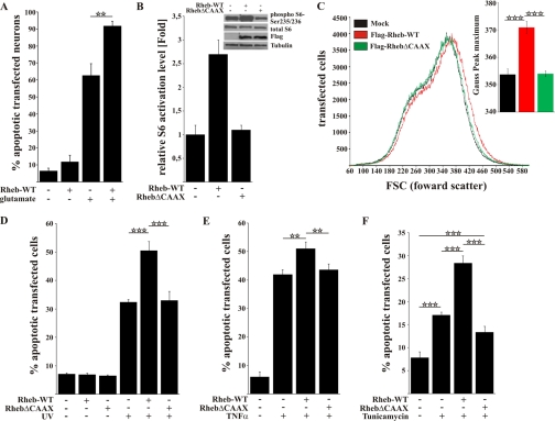
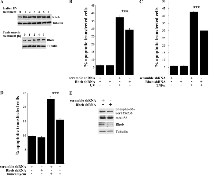
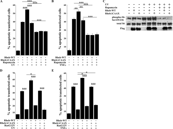
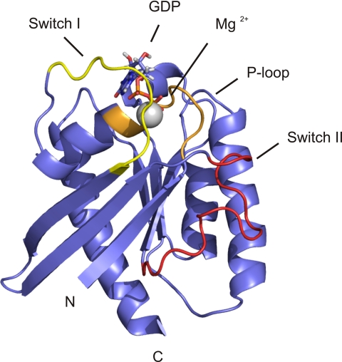
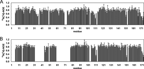
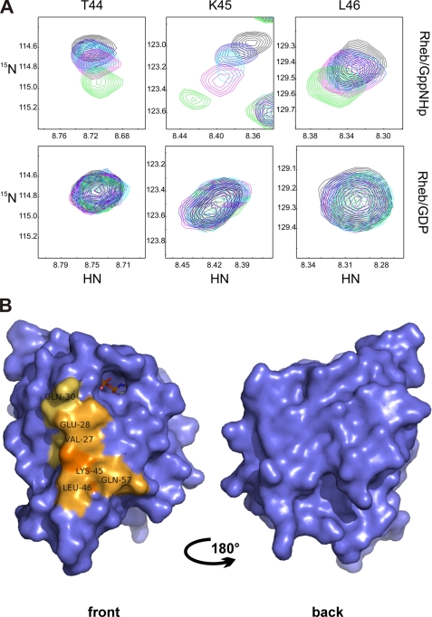
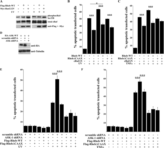
Rheb is a homolog of Ras GTPase that regulates cell growth, proliferation, and regeneration via mammalian target of rapamycin (mTOR). Because of the well established potential of activated Ras to promote survival, we sought to investigate the ability of Rheb signaling to phenocopy Ras. We found that overexpression of lipid-anchored Rheb enhanced the apoptotic effects induced by UV light, TNFα, or tunicamycin in an mTOR complex 1 (mTORC1)-dependent manner. Knocking down endogenous Rheb or applying rapamycin led to partial protection, identifying Rheb as a mediator of cell death. Ras and c-Raf kinase opposed the apoptotic effects induced by UV light or TNFα but did not prevent Rheb-mediated apoptosis. To gain structural insight into the signaling mechanisms, we determined the structure of Rheb-GDP by NMR. The complex adopts the typical canonical fold of RasGTPases and displays the characteristic GDP-dependent picosecond to nanosecond backbone dynamics of the switch I and switch II regions. NMR revealed Ras effector-like binding of activated Rheb to the c-Raf-Ras-binding domain (RBD), but the affinity was 1000-fold lower than the Ras/RBD interaction, suggesting a lack of functional interaction. shRNA-mediated knockdown of apoptosis signal-regulating kinase 1 (ASK-1) strongly reduced UV or TNFα-induced apoptosis and suppressed enhancement by Rheb overexpression. In conclusion, Rheb-mTOR activation not only promotes normal cell growth but also enhances apoptosis in response to diverse toxic stimuli via an ASK-1-mediated mechanism. Pharmacological regulation of the Rheb/mTORC1 pathway using rapamycin should take the presence of cellular stress into consideration, as this may have clinical implications.


Figures

FIGURE 1.
FIGURE 1.
Rheb overexpression enhances the rate of cell death in a CAAX-box-dependent manner after toxic treatment. A, primary cortical neurons transfected with FLAG-Rheb-WT exhibited an increased rate of cell death 24 h after excitotoxic treatment with 1 mm glutamate compared with mock-transfected cells. B, in Rheb-WT- but not RhebΔCAAX-transfected HeLa cells, the mTOR pathway was strongly activated as shown by the activation level of the S6 protein on Western blot. C, the size of Rheb-WT-transfected HeLa cells was increased compared with RhebΔCAAX or mock-transfected HeLa cells. D, HeLa cells transfected with FLAG-Rheb-WT were more sensitive to UV light as measured by quantification of apoptotic cells 4 h after UV light treatment (0.03 J/cm2). Notably, the basal level of apoptotic cells did not change upon transfection with FLAG-Rheb-WT or FLAG-RhebΔCAAX. E, HeLa cells transfected with Rheb-WT had significantly higher rates of cell death after TNFα (10 ng/ml + 2.5 μg/ml cycloheximide) treatment compared with RhebΔCAAX or mock-transfected cells. F, ER stress-induced apoptosis in the presence of tunicamycin was increased in Rheb-WT-transfected cells but not in RhebΔCAAX-transfected cells. All bars represent mean ± S.E. of at least six independent experiments. **, p < 0.01; ***; p < 0.001 (determined by ANOVA followed by a Bonferroni post hoc test).
FIGURE 2.
FIGURE 2.
Knocking down Rheb decreases cell death after toxic treatment. A, Western blot showing that endogenous Rheb expression increased after exposure to UV light or tunicamycin. B–D, HeLa cells were transfected with either a non-silencing scramble shRNA or human-specific Rheb shRNA and incubated for 96 h. The shRNA-mediated knockdown of endogenous Rheb significantly protected against UV light (B), TNFα (C), and tunicamycin-induced apoptosis (D). E, the efficiency of knocking down Rheb and inhibiting mTOR activation was tested by Western blot 96 h after transfection showing a moderate inhibition of S6 phosphorylation. All bars represent mean ± S.E. of at least six independent experiments. ***, p < 0.001 (determined using ANOVA followed by a Bonferroni post hoc test).
FIGURE 3.
FIGURE 3.
Rheb-enhanced apoptosis after toxic treatment is mTOR-dependent. A and B, pretreatment (2 h) of transfected HeLa cells with rapamycin (20 nm) inhibited Rheb-enhanced apoptosis after UV light (A) or TNFα treatment (B). C, Western blot showing a strong induction of the mTOR pathway 4 h after stimulation with UV light in mock-transfected cells, which was comparable with the activation levels of Rheb-WT-transfected cells. Pretreatment with rapamycin blocked activation of the mTOR pathway. D and E, co-expression of permanently activated RasG12V almost completely protects HeLa cells against UV light as well as TNFα-induced cell death but cannot inhibit Rheb-enhanced cell death. All bars represent the mean ± S.E. of at least six independent experiments. * p < 0.05; **, p < 0.01; ***, p < 0.001 (determined using ANOVA followed by a Bonferroni post hoc test).
FIGURE 4.
FIGURE 4.
Structure-based sequence alignment of Rheb from different species, as well as Ras and Rap1A from the rat, and stereoview of the backbone atoms (N, Cα, C′, and O) of the ensemble 〈〈SA〉〉 of 20 rRheb structures. A, invariant residues are highlighted in red-shaded boxes, and open red boxes indicate conserved residues. The secondary structural elements of Rheb are given above the sequence alignment. Residues in blue boxes are discussed in detail throughout the text. B, this superposition shows the lowest root mean square deviation values for the backbone atoms (N, Cα, and C′) of the regions with a regular secondary structure. The unstructured N-terminal residues Ser-1 to Lys-5 and C-terminal residues Ser-175 to Met-184 have been omitted for clarity.
FIGURE 5.
FIGURE 5.
Ribbon drawing of a representative member of the ensemble structures of rRheb. The N and C termini are indicated. The unstructured N-terminal residues Ser-1 to Lys-5 and C-terminal residues Ser-175 to Met-184 have been omitted for clarity. This figure was generated using PyMol (83, 84).
FIGURE 6.
FIGURE 6.
Steady state heteronuclear NOE for the backbone amides of rRheb in its GDP- (A) and Gpp(NH)p-bound (B) states. Residues for which no results are shown correspond to either prolines or residues for which relaxation data could not be extracted. The switch I region extends from residue Asp-33 to Asn-41 and the switch II region from residue Gly-63 to Asn-79. For details, refer to the text under “Experimental Procedures” and “Results.”
FIGURE 7.
FIGURE 7.
Chemical shift differences between rRheb bound to GDP or Gpp(NH)p upon titration with the RBD of c-Raf kinase. A, the upper panel shows representative regions of the 1H-15N HSQC spectra for Thr-44, Lys-45, and Leu-46 of rRheb-GppNHP titrated with c-Raf-RBD (molar ratio of rRheb/c-Raf-RBD ranging from 1:0 (shown in black) to 1:3 (shown in green)). The lower panel shows the corresponding regions of the 1H-15N-HSQC spectra of Rheb-GDP titrated with c-Raf-RBD (molar ration of rRheb/c-Raf-RBD ranging from 1:0 (shown in black) to 1:6 (shown in green)). B, significant chemical shift pertubation for Rheb bound to Gpp(NH)p upon titration with the RBD of c-Raf kinase projected onto the accessible surface of rRheb. The differences are represented in light to dark orange depending on the magnitude of the observed weighted chemical shift differences. For details, refer to the text under “Experimental Procedures” and “Results.” This figure was generated using PyMol (83, 84).
FIGURE 8.
FIGURE 8.
Active c-Raf does not inhibit Rheb-enhanced apoptosis. A, Western blot showing that, in transfected HeLa cells, Rheb expression does not influence the activation level of endogenous c-Raf as indicated by no differences at the Ser-338 phosphorylation site of c-Raf. B and C, co-expression of a constitutively active c-Raf mutant significantly protects against UV light and TNFα-induced apoptosis but is not able to completely inhibit Rheb-enhanced apoptosis. D, representative Western blot illustrating the efficiency of ASK-1 knockdown. E and F, HeLa cells were transfected with scramble or human ASK-1 shRNA plus FLAG-Rheb-WT or FLAG-RhebΔCAAX and incubated for 96 h. ASK-1 knockdown led to a general decrease in cell death and inhibited Rheb-enhanced apoptosis after UV irradiation as well as TNFα stimulation, as shown by a lack of significant differences in cell death. All bars represent mean ± S.E. of at least six independent experiments. *, p < 0.05; ***, p < 0.001 (determined using ANOVA followed by a Bonferroni post hoc test).

References

    1. Reuther G. W., Der C. J. (2000) Curr. Opin. Cell Biol. 12, 157–165 - PubMed
    1. Aspuria P. J., Tamanoi F. (2004) Cell. Signal. 16, 1105–1112 - PubMed
    1. Manning B. D., Cantley L. C. (2003) Trends Biochem. Sci. 28, 573–576 - PubMed
    1. Park K. K., Liu K., Hu Y., Smith P. D., Wang C., Cai B., Xu B., Connolly L., Kramvis I., Sahin M., He Z. (2008) Science 322, 963–966 - PMC - PubMed
    1. Garami A., Zwartkruis F. J., Nobukuni T., Joaquin M., Roccio M., Stocker H., Kozma S. C., Hafen E., Bos J. L., Thomas G. (2003) Mol. Cell 11, 1457–1466 - PubMed

Publication types

MeSH terms

Associated data