. 2010 Oct 15;285(42):32012-9.
doi: 10.1074/jbc.M110.127183. Epub 2010 Aug 4.

Hyaluronan synthesis mediates the fibrotic response of keratocytes to transforming growth factor beta

Affiliations

Hyaluronan synthesis mediates the fibrotic response of keratocytes to transforming growth factor beta

Naxin Guo et al. J Biol Chem. .

Abstract

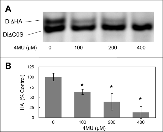
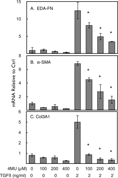
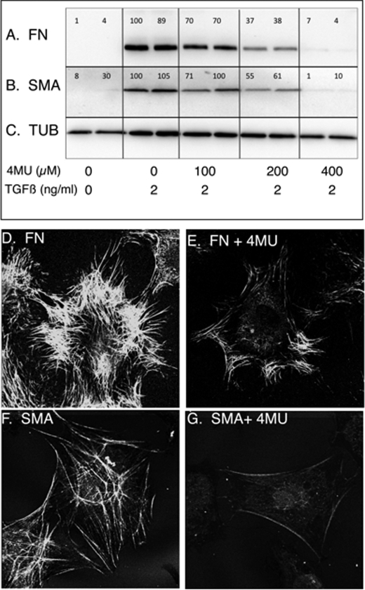
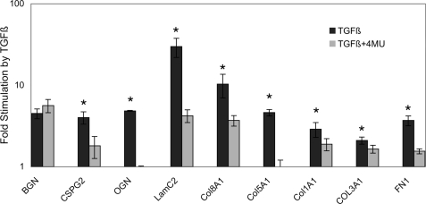
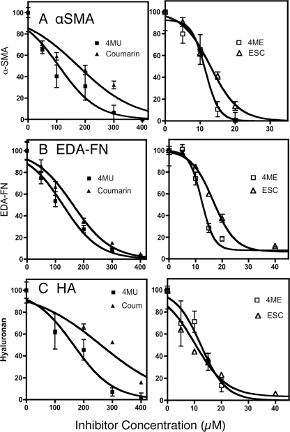
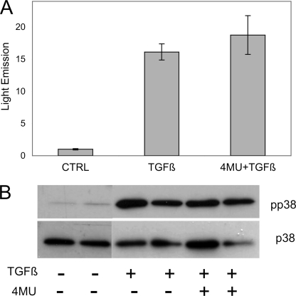
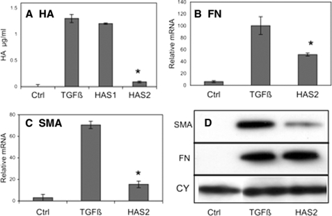
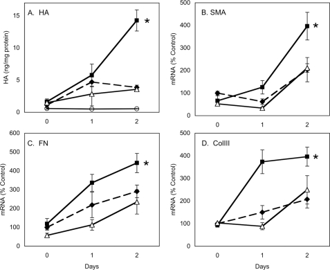
TGFβ induces fibrosis in healing corneal wounds, and in vitro corneal keratocytes up-regulate expression of several fibrosis-related genes in response to TGFβ. Hyaluronan (HA) accumulates in healing corneas, and HA synthesis is induced by TGFβ by up-regulation of HA synthase 2. This study tested the hypothesis that HA acts as an extracellular messenger, enhancing specific fibrotic responses of keratocytes to TGFβ. HA synthesis inhibitor 4-methylumbelliferone (4MU) blocked TGFβ induction of HA synthesis in a concentration-dependent manner. 4MU also inhibited TGFβ-induced up-regulation of α-smooth muscle actin, collagen type III, and extra domain A-fibronectin. Chemical analogs of 4MU also inhibited fibrogenic responses in proportion to their inhibition of HA synthesis. 4MU, however, showed no effect on TGFβ induction of luciferase by the 3TP-Lux reporter plasmid. Inhibition of HA using siRNA to HA synthase 2 reduced TGFβ up-regulation of smooth muscle actin, fibronectin, and cell division. Similarly, brief treatment of keratocytes with hyaluronidase reduced TGFβ responses. These results suggest that newly synthesized cell-associated HA acts as an extracellular enhancer of wound healing and fibrosis in keratocytes by augmenting a limited subset of the cellular responses to TGFβ.


Figures

FIGURE 1.
FIGURE 1.
4MU inhibits secretion of hyaluronan by keratocytes. Primary bovine keratocytes were cultured 4 days in medium containing 1% platelet-poor horse serum and 2 ng/ml TGβ1 in the presence of different concentrations of 4MU. A, HA and chondroitin sulfate from the culture medium from equal numbers of cells were detected by FACE as described under “Materials and Methods.” The HA disaccharide (DiΔHA) was decreased by 4MU but not chondroitin sulfate (DiΔC0S). B, quantification of HA bands from gels similar to that shown in A. The bars show S.D. of triplicate samples. The asterisks indicate significant reduction (p < 0.05) compared with a sample with no 4MU using analysis of variance. No HA was detected in the absence of TGFβ (not shown).
FIGURE 2.
FIGURE 2.
4MU blocks up-regulation of mRNA for fibrotic genes induced by TGFβ. Primary bovine keratocytes were untreated (first four samples) or treated with 2 ng/ml TGFβ in 1% platelet-poor horse serum for 4 days, and mRNA levels were determined by quantitative reverse transcriptase PCR as described under “Materials and Methods.” 4MU was present during treatment at the concentrations noted. A, EDA fibronectin. B, αSMA. C, collagen III α1 chain. The error bars represent S.D. of triplicate analyses. The asterisks show a significant (p < 0.05) decrease in mean values of 4MU+TGFβ-treated samples compared with TGFβ-treated control (Ctrl) as determined by analysis of variance.
FIGURE 3.
FIGURE 3.
Expression and localization of EDA-fibronectin and αSMA protein in response to 4MU. Proteins induced by 4 days of TGFβ treatment were immunodetected in equal amounts of cell lysate protein after separation by SDS-PAGE and electrotransfer to membranes as described under “Materials and Methods.” Results from duplicate cultures are shown. A, EDA-FN. B, αSMA (SMA) in the same cell lysates. C, α-tubulin (TUB) loading control was detected on the same membrane as FN. The numbers above the bands represent a normalized ratio between the FN or SMA and the tubulin bands. In D and E, immunostaining of EDA-fibronectin was photographed after 4 days of TGFβ treatment in the absence (D) or presence (E) of 400 μm 4MU. F, αSMA immunostained as cytoskeletal microfibrils in TGFβ-treated keratocytes. G, little αSMA was detected in cells treated with TGFβ in the presence of 400 μm 4MU.
FIGURE 4.
FIGURE 4.
4MU inhibits TGFβ up-regulation of extracellular matrix proteins in human corneal fibroblasts. Up-regulation of mRNA abundance by 4 days of TGFβ treatment was determined by quantitative reverse transcriptase PCR in low passage human corneal fibroblasts as described under “Materials and Methods.” Expression by untreated cells was set to 1, and the black bars show the relative increase in response to TGFβ treatment (note log scale). The gray bars show the same response in the presence of 400 μm 4MU. The error bars show S.D. of triplicate assays. The differences between the gray and black bars are significant (p < 0.05, by t test) except for biglycan. BGN, biglycan; CSPG2, versican; OGN, osteoglycin; LamC2, laminin gamma 2; Col8A1, collagen type VIII; Col1A1, collagen type I; COL3A1, collagen type III; FN1, fibronectin 1.
FIGURE 5.
FIGURE 5.
Inhibition of TGFβ fibrotic response by coumarin analogs. Expression of cell-associated FN and αSMA in cultures of keratocytes treated with TGFβ for 4 days as in Fig. 3 was detected by cell-based ELISA, and HA was detected by ELISA from culture media. The values were normalized for cell number as determined by Calcein AM staining in parallel plates. Inhibition curves show relative responses compared with noninhibited samples. The error bars are S.D. of six individual sample wells. Coum, coumarin; 4ME, 4-methylesculetin; ESC, esculetin. The lines represent nonlinear curve fitting of data using statistical software as described under “Materials and Methods.”
FIGURE 6.
FIGURE 6.
4MU exerts no effect on initial TGFβ-induced signaling. A, light emission by 3TP-LUX, a luciferase reporter plasmid driven by TGFβ-responsive promoter, after 24 h TGFβ treatment stimulated light emission by ∼15-fold in primary keratocytes (p < 0.001). This reporter activity was not diminished by treatment with 500 μm 4MU. 3TP-Lux activity was normalized by co-transfection with control plasmid as described under “Materials and Methods.” The error bars show S.D. of quadruplicate analyses. The difference between TGFβ and TGFβ+4MU was not significant. B, duplicate cultures of primary keratocytes were treated for 1 h with TGFβ (as in Fig. 2) in the presence or absence of 400 μm 4MU. Equal amounts of protein from cell lysates were immunoblotted for phosphorylated p38 MAPK (pp38) using the nonphosphorylated p38 as a loading control. Coum, coumarin; 4ME, 4-methylesculetin; ESC, esculetin; CTRL, control.
FIGURE 7.
FIGURE 7.
TGFβ stimulation of protein and mRNA for fibrotic markers is reduced by knockdown of HAS2 mRNA. Primary keratocytes were transfected with siRNA for HAS2, HAS1 or vehicle only and cultured 3 days in the absence (control, Ctrl) or presence of TGFβ as described for Fig. 2. A, as previously reported, knockdown of HAS2 mRNA, but not HAS1 RNA, markedly reduced HA expression (p < 0.01). B, EDA-fibronectin mRNA (FN) up-regulation by TGFβ was significantly reduced (p < 0.05) by HAS2 siRNA compared with nontransfected controls. RNA is expressed as fold increase from that in freshly isolated cells. C, stimulation of αSMA mRNA was similarly reduced by HAS2 siRNA transfection (p < 0.01). D, immunoblotting of αSMA and EDA-fibronectin as in Fig. 3 showed a decrease in αSMA protein in response to HAS2 knockdown but no apparent change in cell-associated EDA-FN. CY is a blot for cyclophilin (loading control) on the same membrane as FN. The error bars represent S.D. of triplicate analyses. Coum, coumarin; 4ME, 4-methylesculetin; ESC, esculetin. The asterisks show significant (p < 0.05, by t test) inhibition of TGFβ effect.
FIGURE 8.
FIGURE 8.
PDGF stimulation is eliminated by hyaluronidase. Primary keratocytes were preincubated for 24 h in 10 ng/ml PDGF (solid lines) or in serum-free medium without PDGF (dashed lines). After preincubation (day 0), the cells were washed and treated for 15 min with hyaluronidase as described under “Materials and Methods” (open triangles). The cultures were thoroughly rinsed and incubated for 2 days in the presence of 10 ng/ml PDGF and 2 ng/ml TGFβ. HA was detected in culture media (A), and the relative abundance of mRNA is shown for SMA (B), EDA-FN (C), or COL3A1 (D). The values in A show HA concentration in the culture media normalized to cell protein (ng of HA/mg of protein). The cultures without PDGF and TGFβ served as controls (open circles). B–D show mRNA abundance relative to untreated control cells normalized to 100. The error bars show S.D. of n = 6 for A and n = 3 in B–D. In each case the filled squares (PDGF pretreatment) are significantly greater than the other two values at day 2 (asterisks, p < 0.05, by analysis of variance).

Similar articles

Cited by

References

    1. Fini M. E. (1999) Prog. Retin. Eye Res. 18, 529–551 - PubMed
    1. Funderburgh J. L., Funderburgh M. L., Mann M. M., Corpuz L., Roth M. R. (2001) J. Biol. Chem. 276, 44173–44178 - PMC - PubMed
    1. Funderburgh J. L., Mann M. M., Funderburgh M. L. (2003) J. Biol. Chem. 278, 45629–45637 - PMC - PubMed
    1. Berryhill B. L., Kane B., Stramer B. M., Fini M. E., Hassell J. R. (2003) Exp. Eye Res. 77, 85–92 - PubMed
    1. Cintron C., Hong B. S., Covington H. I., Macarak E. J. (1988) Invest. Ophthalmol. Vis. Sci. 29, 767–775 - PubMed

Publication types

MeSH terms

LinkOut - more resources