

Differential expression profiling analyses identifies downregulation of 1p, 6q, and 14q genes and overexpression of 6p histone cluster 1 genes as markers of recurrence in meningiomas
- PMID: 20685720
- PMCID: PMC3018937
- DOI: 10.1093/neuonc/noq081
Differential expression profiling analyses identifies downregulation of 1p, 6q, and 14q genes and overexpression of 6p histone cluster 1 genes as markers of recurrence in meningiomas
Abstract
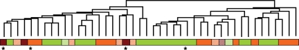
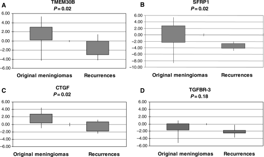
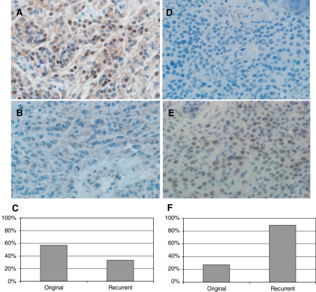
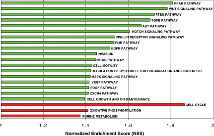
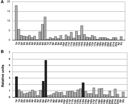
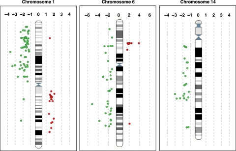
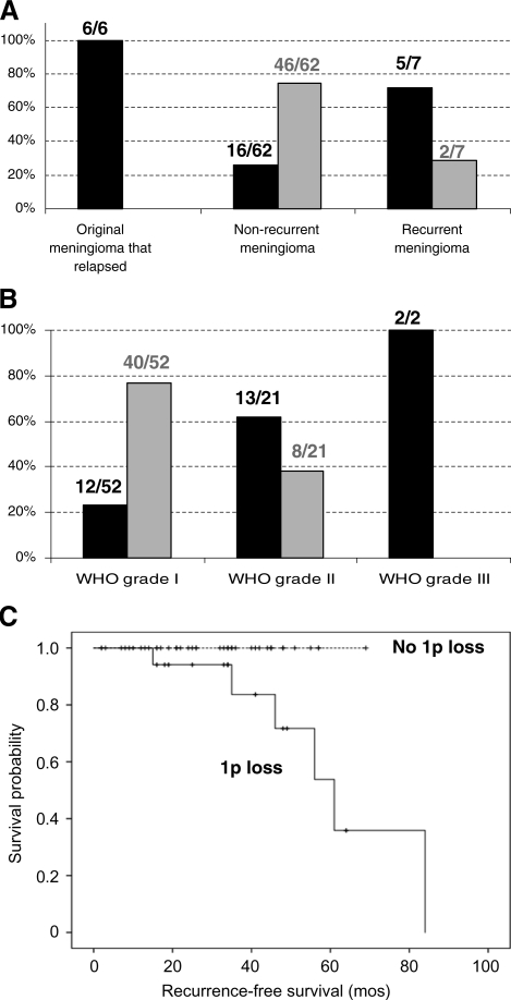
The majority of meningiomas are probably benign but a number of tumors display considerable histological and/or clinical aggressivity, sometimes with unexpectedly high recurrence rates after radical removal. Understanding the potential behavior of these tumors in individual patients is critical for rational therapeutic decision-making. This study aimed to identify gene expression profiles and candidate markers associated with original and recurrent meningiomas. Unsupervised hierarchical clustering of the samples confirmed 2 main groups of meningiomas with distinct clinical behaviors. The gene expression profiling study identified genes and pathways potentially associated with meningioma recurrence, revealing an overall lower level of gene expression. The differential gene expression profiling analyses of original and recurrent meningiomas identified 425 known genes and expressed sequence tags related to meningioma recurrence, with SFRP1 (8p12), TMEM30B (14q23), and CTGF (6q23) showing the most disparate expression. Most of the differentially expressed genes were located at 1p, 6q, and 14q and were underexpressed in recurrences. Loss of such chromosomal regions has previously been associated with a higher risk of meningioma recurrence or malignant progression. Thus, at these locations, we propose the existence of novel candidate genes that could be involved in meningioma recurrence. In addition, the overexpression of genes of histone cluster 1 (6p) in recurrent meningiomas is reported here for the first time. Finally, the altered genes related to meningioma recurrence are involved in pathways such as Notch, TGFβ, and Wnt, as described previously, and in other pathways such as cell cycle, oxidative phosphorylation, PPAR, and PDGF, not related before to meningioma recurrence.
Figures
Similar articles
-
Genetic changes with prognostic value in histologically benign meningiomas.Clin Neuropathol. 2013 Jul-Aug;32(4):311-7. doi: 10.5414/NP300580. Clin Neuropathol. 2013. PMID: 23442303
-
Chromosome 1p and 14q FISH analysis in clinicopathologic subsets of meningioma: diagnostic and prognostic implications.J Neuropathol Exp Neurol. 2001 Jun;60(6):628-36. doi: 10.1093/jnen/60.6.628. J Neuropathol Exp Neurol. 2001. PMID: 11398839
-
Evaluation of 1p and 14q status, MIB-1 labeling index and progesterone receptor immunoexpression in meningiomas: Adjuncts to histopathological grading and predictors of aggressive behavior.Neurol India. 2014 Jul-Aug;62(4):376-82. doi: 10.4103/0028-3886.141248. Neurol India. 2014. PMID: 25237942
-
Recurrence-associated chromosomal anomalies in meningiomas: Single-institution study and a systematic review with meta-analysis.Neurol Neurochir Pol. 2016 Nov-Dec;50(6):439-448. doi: 10.1016/j.pjnns.2016.08.003. Epub 2016 Aug 20. Neurol Neurochir Pol. 2016. PMID: 27575681
-
Two cases of atypical meningioma with pulmonary metastases: a comparative cytogenetic analysis of chromosomes 1p and 22 and a review of the literature.Neuropathology. 2015 Apr;35(2):175-83. doi: 10.1111/neup.12177. Epub 2014 Nov 6. Neuropathology. 2015. PMID: 25376227 Review.
Cited by
-
Genetic profiling by single-nucleotide polymorphism-based array analysis defines three distinct subtypes of orbital meningioma.Brain Pathol. 2015 Mar;25(2):193-201. doi: 10.1111/bpa.12150. Epub 2014 May 21. Brain Pathol. 2015. PMID: 24773246 Free PMC article.
-
Meningiomas and Proteomics: Focus on New Potential Biomarkers and Molecular Pathways.Cancer Genomics Proteomics. 2016 09-10;13(5):369-79. Cancer Genomics Proteomics. 2016. PMID: 27566655 Free PMC article. Review.
-
The immunohistochemical expression of SSTR2A is an independent prognostic factor in meningioma.Neurosurg Rev. 2022 Aug;45(4):2671-2679. doi: 10.1007/s10143-021-01651-w. Epub 2021 Oct 2. Neurosurg Rev. 2022. PMID: 34601710 Free PMC article.
-
Transcriptomic analysis of aggressive meningiomas identifies PTTG1 and LEPR as prognostic biomarkers independent of WHO grade.Oncotarget. 2016 Mar 22;7(12):14551-68. doi: 10.18632/oncotarget.7396. Oncotarget. 2016. PMID: 26894859 Free PMC article.
-
Microarray Expression Data Identify DCC as a Candidate Gene for Early Meningioma Progression.PLoS One. 2016 Apr 20;11(4):e0153681. doi: 10.1371/journal.pone.0153681. eCollection 2016. PLoS One. 2016. PMID: 27096627 Free PMC article.
References
-
- Yee G, Rycroft R, Philips C, et al. Hinsdale, IL: CBTRUS; 2009. CBTRUS Statistical Report: Primary Brain and Central Nervous System Tumors Diagnosed in Eighteen States in 2002–2006.
-
- Rohringer M, Sutherland GR, Louw DF, Sima AA. Incidence and clinicopathological features of meningioma. J Neurosurg. 1989;71:665–672. doi:10.3171/jns.1989.71.5.0665. - DOI - PubMed
-
- Perry A, Louis D, Scheithauer B, Budka H, von Deimling A. World Health Organization Classification of Tumours. Lyon: IARC Press; 2007.
-
- Maillo A, Orfao A, Espinosa A, et al. Early recurrences in histologically benign/grade I meningiomas are associated with large tumors and coexistence of monosomy 14 and del(1p36) in the ancestral tumor cell clone. Neurooncology. 2007;9:438–446. doi:10.1215/15228517-2007-026. - DOI - PMC - PubMed
-
- Mirimanoff RO, Dosoretz DE, Linggood RM, Ojemann RG, Martuza RL. Meningioma: analysis of recurrence and progression following neurosurgical resection. J Neurosurg. 1985;62:18–24. doi:10.3171/jns.1985.62.1.0018. - DOI - PubMed
Publication types
MeSH terms
Substances
LinkOut - more resources
Full Text Sources
Molecular Biology Databases
Miscellaneous
