. 2010 Oct;151(10):4882-93.
doi: 10.1210/en.2009-1483. Epub 2010 Aug 4.

Modulation of gonadotropin-releasing hormone-induced extracellular signal-regulated kinase activation by dual-specificity protein phosphatase 1 in LbetaT2 gonadotropes

Affiliations

Modulation of gonadotropin-releasing hormone-induced extracellular signal-regulated kinase activation by dual-specificity protein phosphatase 1 in LbetaT2 gonadotropes

Kathryn A Nguyen et al. Endocrinology. 2010 Oct.

Abstract

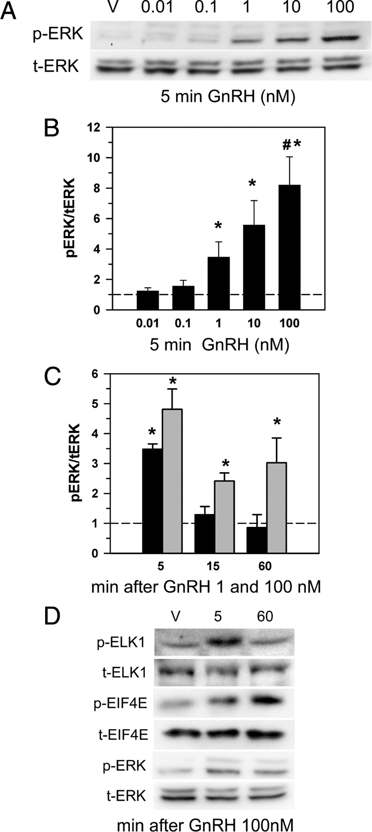
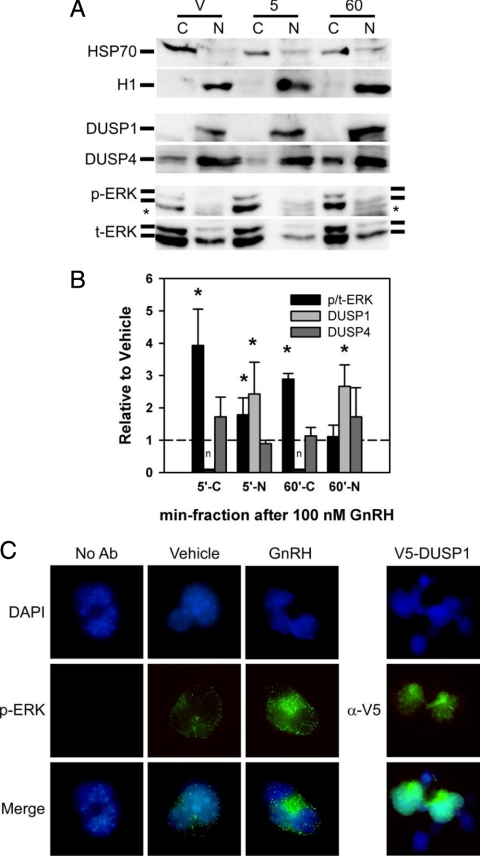
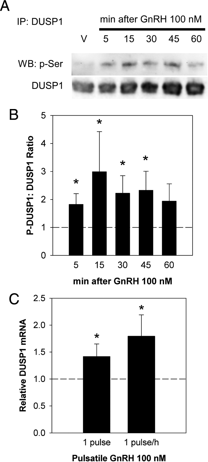
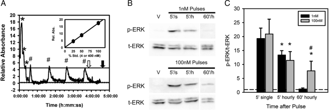
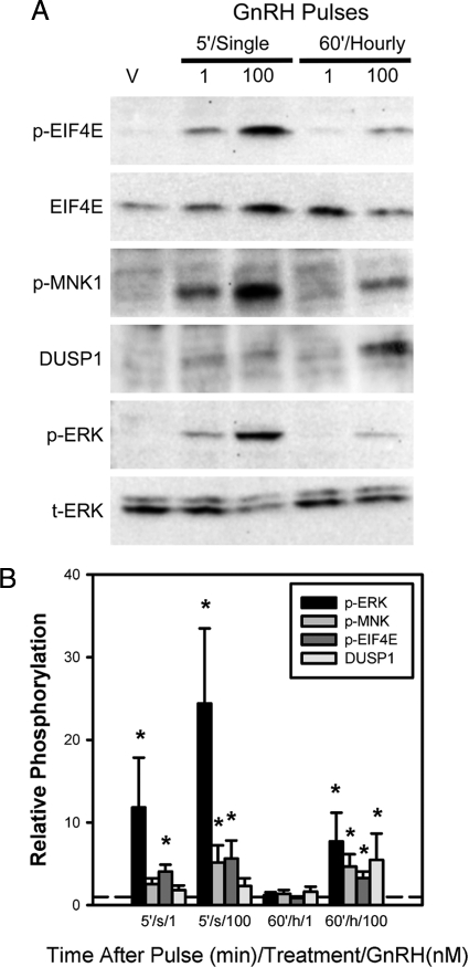
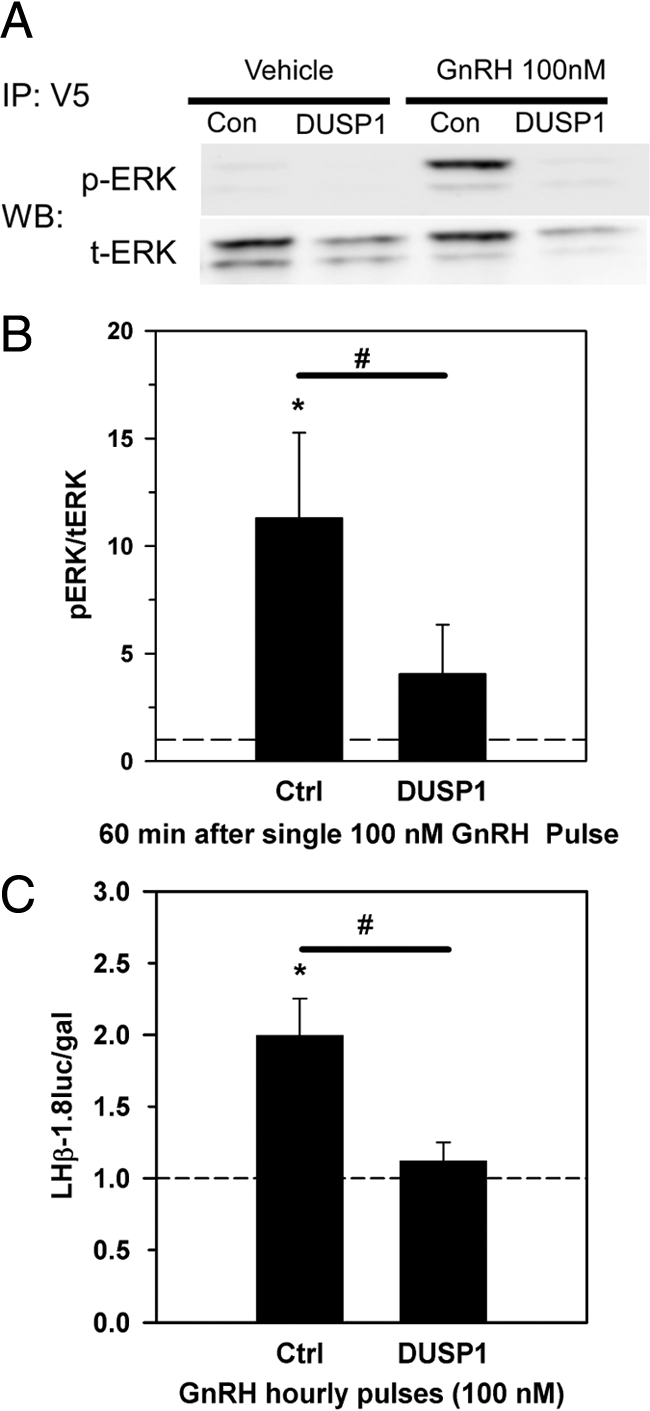
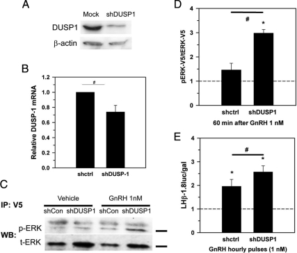
As the regulator of pituitary reproductive hormone synthesis, the hypothalamic neuropeptide GnRH is the central regulator of reproduction. A hallmark of GnRH action is the differential control of gene expression in pituitary gonadotropes through varied pulsatile stimulation. Among other signaling events, GnRH activation of the ERK family of MAPKs plays a significant role in the transcriptional regulation of the luteinizing hormone β-subunit gene and regulation of cap-dependent translation. We evaluated the ERK response to different GnRH pulse amplitudes in the gonadotrope cell line LβT2. We found that low-amplitude stimulation with GnRH invokes a rapid and transient ERK activation, whereas high-amplitude stimulation invokes a prolonged activation specifically in the cytoplasm fraction of LβT2 cells. Nuclear and cytoplasmic targets of ERK, Ets-like gene 1, and eukaryotic initiation factor 4E, respectively, are similarly activated. Feedback control of ERK activation occurs mainly through the dual-specificity protein phosphatases (DUSPs). DUSP1 is localized to the nucleus in LβT2 cells but DUSP4, another member implicated in GnRH feedback, exists in both the nucleus and cytoplasm. Manipulation of nuclear DUSP activity through overexpression or knockdown of Dusp1 modulates the ERK response to low and high GnRH pulse amplitudes and activation of the Lhb promoter. Dusp1 overexpression abolishes sustained ERK activation and inhibits Lhb promoter activity induced by high amplitude pulses. Conversely, Dusp1 knockdown enhances ERK activation by low-amplitude stimulation and increases stimulation of Lhb promoter activity. We conclude that DUSP1 feedback activity modulates ERK activation and the transcriptional response to GnRH.


Figures

Figure 1
Figure 1
Sustained ERK activation in LβT2 cells. LβT2 cells were treated with a 5-min pulse of GnRH or vehicle at indicated concentrations. A, Extracts were harvested and subjected to SDS-PAGE followed by Western blotting with phosphorylated ERK1/2 antibody. Blots were then stripped and reblotted with total ERK1/2 antibody for loading control. Phosphorylation levels were normalized to untreated control. V, Vehicle; p-ERK, phosphorylated ERK; t-ERK, total ERK. The results of three independent determinations are plotted in the histogram ± sem (B). Significant difference from vehicle as determined by Student’s t test is indicated by an asterisk. The 100-nm treatment group was also significantly greater than the 1-nm treatment group (#). C, The histogram represents the mean ratio of phospho-ERK1/2 to total ERK1/2 in LβT2 cells treated for 5 min with GnRH and harvested immediately at 5, at 15, or at 60 min after treatment with 1 nm (black bars) or 100 nm (gray bars) GnRH. Ratios were normalized to vehicle-treated control from quantitative chemiluminescent image analysis of three independent experiments ± sem The asterisk indicates a significant difference from vehicle control designated as V. D, Differential activation of ERK and ERK targets ELK1 and EIF4E. Extracts harvested immediately and 60 min after a 5-min GnRH treatment were examined for activation of ERK, ELK1, and EIF4E. p-ELK1, Phosphorylated ELK1; t-EIF4E, total EIF4E.
Figure 2
Figure 2
Sustained ERK is localized to the cytosolic fraction of LβT2 cells. A, Cytosolic and nuclear fractions were prepared from LβT2 cells stimulated with vehicle or 100 nm GnRH for 5 min, washed, and harvested at the times indicated. Nuclear and cytosolic fractions were Western blotted for HSP70 and H1 to confirm separation. Blots were further probed with antibodies for DUSP1 and DUSP4 as well as phosphorylated and total ERK. The asterisk indicates nonspecific protein detected by the p-ERK antibody. C, Cytosolic fraction; N, nuclear fraction; V, vehicle; HSP70, heat shock protein 70; H1, histone H1; p-ERK, phosphorylated ERK; t-ERK, total ERK. B, Quantitative chemiluminescence was used to compare the levels of phospho-ERK to total-ERK ratio as well as DUSP1 and DUSP4 levels relative to untreated control. The histogram represents mean values ± sem normalized to vehicle control (broken line) of four independent determinations. For ERK quantification, only bands superimposable in both p-ERK and t-ERK images were quantified to avoid inclusion of nonspecific proteins detected in nuclear fractions. The asterisk indicates a significant difference from vehicle control. n, Not detected. All values reported are means ± sem of four independent determinations. C, Immunofluorescent labeling of phospho-ERK in LβT2 cells treated with GnRH for 5 min and fixed at 60 min after treatment and in LβT2 cells transfected with V5-DUSP1. Green indicates phosphor-ERK or V5-DUSP1. Nuclei are visualized in blue using 4′,6-diamidino-2-phenylindole (DAPI). Ab, Antibody; V5-DUSP1, V5 epitope-tagged DUSP1.
Figure 3
Figure 3
DUSP1 is regulated by GnRH in LβT2 cells. A, Immunoprecipitation of DUSP1 from GnRH-treated LβT2 cells harvested at the times indicated after 5 min treatment with 100 nm GnRH. Whole-cell extract was immunoprecipitated with DUSP1 antibody and Western blotted with antiphosphoserine antibody, quantified by chemiluminescent imaging, stripped, and reblotted with a second DUSP1 antibody. IP, Immunoprecipitation; WB, Western blot; p-Ser, phosphoserine antibody. B, Quantization of chemiluminescence from immunoprecipitated DUSP1 in A showing an increase in serine phosphorylation after GnRH treatment. Asterisk indicates a significant difference from control vehicle-treated cells as represented by the reference line at 1 (broken line). C, Results from quantitative PCR of Dusp1 mRNA in LβT2 cells treated with a single pulse or one pulse per hour with GnRH at 100 nm peak amplitude in perifusion chambers and harvested after 4 h as detailed in the text. The asterisk indicates a significant difference from control vehicle-pulsed cells represented as the reference line at 1 (broken line). All values reported are means ± sem of three independent determinations.
Figure 4
Figure 4
High-amplitude but not low-amplitude GnRH pulses invoke sustained ERK activation. LβT2 cells cultured on microcarrier beads were placed into a perifusion column and subjected to four hourly pulses of GnRH at 1 or 100 nm peak pulse amplitude. A representative absorbance profile of a pulsed culture is presented in A. The inset plot is the linear fitting of calibration data of 100, 50, and 25% calibration dye peaks (asterisk) and calculated baseline. Individual pulse peaks are indicated (#). The open arrow indicates the point of harvest 5 min after the final pulse. The solid arrow indicates the time of harvest 60 min after the final pulse. B, Representative Western blot of GnRH pulsed LβT2 cells showing phosphorylation levels of ERK by 1 and 100 nm GnRH 5 min after a single pulse or 5 min after the fourth hourly pulse. The single pulse was administered coincident with the fourth pulse. The 5- and 60-min harvest points are indicated by open arrow and solid arrow on the trace in A, respectively. Cells were also harvested 60 min after the final hourly pulse and examined. V, Vehicle; p-ERK, phosphorylated ERK; t-ERK, total ERK; 5′/s, 5 min after single pulse; 5′/h, 5 min after the fourth hourly pulse; 60′/h, 60 min after the final hourly pulse. Quantization of chemiluminescence is represented in C as p-ERK to t-ERK ratio normalized to vehicle-treated controls shown as the reference line at 1 (broken line). The significant difference from the 5-min, single-pulse p-ERK to t-ERK ratio is indicated by an asterisk. The significant difference between 1 and 100 nm treatment is indicated (#). All except the 60-min hourly 1-nm GnRH pulse group were significantly increased compared with untreated values. All values reported are means ± sem of four independent determinations.
Figure 5
Figure 5
High-amplitude but not low-amplitude GnRH pulses cause sustained activation of cytoplasmic ERK targets. Perifused LβT2 cells pulsed with 1 and 100 nm GnRH were harvested 5min after a single pulse and 60 min after the final of four hourly pulses and phosphorylation levels of pEIF4E (p-EIF4E) and its upstream activating kinase MNK1 (p-MNK1) were determined by Western blot (A). Activation of DUSP1 protein expression and p-ERK to t-ERK ratio (p-ERK) were also determined. V, Vehicle; p-ERK, phosphorylated ERK; t-ERK, total ERK. Quantization of chemiluminescence from multiple trials is represented in B normalized to untreated levels represented by the reference line at 1 (broken line). The significant increase over untreated levels is indicated by an asterisk. All values reported are means ± sem of three independent determinations.
Figure 6
Figure 6
DUSP1 overexpression inhibits ERK phosphorylation and Lhb promoter activation by GnRH. LβT2 cells were cotransfected with a null or DUSP1 expression vector and a V5-epitope-tagged ERK1 cDNA expression plasmid for 48 h before stimulation with 100 nm GnRH for 5 min, washed, and incubated until 60 min after stimulation. Extracts were prepared and immunoprecipitated with anti-V5 antibody. Precipitates were Western blotted for p-ERK and t-ERK levels (A). IP, Immunoprecipitation; WB, Western blot; Con, control; p-ERK, phosphorylated ERK; t-ERK, total ERK. Quantization of phospho-ERK and ERK chemiluminescence is illustrated in B, showing significant suppression of ERK phosphorylation by 100 nm GnRH in the presence of excess DUSP1 (#). A significant difference from control p-ERK to t-ERK ratio marked by the reference line at 1 (broken line) is indicated by an asterisk. LβT2 cells cultured on microcarrier beads were cotransfected with either a null or DUSP1 expression plasmid, a firefly luciferase reporter plasmid under control of the rat 1.8 kb Lhb promoter, and a control β-galactosidase reporter plasmid under the control of the CMV promoter. Cells were placed in a perifusion column and pulsed hourly with 100 nm GnRH for 6 h. Ctrl, Control. Extracts of harvested cells were measured for luciferase and β-galactosidase activity and results are plotted in C. A significant difference in Lhb promoter activity between control (Ctrl) and DUSP1 transfected cells is indicated (#). A significant difference from control luciferase to β-galactosidase ratio marked by the reference line at 1 (broken line) is indicated by an asterisk. All values reported are means ± sem of three independent determinations.
Figure 7
Figure 7
shRNA targeting DUSP1 increases ERK phosphorylation and Lhb promoter activation by GnRH. Knockdown of DUSP1 protein (A) and mRNA (B) in LβT2 cells transfected with a shDUSP1 expression plasmid. LβT2 cells were cotransfected with a null or shDUSP1 expression plasmid and a V5-epitope-tagged ERK1 cDNA expression plasmid for 48 h before stimulation with 1 nm GnRH for 5 min, washed, and incubated until 60 min after stimulation. Extracts were prepared and immunoprecipitated with anti-V5 antibody. Precipitates were Western blotted for p-ERK and t-ERK levels as shown in a representative blot (C). Bars indicate bands identified by both p-ERK and t-ERK antibodies and used for quantification. IP, Immunoprecipitation; WB, Western blot; Con, control; p-ERK, phosphorylated ERK; t-ERK, total ERK. The GnRH-induced phospho-ERK and total ERK chemiluminescence ratio relative to vehicle treatment is illustrated in D, showing a significant increase in ERK phosphorylation by 1 nm GnRH in the presence of shDUSP1 but not shControl (#). A significant difference from the respective vehicle-treated p-ERK to t-ERK ratio, marked by the reference line at 1 (broken line), is indicated by an asterisk. The difference between shControl and shDUSP1 fold change is also indicated (#). LβT2 cells cultured on microcarrier beads were cotransfected with a null or shDUSP1 expression plasmid, a firefly luciferase reporter plasmid under control of the rat 1.8 kb Lhb promoter, and a control β-galactosidase reporter plasmid under the control of the CMV promoter. Cells were placed in a perifusion column and pulsed hourly with 1 nm GnRH for 6 h. Extracts of harvested cells were measured for luciferase and β-galactosidase activity and results are plotted in E. A significant difference in the Lhb promoter activity between control (Ctrl) and shDUSP1 transfected cells is indicated (#). A significant difference from the control luciferase to β-galactosidase ratio marked by the reference line at 1 (broken line) is indicated by an asterisk. All values reported are means ± sem of four independent determinations.

Similar articles

Cited by

References

    1. Knobil E 1974 On the control of gonadotropin secretion in the rhesus monkey. Recent Prog Horm Res 30:1–46 - PubMed
    1. Belchetz PE, Plant TM, Nakai Y, Keogh EJ, Knobil E 1978 Hypophysial responses to continuous and intermittent delivery of hypopthalamic gonadotropin-releasing hormone. Science 202:631–633 - PubMed
    1. Moenter SM, Caraty A, Locatelli A, Karsch FJ 1991 Pattern of gonadotropin-releasing hormone (GnRH) secretion leading up to ovulation in the ewe: existence of a preovulatory GnRH surge. Endocrinology 129:1175–1182 - PubMed
    1. Rippel RH, Johnson ES, White WF 1974 Effect of consecutive injections of synthetic gonadotropin releasing hormone on LH release in the anestrous and ovariectomized ewe. J Anim Sci 39:907–914 - PubMed
    1. Naor Z 2009 Signaling by G-protein-coupled receptor (GPCR): studies on the GnRH receptor. Front Neuroendocrinol 30:10–29 - PubMed

Publication types

MeSH terms

Substances