. 2010 Oct 1;21(19):3304-16.
doi: 10.1091/mbc.E10-04-0364. Epub 2010 Aug 4.

Vestigial is required during late-stage muscle differentiation in Drosophila melanogaster embryos

Affiliations

Vestigial is required during late-stage muscle differentiation in Drosophila melanogaster embryos

Hua Deng et al. Mol Biol Cell. .

Abstract

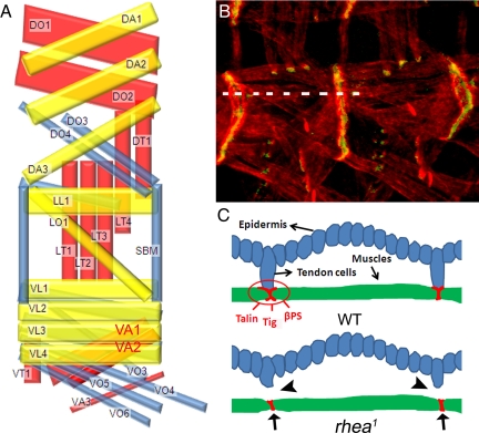
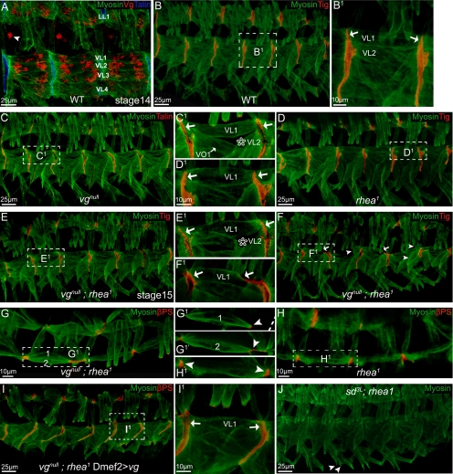
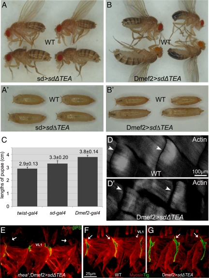
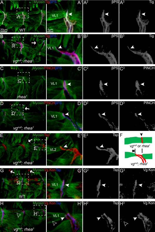
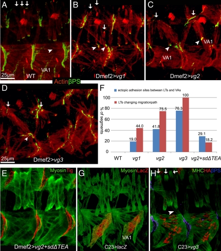
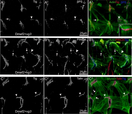
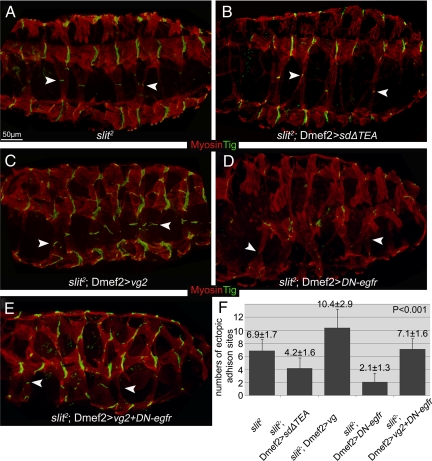
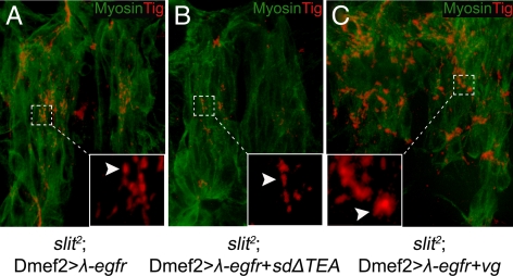
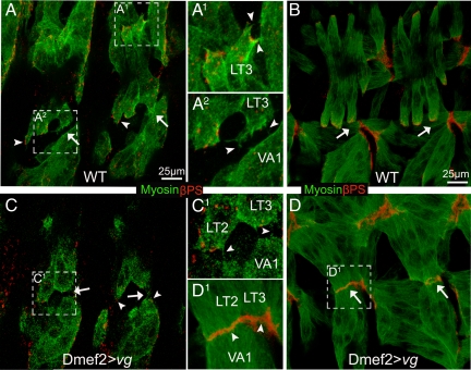
The somatic muscles of Drosophila develop in a complex pattern that is repeated in each embryonic hemi-segment. During early development, progenitor cells fuse to form a syncytial muscle, which further differentiates via expression of muscle-specific factors that induce specific responses to external signals to regulate late-stage processes such as migration and attachment. Initial communication between somatic muscles and the epidermal tendon cells is critical for both of these processes. However, later establishment of attachments between longitudinal muscles at the segmental borders is largely independent of the muscle-epidermal attachment signals, and relatively little is known about how this event is regulated. Using a combination of null mutations and a truncated version of Sd that binds Vg but not DNA, we show that Vestigial (Vg) is required in ventral longitudinal muscles to induce formation of stable intermuscular attachments. In several muscles, this activity may be independent of Sd. Furthermore, the cell-specific differentiation events induced by Vg in two cells fated to form attachments are coordinated by Drosophila epidermal growth factor signaling. Thus, Vg is a key factor to induce specific changes in ventral longitudinal muscles 1-4 identity and is required for these cells to be competent to form stable intermuscular attachments with each other.


Figures

Figure 1.
Figure 1.
(A) Schematic representation of the SMs in each abdominal hemi-segment A2–A7 of the developing embryo (lateral view with anterior left and dorsal up) by using the nomenclature of Bate (1993). Inner, middle, and outer muscle layers are shown in yellow, blue, and red, respectively (Bate, 1993). Dorsal oblique (DO), DA, dorsal transverse (Swan et al., 2004), LL, LO, LT (Cluzel et al., 2005), SBM, VL (Lundstrom et al., 2004), VA, VT, and VO. VA1 and VA2 are highlighted in red. (B) Muscle–muscle and muscle–tendon cell junctions in wild-type embryos visualized by staining developing muscle cells with actin (Tadokoro et al., 2003) and βPS integrin (green). (C) Diagrams showing a cross-sectional view along the broken line in B. The adhesion proteins (talin, βPS, and Tig, etc.) all concentrate at the end of SMs and are involved in forming stable muscle–muscle or muscle–tendon cell adhesions in wild-type embryos. In rhea1 mutant embryos, the muscle–tendon cell connections are broken (arrowheads), but the muscle–muscle connections remain (arrows).
Figure 2.
Figure 2.
SMs were detached in vgnull; rhea1 embryos but not in SD3L; rhea1 embryos. Embryos (stage 16 or a specified stage) are shown as lateral views, with dorsal up, and anterior to the left. Staining is color coded and indicated on each panel. B1–I1 are the close-ups of the framed area in B–I. (A) vg is expressed in muscle LL1 and VL1–4. The arrowhead points to a neuronal cell also expressing vg. Compared with wild-type embryos (B), vgnull (C), or rhea1 single mutation (D), or vgnull; rhea1 double mutant embryos in early stages (before stage 15; E), all produced a muscle pattern similar to wild-type embryos, except that a vgnull mutation caused muscle VL2 to be missing in ∼30% of segments (star in C1 and E1). Notice VL muscles (e.g., VL1) all formed tight adhesions between each other (arrows in B1–E1). (F–F1) By late stage 16 when muscles start to contract, muscle VL1 or other VL muscles detached from the attachment sites only in the vgnull; rhea1 double mutant embryos (arrows in F1). Arrows in F indicate detaching muscles and arrowheads indicate detached muscles. (G–H1) Overview of the vgnull; rhea1 double mutant embryos (stage 16; G–G1′) compared with rhea1 embryos (H–H1). Both VL1 and VL2 are retracting from their normal attachment sites (arrowhead in G1–H1). G1 and G1′ are two different confocal sections, and the broken line in G1 indicates the segment border. (I–I1) The muscle detachment phenotype of vgnull; rhea1 embryos can be rescued by expression of Vg via Dmef2-GAL4. Notice VL1 muscles built tight adhesions between each other (arrows in I1). (J) SD3L; rhea1 embryos did not have a muscle detachment phenotype. Some muscles do not develop well (VO4–6; arrowheads) in these embryos, but this mirrors the phenotype seen in SD3L single mutants.
Figure 3.
Figure 3.
Tissue-specific expression of SDΔTEA interferes with Vg function and produced elongated larvae and adults. (A) Expression of SDΔTEA via SD-GAL4 (SD>SDΔTEA) in the wing disk caused loss of the adult wing by interfering with Vg. However, we also noted that the pupae (A′) and adult flies were elongated compared with wild-type (WT) siblings. (B) This phenotype was caused by interfering with Vg in the muscle cells because these effects were seen in pupae (B′) and adults when UAS-SDΔTEA was expressed exclusively in muscle cells via Dmef2-GAL4 (Dmef2>SDΔTEA). (C) Quantification of pupal length in animals overexpressing SDΔTEA by using the indicated GAL4 drivers (mean ± SD; n = 23). The pupal length of twist-GAL4>SDΔTEA animals was not statistically different from wild type. (D–D′) Larvae expressing SDΔTEA in the muscles (Dmef2-GAL4) (D′) had a larger gap (arrowhead) between DA1 muscles than wild type (D). (E) Expression of SDΔTEA in developing muscle cells in embryos that are homozygous for the rhea1 mutation produced a muscle detachment phenotype in which the majority of the VL1 cells became rounded (arrows). (F) Tig protein localizes to the tips of muscles forming junctions including VL1 (arrows). (G) Embryos expressing SDΔTEA in muscles show the same pattern of Tig localization (arrows). In all panels, ventral muscles in two or three segments are shown in embryos (stage 16) presented as lateral views, with dorsal up, and anterior to left.
Figure 4.
Figure 4.
The muscle detachment phenotype observed in vgnull; rhea1 embryos was not due to lack of localization of integrin or its known ligands, nor to an obvious muscle migration defect. (A) In wild-type embryos, βPS and Tig can be seen localized normally at the junctions between two VL muscles (arrowhead). (B) In vgnull; rhea1 double mutant embryos, the VL muscles were either detaching (arrowheads) or were already detached (arrows). However, βPS and Tig remain concentrated at muscle termini and followed the detaching muscles (arrowheads). (C) In rhea1 mutant embryos, the adhesion proteins PINCH and βPS formed tight junctions between VL muscles (arrowheads). (D) Similar to βPS and Tig, in detaching muscles in vgnull; rhea1 embryos, PINCH and βPS remain concentrated at muscle termini and followed the detaching muscles (arrowheads). Many muscles seemed to be detaching from the posterior border of each segment. (E) In the vgnull; rhea1 embryos, Tsp shows the same localization to the end of detaching muscles as PINCH, βPS, and Tig. (F) A diagram of the localization of adhesion proteins (red; arrowhead) in vgnull or rhea1 mutant embryos and the direction (anterior, arrow) in which VL muscles are moving after they detach. (G) In wild-type embryos, Kon, the major migration guidance protein for VL muscles, normally found at the end of muscle cells (arrowhead). (H) In vgnull; rhea1 embryos, some residual (maternally supplied) Vg protein can still be seen in VL1 muscle (empty arrowheads). These muscles still had a detachment phenotype, but Kon is localized properly (arrowhead). A1–H1 are the close-ups of the framed area in A–H. A2–H2 and A3–H3 show each confocal channel separately.
Figure 5.
Figure 5.
Ectopic expression of Vg in the developing embryonic SMs produced ectopic intermuscular attachments. The transgenic lines vg1, vg2, and vg3 express relatively higher levels of Vg, respectively, as verified by Western blotting. (A) In wild-type embryos, LT1–4 muscles that stain brightly with muscle-specific actin (red; arrows) are seen passing left to right over the VA1 muscle and form βPS-mediated attachments (green) at intrasegmental sites. Normally, no adhesions form where the LT and VA muscles are adjacent (arrowhead). (B–D) Ectopic expression of progressively higher levels of Vg in all muscles via Dmef2-GAL4. (B) Ectopic expression of relatively lower levels of Vg (vg1) in SMs caused the LT muscles to abnormally form attachments at the segment borders. (C) Expression of relatively higher levels of Vg (vg2) cause the formation of abnormal attachments at the segment borders (arrows). Furthermore, ectopic muscle–muscle attachments were observed between LT and VA muscles (arrowheads). In some cases, muscle VA1 was observed deviating from its original position (arrowheads). (D) This number of abnormal and ectopic attachments becomes even more severe when a transgene (vg3) expressing relatively highest levels of Vg is used. (E) Quantification of the percentage of segments having LTs with abnormal migration (red columns) or ectopic adhesion sites between LT and VA muscle cells (blue columns) for each indicated overexpression line (n = 110). (F) Ectopic expression of Vg with SDΔTEA led to a partial rescue of the phenotype caused by ectopic expression of Vg from the UAS-vg2 transgene. (G) The C23-GAL4 line induces expression at high levels in VA1 but relatively low expression in LTs as detected by an UAS-lacZ reporter. (H) Ectopic attachments are not formed when Vg is present at relatively high levels in VA1 cells.
Figure 6.
Figure 6.
Muscle attachments induced by ectopic Vg include βPS integrin and its associated cytoplasmic linker proteins, PINCH and talin. (A) Ectopic expression of Vg by Dmef2-GAL4 caused additional attachments to form between muscles stained with muscle-specific actin (green). These ectopic attachments (arrowheads) contained Tig (red; A1), an extracellular ligand for PS2 integrin (blue; A2). Ectopic muscle attachments were also produced between muscle cells other than LTs and VAs, which also contained Tig (arrows). Individual myofibers were linked to the new adhesion sites through integrin complexes (A4 is a close-up of the boxed area in A3). (B) These ectopic attachments also contain Tsp (blue; B1) and PINCH (red; B2). (C) Talin is also localized to the ectopic muscle attachments (red; C2). Note the processes emerged from the lateral surface of muscle LTs (arrows in C3) and VT1 (empty arrowhead in C3).
Figure 7.
Figure 7.
Altered levels of Vg function regulate ectopic intermuscular attachments independently of signaling from tendon cells in VL muscle cells, and DER mediated cell–cell communication is required for the production of intermuscular attachments between Vg expressing cells in slit2 mutant. (A) In slit2 mutant embryos, VL muscle cells migrate dorsally over the CNS from the lateral sides of the embryo meeting near the midline to form Tig marked muscle–muscle adhesions (arrowheads) in a region of the embryo devoid of tendon cells. (B) Interference with Vg function by expression of SDΔTEA in these slit2 mutant embryos led to fewer and smaller of these midline-located adhesion sites (arrowheads). (C) Overexpression of Vg in slit2 mutant embryos produced more and larger adhesion sites (arrowheads) in the abnormally positioned VL muscles. (D) Expression of a dominant-negative form of DER (DN-egfr) in slit2 mutant embryos strongly reduces the overall size and number of these muscle-muscle adhesion sites (arrowheads in D). (E) The size and number of these ectopic adhesion sites (arrowheads in E) in slit2 mutants that coexpress DN-egfr as well as Vg are reduced when compare with expressing Vg alone (compare E with C) but increased when compared with expressing DN-egfr alone (compare E with D). (F) Quantification of the number of VL cell adhesion sites formed in slit2 mutant embryos with varying levels of Vg activity and/or changing of DER activity (mean ± SD; n ≥ 15).
Figure 8.
Figure 8.
Changes in muscle–muscle adhesion caused by expression of a constitutively active DER (λ-egfr) are sensitive to the presence of Vg. (A) Many small adhesion sites were formed between the midline-crossing muscles in slit2 mutant embryos (arrowhead) when λ-egfr is expressed in developing muscle cells (Dmef2>λ-egfr) (arrowhead, inset). (B) Expression of λ-egfr in developing muscle cells where Vg function was inhibited by SDΔTEA produced fewer of these ectopic adhesions (arrowhead; inset). (C) Relatively more and larger ectopic adhesions were formed when embryonic muscle cells were overexpressing both λ-egfr and Vg (arrowhead, inset). Insets are close-ups of the area framed by the dotted lines.
Figure 9.
Figure 9.
Filopodial contact between muscles forming junctions are not affected by changes in Vg expression. (A) In wild-type embryos (stage 14), the leading edge of LT muscles produce filopodia that contact with corresponding filopodia protruding from the lateral edge of VAs or other muscles (arrows in A and arrowheads in A1–A2). A1 and A2 are magnified photos of the framed areas in A. βPS integrin accumulates at the leading edge of the myotube (arrowhead). (B) At developmental stage 16, LTs normally find their attachment sites and form stable adhesion inside each segment. There are no connections between LTs and VAs (arrows in B). (C) Muscles in embryos that were expressing Vg ectopically (stage 14) produced similar number of filopodia compared with wild type (arrows in C and arrowheads in C1). Also, βPS integrin accumulated at the leading edge of muscles (arrowheads in C) in the same way as wild-type muscles. (D) At stage 16, LT muscle cells have formed stable attachments with muscle VAs (arrows in D and arrowheads in D1).

Similar articles

Cited by

References

    1. Bate M. The Development of Drosophila melanogaster, II. Cold Spring Harbor, NY: Cold Spring Harbor Laboratory Press; 1993. The mesoderm and its derivatives; pp. 1013–1090.
    1. Baylies M. K., Bate M., Ruiz Gomez M. Myogenesis: a view from Drosophila. Cell. 1998;93:921–927. - PubMed
    1. Becker S., Pasca G., Strumpf D., Min L., Volk T. Reciprocal signaling between Drosophila epidermal muscle attachment cells and their corresponding muscles. Development. 1997;124:2615–2622. - PubMed
    1. Bernard F., Dutriaux A., Silber J., Lalouette A. Notch pathway repression by vestigial is required to promote indirect flight muscle differentiation in Drosophila melanogaster. Dev. Biol. 2006;295:164–177. - PubMed
    1. Bernard F., Lalouette A., Gullaud M., Jeantet A. Y., Cossard R., Zider A., Ferveur J. F., Silber J. Control of apterous by vestigial drives indirect flight muscle development in Drosophila. Dev. Biol. 2003;260:391–403. - PubMed

Publication types

MeSH terms

LinkOut - more resources