. 2010 Oct 1;21(19):3459-74.
doi: 10.1091/mbc.E10-02-0133. Epub 2010 Aug 4.

Osmoprotective transcription factor NFAT5/TonEBP modulates nuclear factor-kappaB activity

Affiliations

Osmoprotective transcription factor NFAT5/TonEBP modulates nuclear factor-kappaB activity

Isabelle Roth et al. Mol Biol Cell. .

Abstract

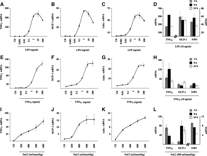
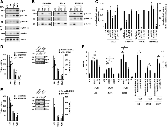
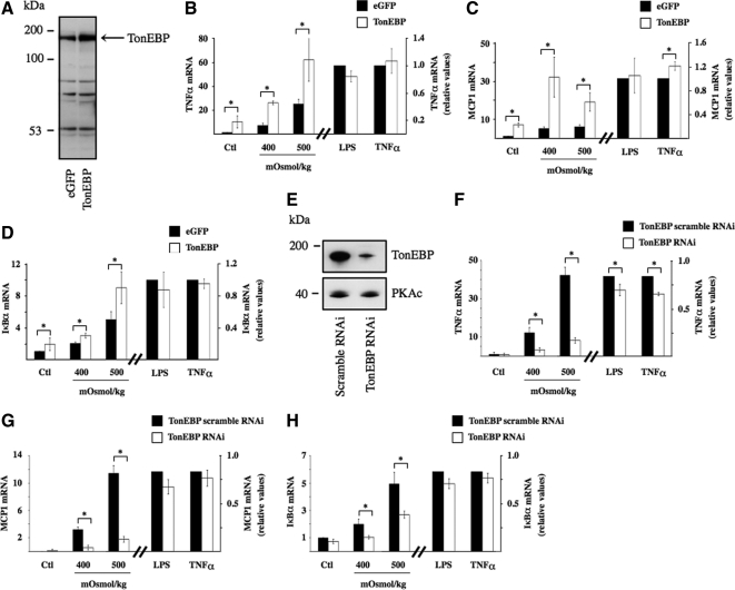
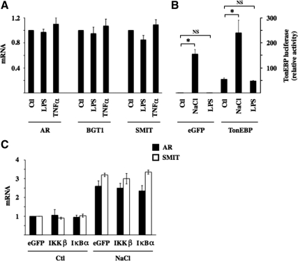
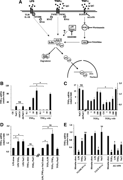
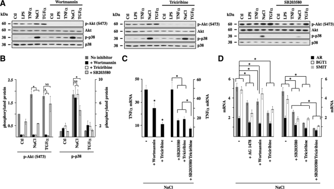
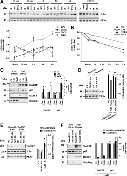
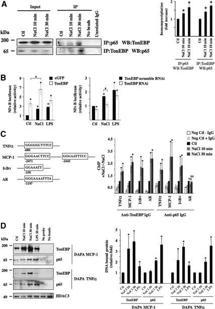
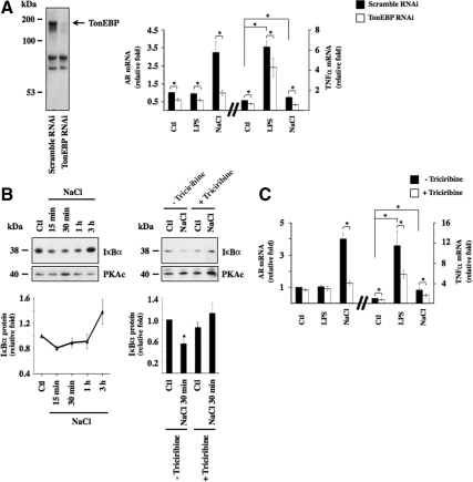
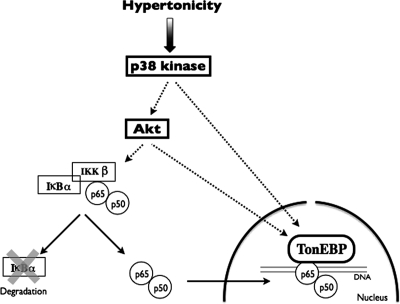
Tonicity-responsive binding-protein (TonEBP or NFAT5) is a widely expressed transcription factor whose activity is regulated by extracellular tonicity. TonEBP plays a key role in osmoprotection by binding to osmotic response element/TonE elements of genes that counteract the deleterious effects of cell shrinkage. Here, we show that in addition to this "classical" stimulation, TonEBP protects cells against hypertonicity by enhancing nuclear factor-κB (NF-κB) activity. We show that hypertonicity enhances NF-κB stimulation by lipopolysaccharide but not tumor necrosis factor-α, and we demonstrate overlapping protein kinase B (Akt)-dependent signal transduction pathways elicited by hypertonicity and transforming growth factor-α. Activation of p38 kinase by hypertonicity and downstream activation of Akt play key roles in TonEBP activity, IκBα degradation, and p65 nuclear translocation. TonEBP affects neither of these latter events and is itself insensitive to NF-κB signaling. Rather, we reveal a tonicity-dependent interaction between TonEBP and p65 and show that NF-κB activity is considerably enhanced after binding of NF-κB-TonEBP complexes to κB elements of NF-κB-responsive genes. We demonstrate the key roles of TonEBP and Akt in renal collecting duct epithelial cells and in macrophages. These findings reveal a novel role for TonEBP and Akt in NF-κB activation on the onset of hypertonic challenge.


Figures

Figure 1.
Figure 1.
Hypertonicity increases NF-κB activity in mpkCCDcl4 cells. Real-time PCR analysis of TNF-α (A, E, and I), MCP-1 (B, F, and J), and IκBα (C, G, and K) transcripts in mpkCCDcl4 cells after 3 h of LPS (1 pg/ml–100 ng/ml), TNF-α (1 pg/ml–100 ng/ml), or NaCl-hypertonic challenge (350–600 mOsmol/kg). Data are normalized to acidic ribosomal phosphoprotein P0 and is represented as fold induction over nonstimulated (Ctl) cells. Also shown are time course expression analysis of each transcript by LPS (10 ng/ml; D), TNF-α (10 ng/ml; H), and NaCl hypertonic challenge (500 mOsmol/kg; L).
Figure 2.
Figure 2.
NF-κB activation by hypertonicity in part depends on p38 kinase activity. (A) Protein lysates from cells challenged or not (Ctl) with either LPS, TNF-α, or hypertonic medium (NaCl) were analyzed by immunoblot for phosphorylated forms of p38 kinase, p38 substrate ATF2, ERK, JNK, and JNK substrate c-Jun. PKAc was used as a loading control. (B) Protein lysates from cells pretreated or not for 30 min with 10 μM SB203580 (a p38 kinase inhibitor), 10 μM U0126 (a MEK inhibitor), or 10 μM SP600125 (a JNK inhibitor) and then challenged or not (Ctl) with hypertonic medium were analyzed by immunoblot for phosphorylated forms of ATF2, ERK, and c-Jun. (C) Fold protein phosphorylation following hypertonic challenge over baseline levels (Ctl) is shown at left. Fold decrease of protein phosphorylation by pharmacological inhibitors in hypertonicity-challenged cells is shown at right. (D and E) Real-time PCR analysis of TNF-α transcript after LPS, TNF-α, or hypertonic challenge in the presence or absence of either SB203580 or U0126 (D, left) or SP600125 (E; left). (D and E) Right, effects of SB203580 and SP600125 on TNF-α transcript expression were echoed by RNAi against p38α (D) or c-Jun (E), respectively. Immunoblots depicting decreased expression levels of target protein by RNAi are also shown. PKAc was used as a loading control. (F) Left, real-time PCR analysis of AR, BGT1, and SMIT transcripts in cells treated or not with a pharmacological inhibitor and challenged or not (Ctl) with hypertonic medium. Data are represented as fold induction over nonstimulated cells in the absence of an inhibitor. Similar to SB203580, RNAi against p38α reduced the enhancer effect of hypertonicity on AR, BGT1, and SMIT transcript expression (right). The effects of pharmacological compounds and RNAi on basal mRNA expression levels for this and all other figures are shown in Supplemental Figure S2.
Figure 3.
Figure 3.
TonEBP mediates NF-κB activation by hypertonicity. (A and E) Immunoblot of protein lysates from cells transfected with cDNA encoding eGFP or TonEBP (A) or transfected with scrambled RNAi or RNAi against TonEBP (E). The arrow in A depicts the band corresponding to TonEBP. In E, PKAc was used as a loading control. (B–D and F–H) real-time PCR analysis of TNF-α, MCP-1, and IκBα transcripts in cells transfected with cDNA encoding eGFP or TonEBP (B–D) or transfected with scrambled RNAi or RNAi against TonEBP (F–H) and challenged or not (Ctl) with hypertonic medium (400 or 500 mOsmol/kg), LPS, or TNF-α. Data are represented as fold induction over nonstimulated cells transfected with eGFP or scrambled RNAi.
Figure 4.
Figure 4.
IKKβ and IκBα do not influence TonEBP activity. (A) Real-time PCR analysis of AR, BGT1, and SMIT transcripts in cells challenged with LPS or TNF-α. Data are represented as fold expression over nonstimulated (Ctl) cells. (B) TonEBP-driven luciferase activity in response to hypertonic (NaCl) or LPS challenge. Cells were transfected with p(kB)3 IFN-Luc plasmid and cotransfected with cDNA encoding either eGFP or TonEBP. Data shown is represented as fold induction over nonstimulated cells transfected with cDNA encoding eGFP. (C) Real-time PCR analysis of AR and SMIT transcripts in cells transfected with cDNA encoding eGFP or constitutively active IKKβ or IκBα and challenged or not (Ctl) with hypertonic medium. Data shown is represented as fold induction over nonstimulated cells transfected with cDNA encoding eGFP.
Figure 5.
Figure 5.
NF-κB signaling by hypertonicity overlaps that elicited by TNF-α and TGF-α. (A) Schematic illustration of NF-κB activation by LPS, TNF-α, and TGF-α. Binding of each ligand to partner receptors initiates distinct transduction cascades that all ultimately lead to IKKβ activation. Under basal states, IκBα interacts with p65, retaining it in the cytoplasm. Once activated, IKKβ typically phosphorylates IκBα, leading to its degradation. This allows nuclear translocation of liberated p65-containing complexes, typically p65-p50 dimers. Tools used in this study to investigate the putative roles of target molecules in hypertonic NF-κB inducibility are shown. Also shown is p38 kinase whose role in NF-κB activation by hypertonicity was investigated. (B and C) Real-time PCR analysis of TNF-α transcript in cells challenged with hypertonic medium (NaCl, 500 mOsmol/kg), TNF-α (0.1–100 ng/ml), or TGF-α (1–1000 ng/ml) in the absence or presence of IgG against TNF-α (B) or TGF-α (C). Data are represented as fold induction over nonstimulated (Ctl) cells. (D) Real-time PCR analysis of TNF-α transcript in cells challenged with LPS, TNF-α, TGF-α (100 ng/ml), or hypertonic medium alone or simultaneously challenged with two stimuli. Data are represented as fold expression over cells challenged with LPS, TNF-α, or hypertonicity alone. (E) Real-time PCR analysis of TNF-α transcript in cells transfected with scrambled RNAi, RNAi against MyD88, or RNAi against TNFR1 and TNFR2 or cells treated with 10 μM of the EGFR antagonist AG1478. Cells were challenged with LPS, TNF-α, TGF-α, or hypertonic medium. Data are represented as fold expression over cells challenged with either stimulus alone.
Figure 6.
Figure 6.
p38 kinase and Akt mediate NF-κB activation by hypertonicity. (A) Immunoblot of protein lysates against Akt phosphorylated at Ser473, nonphosphorylated Akt, and phosphorylated and nonphosphorylated p38 kinase. Cells were treated or not with 100 nM wortmannin (a PI3-kinase antagonist), 30 μM triciribine (an Akt antagonist), or 10 μM SB203580 (a p38 kinase antagonist) and challenged or not (Ctl) with LPS, TNF-α, TGF-α, or hypertonic medium (NaCl). (B) Fold expression of phosphorylated Akt and phosphorylated p38 kinase by either hypertonicity or TGF-α in the presence or absence of a pharmacological inhibitor over that of nonstimulated (Ctl) cells. (C and D) Real-time PCR analysis of TNF-α transcript (C) or AR, BGT1 and SMIT transcripts (D) in cells challenged with hypertonic medium in the presence or absence of wortmannin, triciribine (C) or wortmannin, triciribine or AG1478 (10 μM) (D). The effects of SB203580 and triciribine, applied alone or together, on hypertonic stimulation of TNF-α expression and TonEBP activity is shown at right of each figure. Data are represented as fold decrease of hypertonicity-induced mRNA expression by pharmacological inhibitors.
Figure 7.
Figure 7.
Hypertonicity induces IκBα degradation and p65 nuclear translocation independently of TonEBP. (A) Immunoblot of protein lysates against IκBα or PKAc (used as a loading control) in cells challenged or not (Ctl) with LPS, TNF-α, hypertonic medium (NaCl), or TGF-α for various times. Time-dependent IκBα degradation by each stimulus is represented as fold IκBα expression in stimulated cells at each time point over nonstimulated cells. (B) Real-time PCR analysis of IκBα transcript in cells pretreated 15 min with actinomycin D (10 μM) and then challenged or not (Ctl) with LPS, TNF-α, or hypertonic medium for various times. Data are represented as fold IκBα transcript expression in stimulated cells over that of nonstimulated cells before addition of actinomycin D. (C) Immunoblot of cytoplasmic and nuclear protein lysates against TonEBP, p65, histone deacetylase 3 (HDAC3, (used as a loading control for nuclear extracts), and Tubulinα (used as a loading control for cytosolic extracts) in cells challenged or not (Ctl) with LPS or hypertonic medium for 30 min. Fold TonEBP and p65 expression in cytoplasmic and nuclear extracts over that of nonstimulated cells (Ctl) is shown at right. (D and E) Immunoblot of protein lysates against IκBα, PKAc (used as a loading control) (D) and TonEBP (E) in cells pretreated or not with Akt antagonist triciribine and then challenged or not (Ctl) with hypertonic medium for 15 min (D) or in cells transfected with scrambled RNAi or TonEBP RNAi and then challenged or not (Ctl) with hypertonic medium for 15 min or 1 h (E). IκBα protein expression is represented as fold expression over control cells. (F) Immunoblot of cytoplasmic and nuclear protein lysates against TonEBP, p65, HDAC3, and Tubulinα in cells transfected with scrambled RNAi or TonEBP RNAi and challenged with hypertonic medium for 30 min. Fold TonEBP and p65 expression in cytoplasmic and nuclear extracts over that of cells transfected with TonEBP scrambled RNAi is shown at right.
Figure 8.
Figure 8.
TonEBP associates with p65 on the onset of hypertonic challenge. (A) Immunoblot against p65 and TonEBP immunoprecipitated by anti-TonEBP or anti-p65 IgG, respectively, or unrelated IgG (Na,K-ATPase α subunit) in cells challenged or not (Ctl) with hypertonic medium (NaCl) for 10 or 30 min. Also shown is the absence of bands from lysates precipitated in the absence of either agarose beads or IgG. Similar amounts of p65 and TonEBP between experimental conditions were loaded onto gels before immunoprecipitation (Input, corresponding to 5% of immunoprecipitated protein). Fold immunoprecipitated TonEBP and p65 over that of nonstimulated cells (Ctl) is shown at right. (B) NF-κB–driven luciferase activity in response to hypertonic (NaCl) or LPS challenge. Cells were transfected with a NF-κB-Luc plasmid described in Materials and Methods and cotransfected with cDNA encoding either eGFP or TonEBP (left) or with scrambled RNAi or RNAi against TonEBP (right). Data shown is represented as fold induction over nonstimulated cells transfected either with cDNA encoding eGFP or scrambled RNAi. (C) ChIP analysis. The localization of κB sites of mouse TNF-α, MCP-1, and IκBα promoters as well as the TonE site of mouse AR promoter chosen for analysis is shown. Localization is relative to the AUG start codon. Cells were challenged or not (Ctl) with hypertonic medium for 10 or 30 min before DNA fragmentation and immunoprecipitation using anti-TonEBP or p65 antibodies. Immunoprecipitated DNA was analyzed by real-time PCR using primers flanking κB sites of TNF-α, MCP-1, or IκBα promoters or the TonE site of the AR promoter. Data are represented as fold induction over nonstimulated cells. Negative controls consisted of DNA fragments precipitated in the absence of antibody or with anti- Na,K-ATPase α subunit IgG. (D) DAPA experiments were performed on nuclear extracts of cells challenged or not (Ctl) with hypertonic medium for 10 or 30 min or with LPS for 30 min. Precipitated protein by DAPA probes encompassing κB sites of MCP-1 or TNF-α promoters depicted in C was analyzed by immunoblot using anti-TonEBP or anti-p65 IgG. Equal loading was verified by immunoblot against histone deacetylase 3 (HDAC3). Negative controls consisted of protein precipitated in the absence of either a DAPA probe or beads. Precipitated TonEBP and p65 protein was quantified and is graphically represented as fold expression over control cells.
Figure 9.
Figure 9.
NF-κB activation by hypertonicity in H36.12j cells depends on TonEBP and Akt activity. (A) Left. immunoblot of protein lysates from cells transfected with RNAi against TonEBP or scrambled RNAi. The arrow depicts the band corresponding to TonEBP. Right, real-time PCR analysis of AR and TNF-α transcripts in cells transfected with RNAi against TonEBP or scrambled RNAi and exposed to LPS or hypertonic medium (NaCl). Data are represented as fold induction over nonstimulated cells transfected with scrambled RNAi (Ctl). (B) Left, immunoblot of protein lysates against IκBα or PKAc (used as a loading control) in cells challenged or not (Ctl) with hypertonic medium for various periods. Time-dependent IκBα degradation is graphically represented as fold IκBα expression in stimulated cells at each time point over nonstimulated cells. (B) Right, immunoblot of protein lysates against IκBα or PKAc (used as a loading control) in cells pre-treated or not with Akt antagonist triciribine and challenged or not (Ctl) with hypertonic medium for 30 min. IκBα protein expression is graphically represented as fold expression over control cells. (C) Real-time PCR analysis of AR and TNF-α transcripts in cells treated or not with triciribine and challenged with either LPS or hypertonic medium for 3 h. Data are represented as fold induction over nonstimulated (Ctl) cells.
Figure 10.
Figure 10.
Proposed mechanism for NF-κB activation by hypertonicity. Hypertonicity activates NF-κB in a two-step process. In a first step, hypertonicity stimulates p38 kinase, which enhances the activities of both Akt and TonEBP. Increased Akt activity in turn not only contributes to enhancing TonEBP activity but also induces IκBα to dissociate from cytosolic p65. This process depends on IKKβ activity. Unconjugated IκBα is degraded allowing free p65, associated with other NF-κB binding partners such as p50, to translocate across the nuclear membrane. In a second step, TonEBP associates with p65-containing complexes bound to DNA, enhancing NF-κB transcriptional activity. NF-κB activation by hypertonicity is transient and decreases as IκBα protein expression increases in response to increased IκBα transcriptional activity.

Similar articles

Cited by

References

    1. Andrieu N., Salvayre R., Jaffrezou J. P., Levade T. Low temperatures and hypertonicity do not block cytokine-induced stimulation of the sphingomyelin pathway but inhibit nuclear factor-kappa B activation. J. Biol. Chem. 1995;270:24518–24524. - PubMed
    1. Biswas D. K., Iglehart J. D. Linkage between EGFR family receptors and nuclear factor kappaB (NF-kappaB) signaling in breast cancer. J. Cell. Physiol. 2006;209:645–652. - PubMed
    1. Bonnard M., et al. Deficiency of T2K leads to apoptotic liver degeneration and impaired NF-kappaB-dependent gene transcription. EMBO J. 2000;19:4976–4985. - PMC - PubMed
    1. Burg M., Ferraris J., Dmitrieva N. Cellular response to hypertonic stress. Physiol. Rev. 2007;87:1441–1474. - PubMed
    1. Canono B. P., Campbell P. A. Production of mouse inflammatory macrophage hybridomas. J. Tissue Cult. Methods. 1992;14:3–8.

Publication types

MeSH terms

LinkOut - more resources