. 2010 Aug 4;30(31):10263-71.
doi: 10.1523/JNEUROSCI.1125-10.2010.

Fragile X mental retardation protein is required for rapid experience-dependent regulation of the potassium channel Kv3.1b

Affiliations

Fragile X mental retardation protein is required for rapid experience-dependent regulation of the potassium channel Kv3.1b

John G Strumbos et al. J Neurosci. .

Abstract

Fragile X mental retardation protein (FMRP) is an RNA-binding protein that regulates synaptic plasticity by repressing translation of specific mRNAs. We found that FMRP binds mRNA encoding the voltage-gated potassium channel Kv3.1b in brainstem synaptosomes. To explore the regulation of Kv3.1b by FMRP, we investigated Kv3.1b immunoreactivity and potassium currents in the auditory brainstem sound localization circuit of male mice. The unique features of this circuit allowed us to control neuronal activity in vivo by exposing animals to high-frequency, amplitude-modulated stimuli, which elicit predictable and stereotyped patterns of input to the anterior ventral cochlear nucleus (AVCN) and medial nucleus of the trapezoid body (MNTB). In wild-type (WT) animals, Kv3.1b is expressed along a tonotopic gradient in the MNTB, with highest levels in neurons at the medial, high-frequency end. At baseline, Fmr1(-/-) mice, which lack FMRP, displayed dramatically flattened tonotopicity in Kv3.1b immunoreactivity and K(+) currents relative to WT controls. Moreover, after 30 min of acoustic stimulation, levels of Kv3.1b immunoreactivity were significantly elevated in both the MNTB and AVCN of WT, but not Fmr1(-/-), mice. These results suggest that FMRP is necessary for maintenance of the gradient in Kv3.1b protein levels across the tonotopic axis of the MNTB, and are consistent with a role for FMRP as a repressor of protein translation. Using numerical simulations, we demonstrate that Kv3.1b tonotopicity may be required for accurate encoding of stimulus features such as modulation rate, and that disruption of this gradient, as occurs in Fmr1(-/-) animals, degrades processing of this information.


Figures

Figure 1.
Figure 1.
FMRP binds Kv3.1b mRNA in brainstem-enriched synaptosomes. a, FMRP was immunoprecipitated from synaptosomes prepared from either WT or Fmr1 −/− mice followed by isolation of RNA and RT-PCR with primers designed to either detect the 5′ region of Kv3.1b mRNA (lanes 1–4) or Map1b mRNA (lanes 5–8), a known mRNA target of FMRP. We detected both Kv3.1b mRNA and Map1b mRNA bound to FMRP in WT mouse brain synaptosomal samples (lanes 3, 7). As negative controls, no Kv3.1b or Map1b mRNA was detected in FMRP immunoprecipitates from synaptosomes prepared from Fmr1 −/− mice (lanes 4, 8). b, Total levels of Kv3.1b protein in brainstem-enriched homogenates were not significantly different in WT versus Fmr1 −/− mice, suggesting that FMRP-dependent regulation of Kv3.1b translation is restricted to specific pools of Kv3.1b mRNA that may account for only a small proportion of total brainstem Kv3.1b. For statistical comparisons, GAPDH was used as a loading control and OD measurements for Kv3.1b bands were normalized to the GAPDH bands (WT, 1.09 ± 0.13; Fmr1 −/−, 1.13 ± 0.11; n = 5, p = 0.78, unpaired t test). IP, Immunoprecipitated.
Figure 2.
Figure 2.
Kv3.1b tonotopicity is flattened in Fmr1 −/− mice. a, Representative MNTB sections from a single WT and Fmr1 −/− mouse demonstrating the flattened pattern of Kv3.1b immunoreactivity across the rostrocaudal and mediolateral axes of the MNTB. Scale bar, 20 μm. b, Representative three-dimensional plots of average Kv3.1b immunoreactivity (OD) in each of 25 stereotaxic zones. Plots correspond to the sections shown on left. Insets, Plots of relative OD across the mediolateral axis for each control animal used in the study (n = 9). Data points represent the average and SE of 5 sections for each animal. To facilitate comparisons across animals, the OD in each section was normalized to the darkest cell in the section. A linear regression for the WT plots was statistically significant (p = 0.01, R 2 = 0.86; dotted line), whereas a linear regression for the gradient in Fmr1 −/− mice was not statistically significant (p = 0.08, R 2 = 0.83). Lat, Lateral; Med, medial; Post, posterior; Ant, anterior; freq, frequency.
Figure 3.
Figure 3.
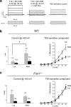
Kv3.1-like currents do not vary between lateral and medial MNTB neurons in Fmr1 −/− mice. a, Representative traces demonstrating the technique for isolating the Kv3.1-like current in MNTB neurons, which mainly represents the activity of Kv3.1b homomers (Wang et al., 1998b; Macica et al., 2003). Control traces are recorded in the presence of TTX (1 μm) and ZD-7288 (20 μm) to block Na+ and h-currents, respectively. Cells were held at a resting potential of −40 mV for at least 2 min before all recordings to ensure complete inactivation of low-threshold, voltage-activated K+ currents (left). Application of 1 mm TEA selectively blocks the high-threshold Kv3.1-like current (center). The TEA-sensitive current is shown in gray (right). A schematic of the pulse protocol is shown below traces. b, c, Consistent with our densitometric analysis, the proportion of K+ current that was blocked by 1 mm TEA when neurons were stepped from −40 to +60 mV varied significantly (**) between medially and laterally located neurons in WT mice (b, right; n = 8, p = 0.009, unpaired t test), but not in Fmr1 −/− mice (c, right; n = 8, p = 0.84, unpaired t test). The amplitude of total K+ current at +60 mV also differed significantly (*) across the tonotopic axis in WT mice (b, left; n = 8, p = 0.03, unpaired t test), but not in Fmr1 −/− mice (c, left; n = 8, p = 0.56, unpaired t test). Lat, Lateral; Med, medial.
Figure 4.
Figure 4.
Representative sections and 3D tonotopic curve-fits. All sections shown were immunolabeled simultaneously on the same slide (above). The WT and Fmr1 −/− control sections are from the same animals shown in Figure 2. Representative three-dimensional curve-fits (below) are shown to highlight the flattening of the tonotopic gradient after 30 min high-frequency acoustic stimulation in WT, but not Fmr1 −/−, mice. Curves were constructed from raw data (e.g., those shown in Fig. 2) using a thin-plate spline algorithm. Lat, Lateral; Med, medial; Post, posterior; Ant, anterior.
Figure 5.
Figure 5.
Rapid effects of acoustic stimulation on Kv3.1b tonotopicity and total levels of Kv3.1b immunoreactivity in the MNTB. a, Gradient strength (curvature) was quantified for each animal (n = 25) by calculating the ratio of immunoreactivity in zone 5 to the average immunoreactivity in the zones 1–4 (Z5 ratio). Box plot shows the full range of Z5 ratios from each treatment group. Maximum and minimum (error bars), 99 and 1% (box), mean (circle), and median (horizontal line) values are shown for each group. We observed a significant difference (*) in the Z5 ratios between WT control mice and WT mice receiving acoustic stimulation (n = 13, p = 0.02, unpaired t test). In Fmr1 −/− mice, we observed no significant effect of stimulation on the Z5 ratio (n = 12, p = 0.33, unpaired t test). b, Total levels of MNTB Kv3.1b immunoreactivity in each group were determined by measuring the mean OD of each section (N = 365 sections). Kv3.1b levels were significantly elevated (*) in sound-stimulated WT mice relative to WT controls (N = 162 sections, p = 0.02, unpaired t test). By contrast, sound stimulation did not significantly influence total Kv3.1b levels in Fmr1 −/− mice (N = 203 sections, p = 0.17, unpaired t test). Ctrl, Control; Stim, stimulated.
Figure 6.
Figure 6.
Rapid effects of acoustic stimulation on total levels of Kv3.1b immunoreactivity in the AVCN. a, Low-magnification image of a representative WT section showing the auditory brainstem circuit in relation to the cerebellum. Scale bar, 200 μm. b, c, Total levels of AVCN Kv3.1b immunoreactivity in each group were determined by measuring the mean OD of cells in each section (N = 149 sections). Representative sections from each group are shown (b; scale bar, 40 μm). Kv3.1b levels were significantly elevated (*) in sound-stimulated WT mice relative to WT controls (N = 81 sections, p = 0.02, unpaired t test). By contrast, sound stimulation did not significantly influence total Kv3.1b levels in Fmr1 −/− mice (N = 68 sections, p = 0.68, unpaired t test). Both groups of Fmr1 −/− mice displayed significantly elevated (*) Kv3.1b levels relative to WT controls [control (Ctrl), p = 0.04; stimulated (Stim), p = 0.04, ANOVA followed by Tukey test].
Figure 7.
Figure 7.
Acoustic stimulation has no effect on total levels of Kv3.1b immunoreactivity in the molecular layer of the cerebellum. a, As a negative control for the effects of sound stimulation, we measured Kv3.1b levels in the molecular layer of the cerebellum, where Kv3.1b is highly expressed in the parallel fibers of cerebellar granule cells (Weiser et al., 1995). Scale bar, 40 μm. b, We observed no effect of sound stimulation on Kv3.1b levels in WT mice (N = 65 sections, p = 0.56, unpaired t test) or Fmr1 −/− mice (N = 60 sections, p = 0.16, unpaired t test). Ctrl, Control; Stim, stimulated.
Figure 8.
Figure 8.
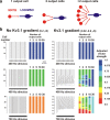
The presence of a tonotopic gradient in Kv3.1 conductance may be required for accurate ensemble encoding of sound stimuli. a, Numerical simulations using a previously developed computer model of MNTB neurons illustrate the inherent advantage for auditory processing when a range of Kv3.1 conductances are present across the tonotopic axis. Model MNTB neurons (output neurons, red) are shown as projecting to only 1 (left), 2 (center), or multiple (right) postsynaptic target neurons [lateral superior olive (LSO) or medial superior olive (MSO) neurons, blue]. b, Two sets of 30 neurons are shown, representing either uniform levels of Kv3.1 conductance (0.2 μS; left) or a gradient in Kv3.1 levels across the tonotopic axis (0.02–0.6 μS; right). These model neurons were stimulated for 100 ms at 200, 400, or 700 Hz, and the evoked patterns of firing are shown in the raster plots, with the time of each evoked action potential represented by a vertical line. In each case, the strength of the phase vector was calculated for the output of each neuron alone or for the combined output of 1, 2, 5, 10, or 30 neurons. Colored bars encode the adjusted vector strength, a measure of phase-locking fidelity, for each cell individually and for successively larger combinations of adjacent neurons. Although both groups of cells respond in the same fashion to a 200 Hz stimulus, they respond differently to higher rates of stimulation, such as 400 or 700 Hz. Whereas the 400 and 700 Hz stimuli each produce an identical pattern of firing in the group of neurons with fixed levels of Kv3.1 conductance, these two stimuli produce very different patterns of activity across the tonotopic axis when a gradient is present (right). With a gradient, the integrated phase vector improves as the number of cells contributing to encoding increases, whereas with uniform Kv3.1 levels it does not.

Similar articles

Cited by

References

    1. Ashley CT, Jr, Wilkinson KD, Reines D, Warren ST. FMR1 protein: conserved RNP family domains and selective RNA binding. Science. 1993;262:563–566. - PubMed
    1. Baranauskas G, Tkatch T, Nagata K, Yeh JZ, Surmeier DJ. Kv3.4 subunits enhance the repolarizing efficiency of Kv3.1 channels in fast-spiking neurons. Nat Neurosci. 2003;6:258–266. - PubMed
    1. Barnes-Davies M, Forsythe ID. Pre- and postsynaptic glutamate receptors at a giant excitatory synapse in rat auditory brainstem slices. J Physiol. 1995;488:387–406. - PMC - PubMed
    1. Bassell GJ, Warren ST. Fragile X syndrome: loss of local mRNA regulation alters synaptic development and function. Neuron. 2008;60:201–214. - PMC - PubMed
    1. Brew HM, Forsythe ID. Systematic variation of potassium current amplitudes across the tonotopic axis of the rat medial nucleus of the trapezoid body. Hear Res. 2005;206:116–132. - PubMed

Publication types

LinkOut - more resources