. 2010 Aug 4;30(31):10391-406.
doi: 10.1523/JNEUROSCI.0381-10.2010.

Centrosome motility is essential for initial axon formation in the neocortex

Affiliations

Centrosome motility is essential for initial axon formation in the neocortex

Froylan Calderon de Anda et al. J Neurosci. .

Abstract

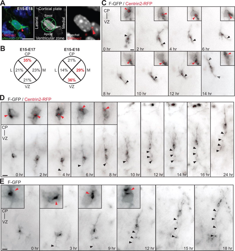
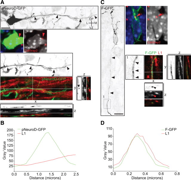
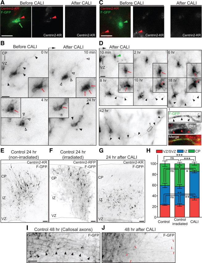
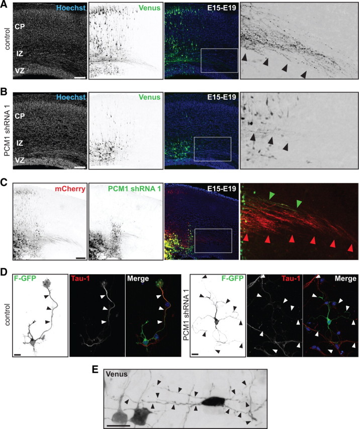
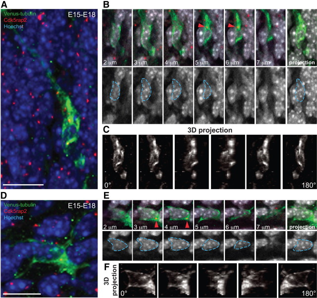
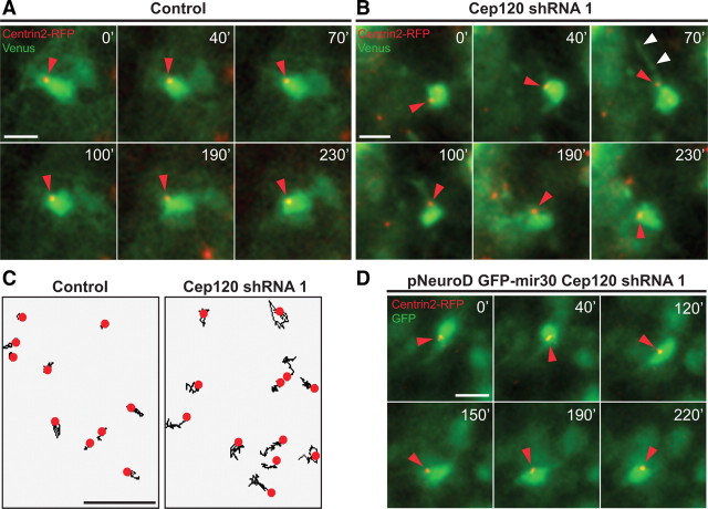
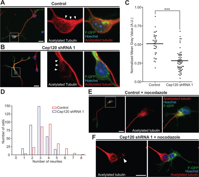
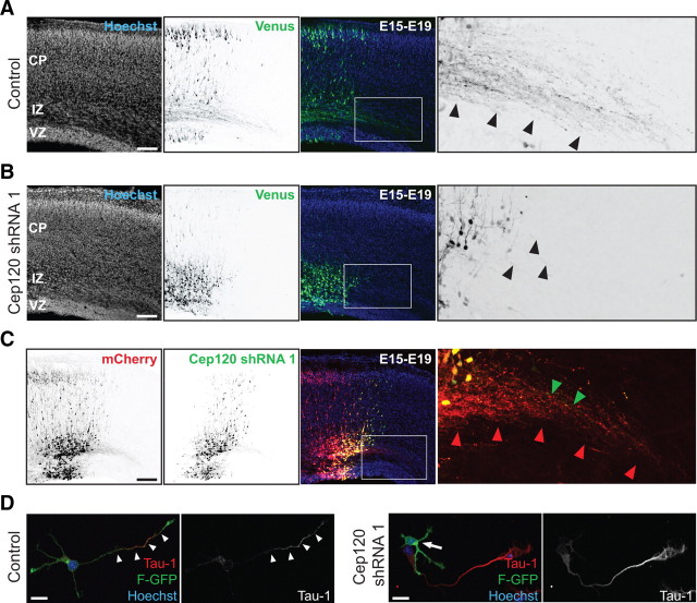
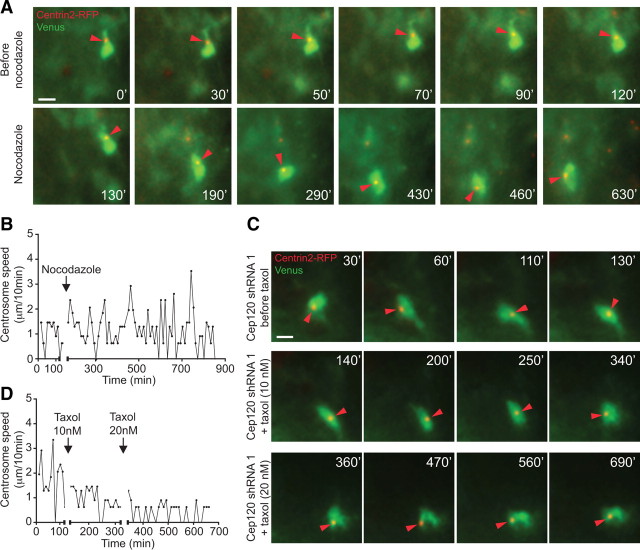
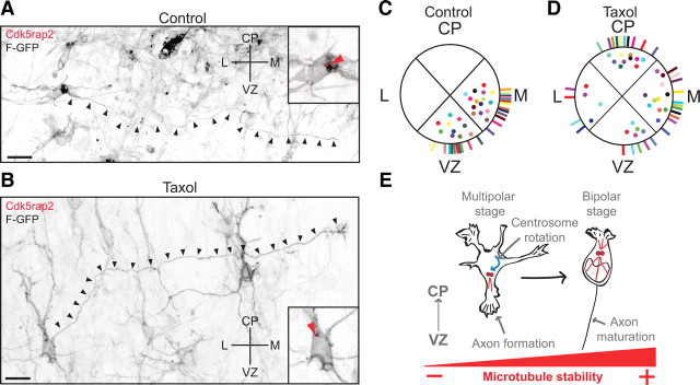
The mechanisms underlying the normal development of neuronal morphology remain a fundamental question in neurobiology. Studies in cultured neurons have suggested that the position of the centrosome and the Golgi may predict the site of axon outgrowth. During neuronal migration in the developing cortex, however, the centrosome and Golgi are oriented toward the cortical plate at a time when axons grow toward the ventricular zone. In the current work, we use in situ live imaging to demonstrate that the centrosome and the accompanying polarized cytoplasm exhibit apical translocation in newborn cortical neurons preceding initial axon outgrowth. Disruption of centrosomal activity or downregulation of the centriolar satellite protein PCM-1 affects axon formation. We further show that downregulation of the centrosomal protein Cep120 impairs microtubule organization, resulting in increased centrosome motility. Decreased centrosome motility resulting from microtubule stabilization causes an aberrant centrosomal localization, leading to misplaced axonal outgrowth. Our results reveal the dynamic nature of the centrosome in developing cortical neurons, and implicate centrosome translocation and microtubule organization during the multipolar stage as important determinants of axon formation.


Figures

Figure 1.
Figure 1.
The centrosomes of multipolar neurons translocate toward the VZ in advance of axon formation. A, Multipolar neurons in the lower IZ, identified by pNeuroD-GFP expression, are labeled with Cdk5rap2 (red arrowhead) and Hoechst. Cell body centroid analysis is used to determine centrosome position (middle panel). B, Quantification of centrosome position at E17 and E18 in pNeuroD-GFP-positive multipolar neurons from E15 in utero electroporation. C, Time-lapse imaging of a multipolar cell expressing F-GFP and Centrin2-RFP (insets, red arrowhead). At 0 h, centrosome is oriented toward the CP. From 2 to 12 h, the centrosome orients toward the VZ and midline (6 h). The cell assumes a bipolar morphology (open arrowheads) with the centrosome toward the leading edge and the CP at 14 h. The apical neurite accumulates more membrane components when the centrosome is located at its base (black arrowhead). D, E, Multipolar cells form an axon before migration into the CP. D, Time-lapse imaging of a cell expressing F-GFP and Centrin2-RFP, with the centrosome toward the VZ before elongation of the axon (insets in 0 h-4 h, red arrowheads). The centrosome translocates toward the CP (red arrowheads, inset in 6 h-8 h) and the cell reassumes a bipolar morphology, leaving the growing axon behind (black arrowheads, 6 h-24 h). E, Inversion of the polarized cytoplasm (red arrowheads, inset in 0–12 h) in a cell expressing F-GFP toward the VZ before elongation of the axon (black arrowhead). Scale bars: A, CE, 10 μm.
Figure 2.
Figure 2.
Temporal and spatial relationship between centrosome relocation and axonal specification in multipolar cells. A, The centrosome (red arrowhead) is oriented toward the neurite that will form the axon (black arrowheads), and the neurite is L1 negative. C, Migrating neuron with the centrosome oriented toward the leading edge (red arrowhead) and the trailing process positive for L1 (black and red arrowheads, inset 1 from left panel). B, GFP and L1 intensity profiles from the selected place for y–z scan in A. D, GFP and L1 intensity profiles from the selected place for x–z scan in C. The profile in B and D was obtained by drawing a line that crossed the middle of the GFP signal. Scale bars: A, C, 10 μm.
Figure 3.
Figure 3.
Light-induced inactivation of Centrin2 disrupts axon formation and neuronal migration in situ. A, Centrin2-KR fusion protein (left panel, red arrowhead) localized with the polarized F-GFP signal (left panel, green arrow). B, Before CALI, the cell displayed several neurites (black arrowheads), and a polarized cytoplasm (inset, red arrow). CALI of Centrin2-KR induces neurite retraction (10 min, 4 h, open arrowheads) and redistribution of the polarized F-GFP signal (10 min, inset, red arrows). Eventually, neurites regrew (24 h, black arrowheads) and the cytoplasm repolarized (4 h, 24 h, inset, red arrow). C, Control cells (left panel, green arrowhead) did not express Centrin2-KR (left panel, red arrowhead) but were irradiated with green light. D, The same cell as C (green arrowhead) developed an axon (8 h-42 h, black arrowheads) and markedly polarized cytoplasm (8–18 h, inset, red arrow). The neurite is L1 positive (left bottom, Inset 1 in 42 h, black and red arrowheads). E, F, 24 h after CALI, control slices show labeled cells migrating toward the CP. G, 24 h after CALI, irradiated slices have the majority of the labeled cells in the IZ. H, Quantification of cell distribution in control and irradiated slices 24 h after CALI (mean ± SEM; CP: ***p < 0.0001; IZ: *p = 0.0109; VZ: **p = 0.0043 by one-way ANOVA). I, 48 h after CALI, control slices have a robust callosal axonal tract (black arrowheads). J, 48 h after CALI, treated slices have few F-GFP-positive callosal axons (red arrows). Scale bars: A–D, 10 μm; E–G, I, J, 200 μm.
Figure 4.
Figure 4.
PCM-1 downregulation disrupts axon formation and neuronal migration. A, In utero electroporation of control shRNA at E15 labels transfected cells that form a callosal axonal tract (inset, black arrowheads) at E19. B, PCM-1 shRNA expression resulted in migration defect (middle panel) and failure to form a callosal axonal tract (inset, black arrowheads). C, The cell-autonomous phenotype of PCM-1 shRNA was demonstrated by sequential electroporation of mCherry and PCM-1 shRNA/Venus. Cells with only mCherry expression migrate toward the CP and project callosal axons. PCM-1 shRNA-transfected cells failed to migrate (middle panel) and have limited callosal axonal projections (right inset, green arrowheads). D, PCM-1 downregulation inhibits axon formation in vitro. Polarized control neuron extending a long neurite with typical Tau-1 gradient (black and white arrowheads). PCM-1 downregulation disrupts axon formation but cells have several long (> 40 mm) and thin Tau-1 negative neurites (black and white arrowheads). E, Multipolar cells in the IZ displayed several long and thin neurites after PCM-1 shRNA. Scale bars: AC, 200 μm; D, E, 10 μm.
Figure 5.
Figure 5.
Differential microtubule organization in bipolar and multipolar cells. A, A characteristic Venus-tubulin-expressing bipolar neuron in the lower CP labeled with the centrosomal marker Cdk5rap2 and Hoechst. B, Top: inset from cell in A showing different stacks (2–7 μm) and the maximal intensity projection (right) of the Venus-tubulin, Cdk5rap2 and Hoechst signal. The centrosome from cell in A is shown (red arrowhead). Bottom: different stacks (2–7 μm) and the maximal intensity projection of the Hoechst signal. The nucleus from cell in A is delineated with blue dashed lines. C, 3D reconstruction and rotation (0–180°) from the Venus-tubulin signal of the cell in A. Note the complex microtubule network that surrounds the nucleus and converge at the centrosome. D, Multipolar neuron in the IZ with Venus-tubulin expression labeled with Cdk5rap2 and Hoechst. E, Top: inset from the cell in D showing different stacks (2–7 μm) and the maximal intensity projection (right) of the Venus-tubulin, Cdk5rap2, and Hoechst signals. The centrosome from cell in D is shown (red arrowhead). Bottom: different stacks (2–7 μm) and the maximal intensity projection of the Hoechst signal. The nucleus from cell in D is delineated with blue dashed lines. F, 3D reconstruction and rotation (0–180°) of the Venus-tubulin signal in the cell from D. Note the lower abundance of the microtubule network surrounding the nucleus compared with C. Scale bars: A, D, 10 μm.
Figure 6.
Figure 6.
Cep120 silencing increases centrosome motility in multipolar cells. A, Time-lapse imaging of a multipolar control cell from the IZ. B, Transfection with Cep120 shRNA increases centrosome motility (red arrowheads), and the frequent extension and retraction of neurites (white arrowheads: neurite extension) (supplemental Video 2, available at www.jneurosci.org as supplemental material). C, Centrosome trajectories in control and Cep120 downregulated cells (4 h time lapse). D, Time-lapse imaging of a multipolar cell from the IZ expressing pNeuroD GFP-mir30 Cep120 shRNA. Neuronal-specific Cep120 silencing increases centrosome motility (red arrowheads). Supplemental Video 2 (available at www.jneurosci.org as supplemental material) corresponds to A, B. Time: minutes. Scale bars, 10 μm.
Figure 7.
Figure 7.
Cep120 silencing decreases microtubules stability. A–C, The acetylated microtubule content diminishes in the soma of neurons with Cep120 suppression. A, Example of a control neuron (left). Inset of cell body (white box from left panel): the cell body is rich in stable microtubules (white arrowheads). B, Example of a neuron transfected with Cep120 shRNA (left). Inset of cell body (white box from left panel): Cep120 downregulation decreased the stable microtubules in the cell body (white arrows). C, Silencing Cep120 reduced the acetylated microtubule content in the cell body (mean ± SEM; ***p < 0.0001 by t test). D, Frequency distribution of the number of neurites in Cep120-silenced cells and control cells. E, Control neuron transfected with F-GFP and shRNA control plasmid and treated with nocodazole (6 μm, 30 min). Stable microtubules remain in long processes and in the cell body. F, A neuron transfected with F-GFP and Cep120 shRNA. Nocodazole treatment depolymerizes microtubules completely (white arrowhead). Scale bars: A, B, E, F, 10 μm.
Figure 8.
Figure 8.
Cep120 silencing affects axon formation. A, In control brains long callosal projections are evident at E19 four days after in utero electroporation. The majority of transfected neurons are located in the CP, extending long axon projections (right inset from white box from middle panel, black arrowheads). B, Silencing of Cep120 resulted in cell mispositioning at deep layers of the neocortex (middle), and transfected neurons failed to form a robust callosal axonal tract (right inset from white box from middle panel, black arrowheads). C, Sequential transfection of mCherry–Cep120 shRNA/Venus confirm that the axon formation and migration defects after Cep120 downregulation is cell autonomous. Cells expressing just mCherry migrate toward the CP (left) and project callosal axons (right inset from white box from middle panel, red arrowheads). Cep120-silenced cells failed to migrate toward the CP (middle), remaining in the IZ, and the callosal axonal tract was not well developed (right inset from white box from middle panel, green arrowheads). D, Cep120 downregulation precludes axon formation in vitro. Left: polarized control neuron extending a long neurite with the typical Tau-1 gradient toward its distal part (white arrowheads). Right: Cep120 downregulation precludes the formation of a single Tau-1-positive neurite. Scale bars: A–C, 200 μm; D, 10 μm.
Figure 9.
Figure 9.
Microtubule dynamics control centrosome motility in multipolar cells. A, Time-lapse imaging of a multipolar cell in the IZ before and after nocodazole. Nocodazole (2 μm) increases centrosome motility (red arrowheads, bottom). B, Centrosome velocity changes in cell in A before and after nocodazole (black arrow). C, Time-lapse imaging of a multipolar cell with Cep120 shRNA expression before and after taxol. Taxol (10 nm) decreased centrosome movement (red arrowheads, intermediate panel); 20 nm taxol further inhibited centrosome motility (red arrowheads, bottom). D, Profile of centrosome velocity in cell in C before and after taxol (black arrows). Supplemental Videos 3 and 4 (available at www.jneurosci.org as supplemental material) correspond to A and C, respectively. Time: minutes. Scale bars: A, C, 10 μm.
Figure 10.
Figure 10.
Microtubule stabilization changes the position of axon outgrowth. A, The axon (black arrowheads) grows proximal to the centrosomal pole (inset at right, red arrowhead) in control cells. B, Microtubule stabilization with taxol (10 nm) changes the axon outgrowth position in some multipolar cells. Aberrant initial trajectory of the axon that is proximal to the centrosomal pole (inset at right, red arrowhead). Distal parts of the axon project properly. C, D, Quantification of the centrosome (dots) and axon outgrowth position (lines) in control cells (C) and after taxol treatment (D). In each quadrant, dots and lines with unique colors represent a single cell. E, Model of microtubule dynamics in multipolar and bipolar neurons. Microtubule dynamics regulate the rotation of the centrosome (red dots) toward the VZ in multipolar cells; this centrosomal positioning underlies axon formation. Subsequently, stable microtubules form a microtubule aster in bipolar migrating neurons (red lines), to promote nuclear translocation and axon elongation. Scale bars: A, B, 10 μm.

Similar articles

Cited by

References

    1. Adler CE, Fetter RD, Bargmann CI. UNC-6/Netrin induces neuronal asymmetry and defines the site of axon formation. Nat Neurosci. 2006;9:511–518. - PMC - PubMed
    1. Arimura N, Kaibuchi K. Neuronal polarity: from extracellular signals to intracellular mechanisms. Nat Rev Neurosci. 2007;8:194–205. - PubMed
    1. Asada N, Sanada K, Fukada Y. LKB1 regulates neuronal migration and neuronal differentiation in the developing neocortex through centrosomal positioning. J Neurosci. 2007;27:11769–11775. - PMC - PubMed
    1. Baas PW, Yu W. A composite model for establishing the microtubule arrays of the neuron. Mol Neurobiol. 1996;12:145–161. - PubMed
    1. Bai J, Ramos RL, Ackman JB, Thomas AM, Lee RV, LoTurco JJ. RNAi reveals doublecortin is required for radial migration in rat neocortex. Nat Neurosci. 2003;6:1277–1283. - PubMed

Publication types

MeSH terms