. 2010 Aug 4;30(31):10441-7.
doi: 10.1523/JNEUROSCI.5943-09.2010.

Lipoprotein particles cross the blood-brain barrier in Drosophila

Affiliations

Lipoprotein particles cross the blood-brain barrier in Drosophila

Marko Brankatschk et al. J Neurosci. .

Abstract

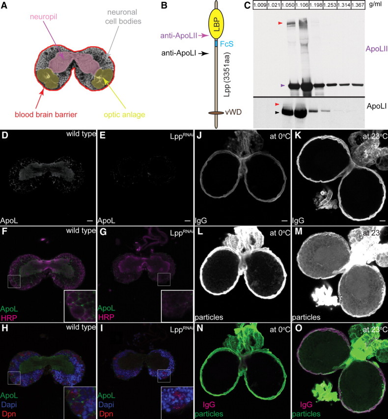
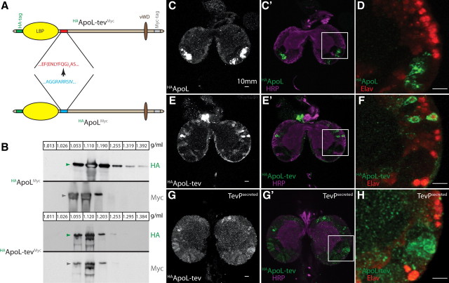
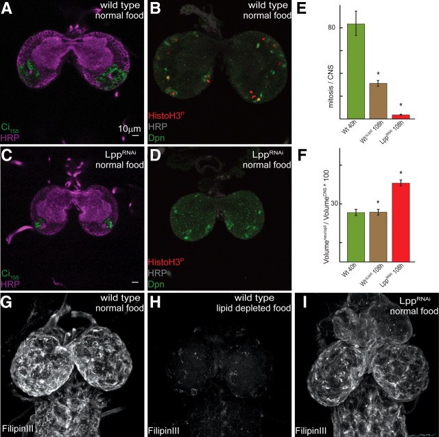
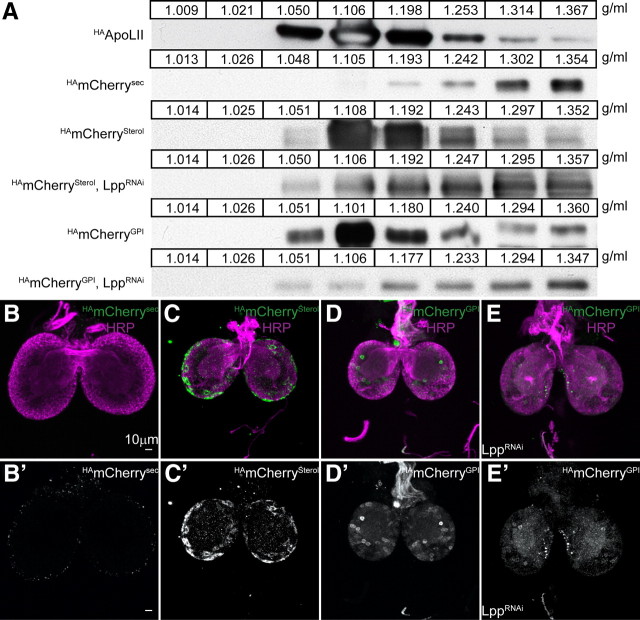
The blood-brain barrier (BBB) regulates passage of nutrients and signaling molecules from the circulation into the brain. Whether lipoproteins cross the BBB in vivo has been controversial, and no clear requirement for circulating lipoproteins in brain development has been shown. We address these issues in Drosophila, which has an functionally conserved BBB, and lipoproteins that resemble those of vertebrates. We show that the Drosophila lipoprotein lipophorin exists in two isoforms. Both isoforms cross the BBB, but accumulate on distinct subsets of cells within the brain. In addition to acting as a lipid carrier, lipophorin carries both sterol-linked and GPI-linked proteins into the circulation and transports them across the BBB. Finally, lipophorin promotes neuroblast proliferation by a mechanism that does not depend on delivery of dietary lipids. Transport of lipophorin and its cargo across the BBB represents a novel mechanism by which peripherally synthesized proteins might enter the brain and influence its development. Furthermore, lipid-linkage may be an efficient method to transport therapeutic molecules across the BBB.


Figures

Figure 1.
Figure 1.
Lpp particles cross the larval BBB. A, Scheme depicts the larval CNS at the level of the commissure: neuropil (magenta), optic anlage (yellow), cell bodies (gray) and the BBB (red). B, Motifs within apolipophorin. Lipid-binding pocket (LBP) (yellow oval), Furin cleavage site (FcS) (blue line), von Willebrand factor D domain (vWD) homology region (brown oval). Arrows indicate the position of peptides used to generate anti-ApoLII (magenta) and anti-ApoLI (black). C, Western blots of larval hemolymph iodixanol density gradients probed with anti-ApoLII and anti-ApoLI. Arrowheads indicate ApoLII (magenta), ApoLI (black) and ApoFL (red). Fraction densities in g/ml. D–I, Confocal section at the level of the commissures from wild-type (D, F, H) and LppRNAi (E, G, I) CNS stained with anti-Lpp (D, E; F–I; green), anti-HRP (F, G; magenta), anti-Dpn (H, I; red) and DAPI (4′,6′-diamidino-2-phenylindole dihydrochloride) (H, I; blue). J–O, Wild-type larval brains incubated at 0°C (J, L, N) or 23°C (K, M, O) with labeled Lpp (L, M; N, O, in green) and labeled IgG (J, K; N, O; magenta). Ten micrometer projections at the level of the commissure.
Figure 2.
Figure 2.
HAApoL-tevMyc and HAApoLII localize differently within the CNS. A, Replacement of the apolipophorin furin cleavage site (blue) with TEV-protease cleavage sequences (red). HA (green) and Myc (gray) tags are shown. Other motifs as in Figure 1B. B, Western blots of hemolymph iodixanol density gradients from larvae expressing either HAApoLMyc or HAApoL-tevMyc. Blots are probed with anti-HA (green arrowheads) and anti-Myc (gray arrowheads). Size heterogeneity of HAApoL-tevMyc detected with both N-terminal HA and C-terminal Myc tags suggests alternative splicing may generate different ApoLFL isoforms. Fraction densities in g/ml. C–H, Ten micrometer projections of second instar brains from larvae expressing either HAApoLMyc (C, C′, D), HAApoL-tevMyc (E, E′, F), or HAApoL-tevMyc + TevPsecreted (G, G′, H) in the fat body. Brains are stained with anti-HA (C, E, G; C′–H; green), anti-HRP (C′, E′, G′; magenta) and anti-Elav (D, F, H; red). Staining was observed only in the optic anlage for three of three brains expressing HAApoLMyc. Both neuronal and optic anlage staining was observed for five of six brains from animals expressing HAApoL-tevMyc + TevPsecreted. Optic anlage staining was never observed in five of five brains from animals expressing either HAApoL-tevMyc alone or + TevPcytoplasmic.
Figure 3.
Figure 3.
Lpp loss of function blocks neuroblast proliferation. A–D, Confocal sections of second instar brains from wild-type (A, B) or LppRNAi (C, D) larvae, stained with anti-HRP (A, C; magenta), anti-Ci155 (A, C; green), anti-HRP (B, D; gray), anti-Dpn (B, D; green) or anti-HistoH3P (B, D; red). Ci-155 labels the optic anlage, Dpn marks neuroblasts, and phospho-histone H3 indicates mitotic cells. E, Quantification of phospho-histone H3-positive cells within second instar brains. Green bar, Wild-type larvae fed normal food (n = 5, 83.4 ± 21.5); brown bar, wild-type larvae fed lipid-depleted food (n = 5, 31.2 ± 5.2); red bar, LppRNAi larvae fed normal food (n = 6, 3.5 ± 1.0). Asterisks indicate significant differences from wild-type larvae fed normal food (p < 0.001). Error bars are SDs. F, Neuropil/CNS volume ratio of wild-type larvae raised on normal food (green bar) or lipid-depleted food (brown bar) and LppRNAi larvae raised on normal food (red bar). Asterisks indicate significant differences from wild-type larvae fed normally (p < 0.0001). Error bars are SDs. G–I, Projections of whole brains stained with FilipinIII from wild-type larvae raised on normal food (G) or lipid-depleted food (H) and LppRNAi larvae raised on normal food (I).
Figure 4.
Figure 4.
HAmCherryGPI crosses the BBB on Lpp. A, Iodixanol density gradients of hemolymph from third instar wild-type (rows 1, 2, 3, 5) and LppRNAi larvae (rows 4, 6) expressing HAmCherrysec, HAmCherrysterol or HAmCherryGPI in the fat body. Western blots are probed with anti-ApoLII (first row) or anti-HA (rows 2–6). Fraction densities are shown in g/ml. B–D′, Forty micrometer projection at the level of the commissures from wild-type (B, C′) and LppRNAi (D, D′) second instar brains expressing HAmCherrysec (B, B′), HAmCherrysterol (C, C′), or HAmCherryGPI (D, E′) in the fat body stained with anti-HA (B′, C′, D′; B–D; green) and anti-HRP (B–D, magenta). HAmCherrysec is not found within the CNS (0 HAmCherry-positive neurons in 10 brains examined). HAmCherryGPI accumulates in the CNS (>50 HAmCherry-positive neurons in all 5 brains examined). HAmCherrysterol accumulates weakly in the CNS (5, 13, and 22 HAmCherry-positive neurons in 3 brains examined).

References

    1. Awasaki T, Lai SL, Ito K, Lee T. Organization and postembryonic development of glial cells in the adult central brain of Drosophila. J Neurosci. 2008;28:13742–13753. - PMC - PubMed
    1. Balazs Z, Panzenboeck U, Hammer A, Sovic A, Quehenberger O, Malle E, Sattler W. Uptake and transport of high-density lipoprotein (HDL) and HDL-associated alpha-tocopherol by an in vitro blood-brain barrier model. J Neurochem. 2004;89:939–950. - PubMed
    1. Banerjee S, Bhat MA. Neuron-glial interactions in blood-brain barrier formation. Annu Rev Neurosci. 2007;30:235–258. - PMC - PubMed
    1. Boone JQ, Doe CQ. Identification of Drosophila type II neuroblast lineages containing transit amplifying ganglion mother cells. Dev Neurobiol. 2008;68:1185–1195. - PMC - PubMed
    1. Brankatschk M, Dickson BJ. Netrins guide Drosophila commissural axons at short range. Nat Neurosci. 2006;9:188–194. - PubMed

Publication types

LinkOut - more resources