. 2010 Aug 4;30(31):10484-92.
doi: 10.1523/JNEUROSCI.4721-09.2010.

Notch1 is required for maintenance of the reservoir of adult hippocampal stem cells

Affiliations

Notch1 is required for maintenance of the reservoir of adult hippocampal stem cells

Jessica L Ables et al. J Neurosci. .

Abstract

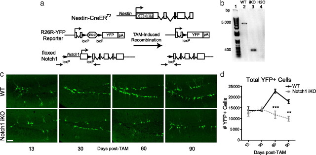
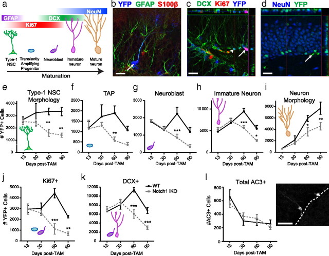
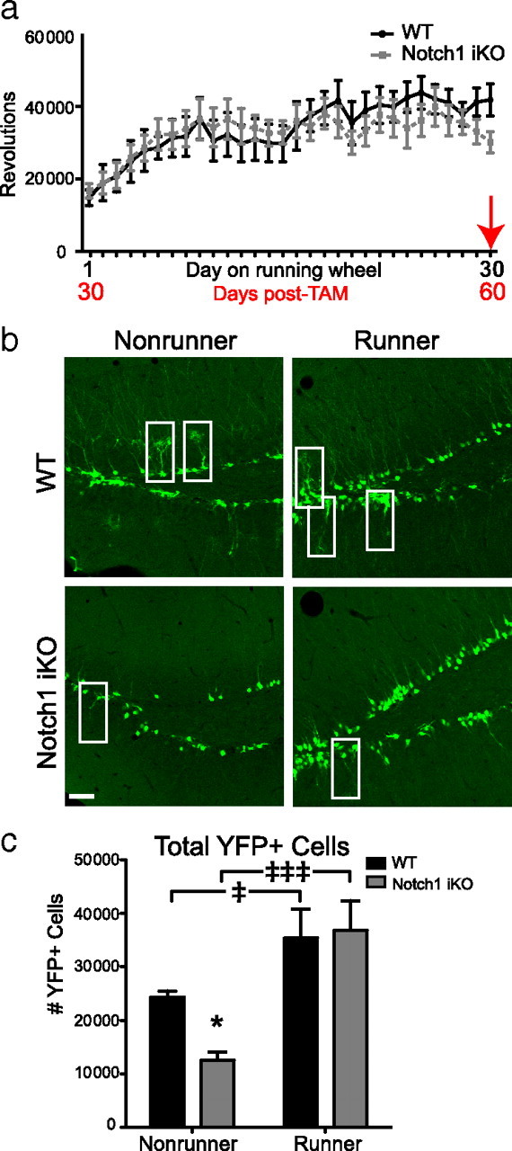
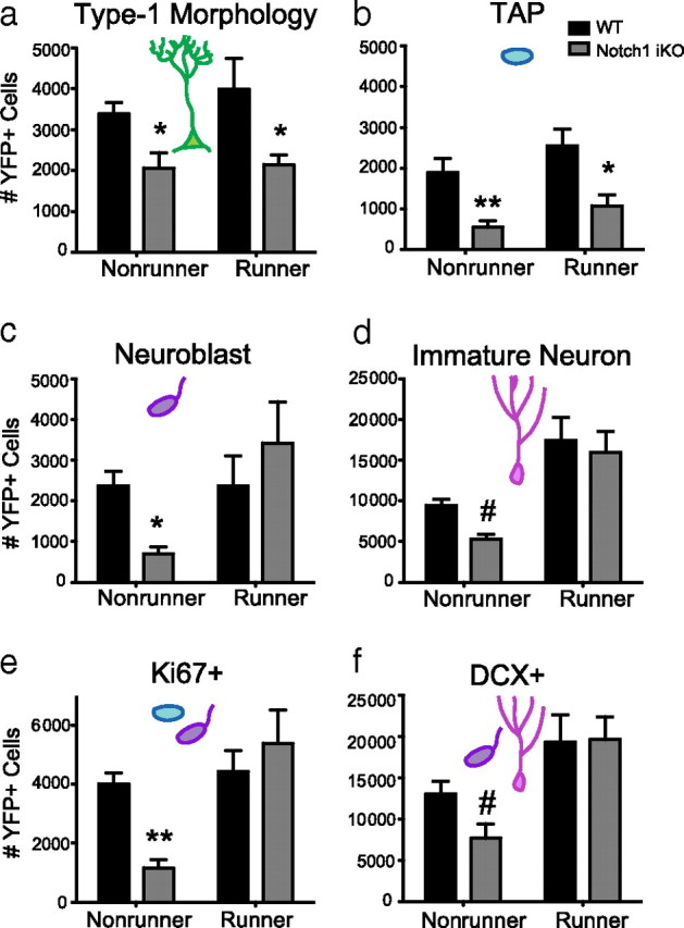
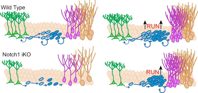
Notch1 regulates neural stem cell (NSC) number during development, but its role in adult neurogenesis is unclear. We generated nestin-CreER(T2)/R26R-YFP/Notch1(loxP/loxP) [Notch1inducible knock-out (iKO)] mice to allow tamoxifen (TAM)-inducible elimination of Notch1 and concomitant expression of yellow fluorescent protein (YFP) in nestin-expressing Type-1 NSCs and their progeny in the adult hippocampal subgranular zone (SGZ). Consistent with previous research, YFP+ cells in all stages of neurogenesis were evident in the subgranular zone (SGZ) of wild-type (WT) mice (nestin-CreER(T2)/R26R-YFP/Notch1(w/w)) after tamoxifen (post-TAM), producing adult-generated YFP+ dentate gyrus neurons. Compared with WT littermates, Notch1 iKO mice had similar numbers of total SGZ YFP+ cells 13 and 30 d post-TAM but had significantly fewer SGZ YFP+ cells 60 and 90 d post-TAM. Significantly fewer YFP+ Type-1 NSCs and transiently amplifying progenitors (TAPs) resulted in generation of fewer YFP+ granule neurons in Notch1 iKO mice. Strikingly, 30 d of running rescued this deficit, as the total YFP+ cell number in Notch iKO mice was equivalent to WT levels. This was even more notable given the persistent deficits in the Type-1 NSC and TAP reservoirs. Our data show that Notch1 signaling is required to maintain a reservoir of undifferentiated cells and ensure continuity of adult hippocampal neurogenesis, but that alternative Notch- and Type-1 NSC-independent pathways compensate in response to physical activity. These data shed light on the complex relationship between Type-1 NSCs, adult neurogenesis, the neurogenic niche, and environmental stimuli.


Figures

Figure 1.
Figure 1.
Notch1 iKO mice have fewer YFP+ cells in the SGZ. a, Nestin-CreERT2 and R26R-YFP mice were crossed with floxed Notch1 mice to generate Notch1 iKO mice. A large portion of the rat nestin gene drives expression of a fusion protein of Cre recombinase and the modified estrogen receptor (CreERT2). Administration of tamoxifen (TAM) results in removal of the promoter and first exon of notch1, and the “stop” signal of R26R-YFP. This leads to elimination of Notch1 and to expression of YFP in nestin-expressing cells and their progeny. b, PCR of neurospheres isolated 40d post-TAM confirmed that the notch1 locus was recombined after TAM in Notch1 iKO mice but not WT littermates. Primers (indicated by arrows in a) were designed against regions outside the floxed portion of notch1. c, YFP+ SGZ cells in WT and Notch1 iKO mice. d, YFP+ SGZ cell number. **p < 0.01, ***p < 0.001 vs WT, Bonferroni post hoc; n = 5–11/group. Scale bar: (c), 50 μm.
Figure 2.
Figure 2.
Elimination of Notch1 from nestin-expressing cells decreases YFP+ SGZ NSC number and neurogenesis. a, YFP+ SGZ cells express markers across stages of neurogenesis. b, YFP+GFAP+S100β− Type-1 NSC (arrow). c, YFP+Ki67+DCX− (arrow) and YFP+Ki67−DCX+ (arrowhead) cells. d, YFP+NeuN+ neurons (YFP+NeuN− cell, arrow). e, Notch1 iKO mice have significantly fewer Type-1 NSCs at extended times post-TAM. f, There are fewer TAPs (YFP+Ki67+DCX− cells) in Notch1 iKO mice regardless of time post-TAM. g, Notch1 iKO mice have significantly fewer neuroblasts (YFP+Ki67+DCX+ cells) at 60 d post-TAM. h, Notch1 iKO mice have significantly fewer immature neurons (YFP+Ki67−DCX+ cells) at extended times post-TAM. i, Notch1 iKO mice have significantly fewer YFP+ neurons at extended times post-TAM. j, Notch1 iKO mice have significantly fewer proliferating (Ki67+) YFP+ cells at extended times post-TAM. k, The number of YFP+ cells that are becoming neurons (DCX+) is significantly decreased in Notch1 iKO mice at extended times post-TAM. l, Apoptosis is not increased in the SGZ of Notch1 iKO mice compared with WT littermates. *p < 0.05, **p < 0.01, ***p < 0.001 vs WT, Bonferroni post hoc; n = 3–7/group. Scale bars: (b–d, l), 20 μm.
Figure 3.
Figure 3.
Dendritic morphology is decreased in Notch1 iKO mice. a, The number of dendrites per cell body in YFP+DCX+ cells in the SGZ did not differ between the two genotypes at 90 d post-TAM. b, Sholl analysis of YFP+DCX+ cells revealed significantly fewer dendritic intersections in Notch1 iKO mice, specifically at a radius range of 960–1060 μm. c, Representative YFP+DCX+ neuron tracings. *p < 0.05 vs WT, Bonferroni post hoc; n = 3–4/group.
Figure 4.
Figure 4.
Elimination of Notch1 from nestin-expressing Type-1 NSCs in vivo decreases neurosphere formation in vitro. a, Number of secondary spheres formed from cells isolated 40 d post-TAM from the subventricular zone of WT and Notch1 iKO mice. b, Representative photomicrographs of passage 6 neurospheres from WT and Notch1 iKO mice. Scale bars, 100 μm. *p < 0.05, t test; n = 3–4 replicates per group.
Figure 5.
Figure 5.
Physical activity rescues total YFP+ cell number in Notch1 iKO runner mice. a, Beginning 30 d post-TAM, animals were given free access to a running wheel for 30 d. WT and Notch1 iKO mice did not differ in the amount they ran in any given day. b, YFP+ SGZ cells in WT and Notch1 iKO nonrunners and runners; white boxes, Type-1 NSCs. c, Total YFP+ SGZ cells. Scale bar, 20 μm. *p < 0.05 vs WT and p < 0.05, ‡‡‡p < 0.001 vs nonrunner, Bonferroni post hoc; n = 4–11/group.
Figure 6.
Figure 6.
Physical activity rescues YFP+ neurogenesis but not YFP+ Type-1 NSCs or TAPs in Notch1 iKO mice. a, Thirty days of running did not affect the number of YFP+ Type-1 NSCs. b, Thirty days of running did not affect the number of TAPs (YFP+Ki67+DCX− cells). c, Running rescued the number of neuroblasts (YFP+Ki67+DCX+ cells) in Notch1 iKO mice. d, Running rescued the number of immature neurons (YFP+Ki67−DCX+ cells) in iKO mice. e, Thirty days of running was sufficient to normalize the number of proliferating (Ki67+) YFP+ cells in Notch1 iKO mice to WT levels. f, The number of DCX+YFP+ cells was increased in both WT and iKO mice after running. #p < 0.1, *p < 0.05, **p < 0.01 vs WT, Bonferroni post hoc; n = 3–8/group.
Figure 7.
Figure 7.
Proposed model of Notch1 in regulating adult neurogenesis under basal conditions and after physical activity. Without Notch1, self-renewal and expansion of nestin-expressing cells is disrupted and the net number of adult-generated dentate gyrus neurons is decreased. Physical activity increases adult-generated neurons in WT and Notch1 iKO mice by increasing neuroblast proliferation. However, physical activity does not rescue Type-1 NSC or TAP number in Notch1 iKO mice.

References

    1. Androutsellis-Theotokis A, Leker RR, Soldner F, Hoeppner DJ, Ravin R, Poser SW, Rueger MA, Bae SK, Kittappa R, McKay RD. Notch signalling regulates stem cell numbers in vitro and in vivo. Nature. 2006;442:823–826. - PubMed
    1. Annenkov A. The insulin-like growth factor (IGF) receptor type 1 (IGF1R) as an essential component of the signalling network regulating neurogenesis. Mol Neurobiol. 2009;40:195–215. - PubMed
    1. Artavanis-Tsakonas S, Rand MD, Lake RJ. Notch signaling: cell fate control and signal integration in development. Science. 1999;284:770–776. - PubMed
    1. Basak O, Taylor V. Stem cells of the adult mammalian brain and their niche. Cell Mol Life Sci. 2009;66:1057–1072. - PMC - PubMed
    1. Bednarczyk MR, Aumont A, Décary S, Bergeron R, Fernandes KJ. Prolonged voluntary wheel-running stimulates neural precursors in the hippocampus and forebrain of adult CD1 mice. Hippocampus. 2009;19:913–927. - PubMed

Publication types

Substances