. 2010 Aug 4;30(31):10551-62.
doi: 10.1523/JNEUROSCI.5563-09.2010.

LIM-homeobox gene Lhx5 is required for normal development of Cajal-Retzius cells

Affiliations

LIM-homeobox gene Lhx5 is required for normal development of Cajal-Retzius cells

Amaya Miquelajáuregui et al. J Neurosci. .

Abstract

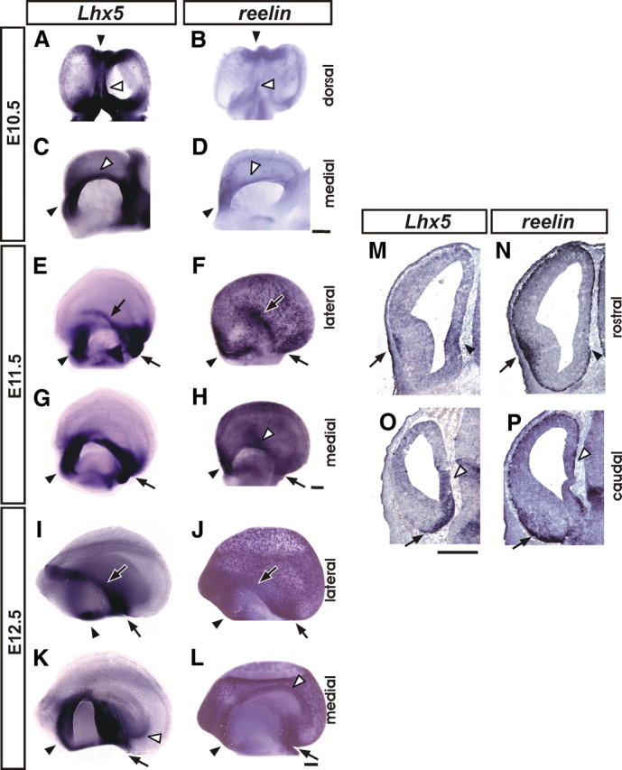
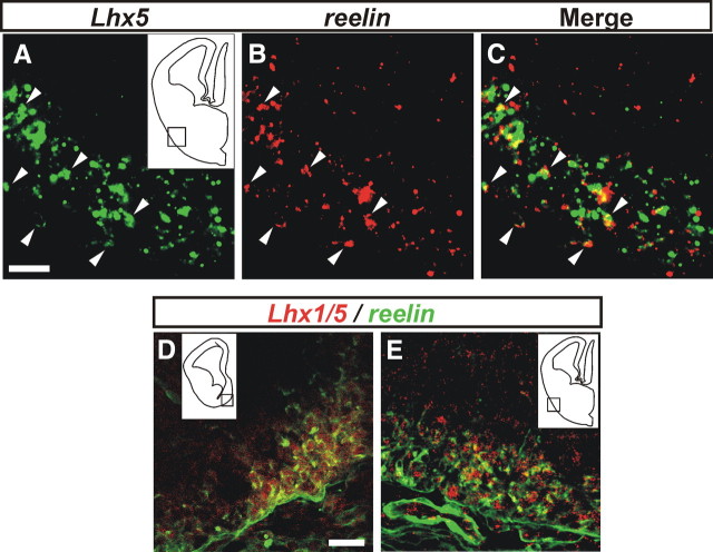
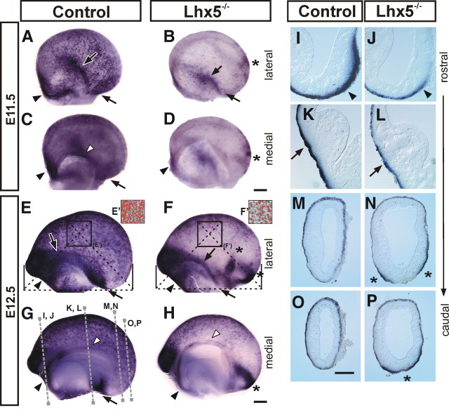
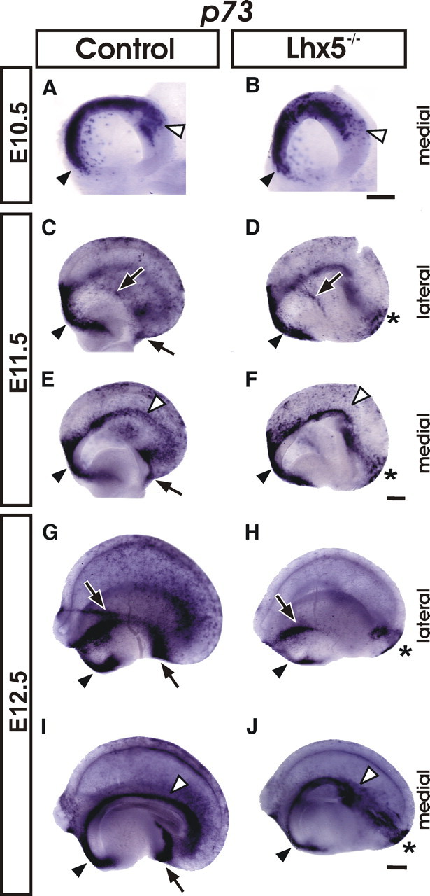
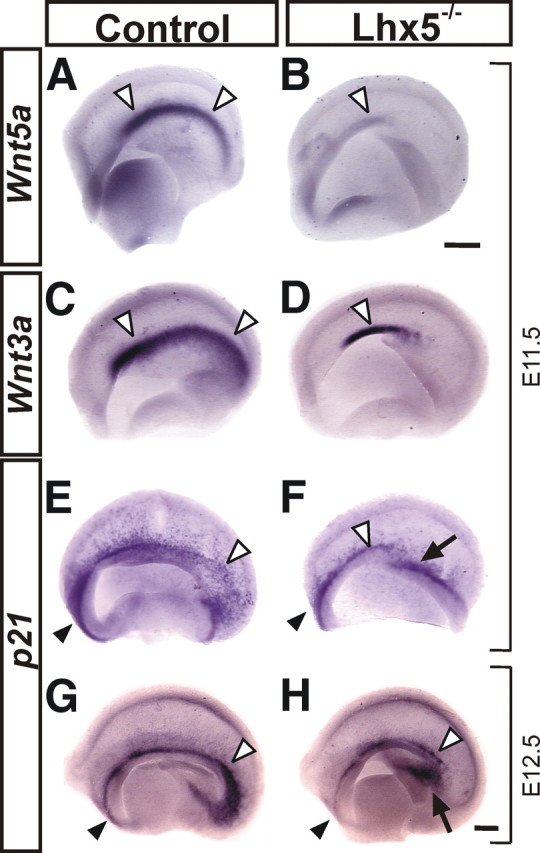
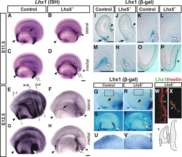
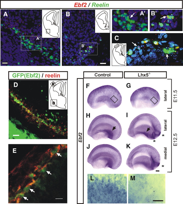
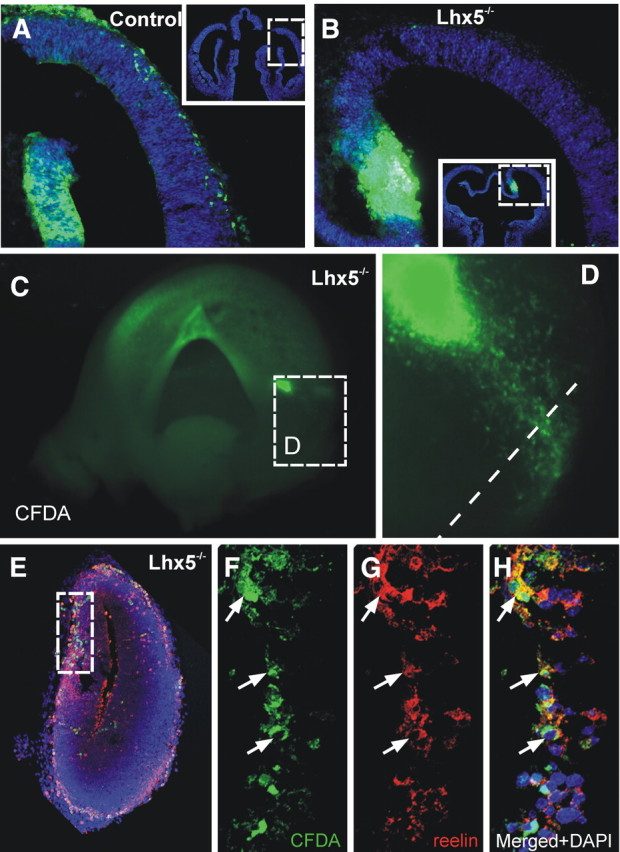
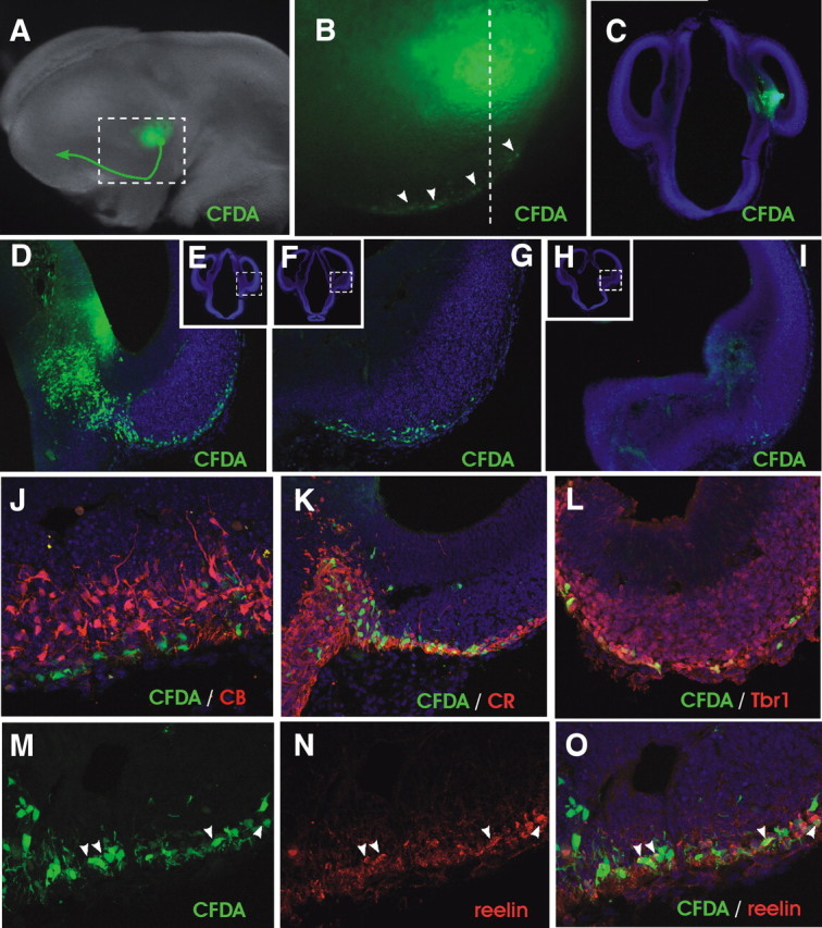
Cajal-Retzius (C-R) cells play important roles in the lamination of the mammalian cortex via reelin secretion. The genetic mechanisms underlying the development of these neurons have just begun to be unraveled. Here, we show that two closely related LIM-homeobox genes Lhx1 and Lhx5 are expressed in reelin+ cells in various regions in the mouse telencephalon at or adjacent to sites where the C-R cells are generated, including the cortical hem, the mantle region of the septal/retrobulbar area, and the ventral pallium. Whereas Lhx5 is expressed in all of these reelin-expressing domains, Lhx1 is preferentially expressed in the septal area and in a continuous domain spanning from lateral olfactory region to caudomedial territories. Genetic ablation of Lhx5 results in decreased reelin+ and p73+ cells in the neocortical anlage, in the cortical hem, and in the septal, olfactory, and caudomedial telencephalic regions. The overall reduction in number of C-R cells in Lhx5 mutants is accompanied by formation of ectopic reelin+ cell clusters at the caudal telencephalon. Based on differential expression of molecular markers and by fluorescent cell tracing in cultured embryos, we located the origin of reelin+ ectopic cell clusters at the caudomedial telencephalic region. We also confirmed the existence of a normal migration stream of reelin+ cells from the caudomedial area to telencephalic olfactory territories in wild-type embryos. These results reveal a complex role for Lhx5 in regulating the development and normal distribution of C-R cells in the developing forebrain.


Figures

Figure 1.
Figure 1.
Lhx5 and reelin expression in the developing mouse telencephalon. Expression was analyzed by whole-mount in situ hybridization on telencephalic vesicles from E10.5 (A–D), E11.5 (E–H), and E12.5 (I–L), shown in dorsal, medial, and lateral views as indicated (rostral is to the top in A and B and to the left in C–L). M–P, E12.5 coronal telencephalic sections of comparable rostral (M, N) and caudal (O, P) levels were analyzed by ISH for Lhx5 (M, O) and reelin (N, P) (medial to the right). The black arrowheads point to the septal domain; the black arrows indicate the location of the pOC domain, and the white arrowheads mark the location of the cortical hem. Scale bars: A–D, 400 μm; E–P, 200 μm.
Figure 2.
Figure 2.
Expression of Lhx5/Lhx1 in reelin+ cells. A–C, Double fluorescent in situ hybridization on E12.5 coronal sections showing cellular colocalization of Lhx5 (A) and reelin (B) transcripts on a 3 μm optical section acquired from the pOC domain, as indicated in the diagram in A. C, Merged images (A, B). The arrowheads point to examples of colocalization of the Lhx5/reelin transcripts. D, E, Double immunostaining of Lhx5/1 (red) and reelin (green) of coronal brain sections from E12.5 embryos. Location of images from septal (D) and pOC (E) domains are indicated in insets. Scale bar, 20 μm.
Figure 3.
Figure 3.
Abnormal development of Cajal–Retzius cells in Lhx5-null mutant embryos. Whole-mount reelin in situ hybridization on E11.5 (A–D) and E12.5 (E–H) telencephalic vesicles from control and mutant (Lhx5−/−) littermates. As indicated, lateral and medial views of the telencephalon are shown (rostral is to the left). E′, F′, Overthreshold reelin+ signal (red overlay) in control and mutant embryos, respectively, obtained from equivalent regions in the lateral cortex (E, F, squared boxes) (for details, see Materials and Methods). I–P, Coronal sections showing expression of reelin in control and Lhx5−/− telencephalic hemispheres at the approximate rostrocaudal levels indicated by dotted lines in G (medial is to the right). For all panels, Black arrowheads, Septal domain; black arrows, pOC domain; white arrowheads, cortical hem; asterisks, ectopic reelin+ cell clusters. Scale bar, 200 μm.
Figure 4.
Figure 4.
p73 expression is altered in Lhx5 mutants. Whole-mount in situ hybridization on telencephalic vesicles from control and mutant embryos at E10.5 (A, B), E11.5 (C–F), and E12.5 (G–J) (rostral is to the left). The black arrowheads point to the septal domain; the black arrows indicate the location of the pOC domain; the white arrowheads mark the location of the cortical hem, and the asterisks indicate ectopic cell clusters. Scale bar, 200 μm.
Figure 5.
Figure 5.
Cortical hem alterations in Lhx5 mutants. Medial views of telencephalic vesicles from control and mutant embryos showing the expression of the hem-specific markers Wnt5a (A, B) and Wnt3a (C, D) at E11.5. E–H, Hem-derived C-R cells labeled by p21 expression at E11.5 and E12.5. Note the shortening of the cortical hem in Lhx5 mutants by Wnt3a expression, the dramatic reduction of Wnt5a expression levels, and the decreased numbers of p21+ cells in the medial telencephalon of Lhx5 mutants. In all frames, rostral is to the left. The white arrowheads point to the cortical hem, the black arrowheads to the septal region, and the arrows indicate the approximate site of origin of the ectopic cell clusters. Scale bar, 200 μm.
Figure 6.
Figure 6.
Lhx1 labels specific C-R cell subpopulations affected by Lhx5 mutation. A–H, Whole-mount Lhx1 in situ hybridization of control and Lhx5 mutants at E11.5 (A–D) and E12.5 (E–H). Medial and lateral views of the telencephalon are shown (rostral is to the left). I–V, Detection of LacZ activity from the Lhx1tlz allele by X-gal staining on coronal sections (I–P; approximate locations of the sections are indicated by dotted lines in E) and whole telencephalic preparations (Q–V) from E12.5 control and Lhx5 mutant embryos. U and V show higher magnification views of the dorsal cortex from boxed areas in Q and R, respectively. W, X, Double immunostaining of reelin (red) and Lhx1 (green) in a caudal telencephalic coronal section. The diagram in Y indicates location of ectopic clusters shown in W (lateral) and X (medial). For all panels: Black arrowheads, Septal region; white arrowheads, ectopic Lhx1+ cells; black arrows, pOC domain; white arrows, medial expression domain at the vCMTW; asterisks, ectopic cell clusters. Scale bar, 200 μm.
Figure 7.
Figure 7.
Expression of Ebf2 in C-R cells is affected in Lhx5 mutants. A–C, Double FISH of Ebf2 (red) and reelin (green) showing cellular colocalization at E12.5. DAPI (blue). A, pOC. B, vCMTW. A′ and B′ are magnified views of the boxed areas in A and B, respectively. C, Colocalization in mantle regions of the lateral pallium. Examples of cellular colocalization are indicated by arrows. D, E, Confocal images of the pOC domain and marginal zone of the lateral pallium from E12.5 embryos, respectively, showing that Ebf2-GFP+ cells (green) coexpress reelin (red). The insets in A–D indicate the location of the fields shown in each image. F–M, E11.5 and E12.5 telencephalic vesicles from control (F, H, J) and Lhx5 mutant (G, I, K) littermates analyzed by whole-mount in situ hybridization (rostral to the left; medial and lateral views as indicated). L, M, High magnification of lateral cortex at E11.5 as indicated by insets in F and G, respectively. Black arrows, pOC domain; asterisks, ectopic reelin+ cell clusters. Scale bar, 200 μm.
Figure 8.
Figure 8.
Reelin-expressing cells migrate aberrantly in Lhx5 mutants. A, B, Coronal sections of E11.5 control and mutant embryos injected with CFDA (green) in the cortical hem and cultured for 24 h. Migrating cells in the mutant migrate scarcely toward the dorsal telencephalon (insets show location of images in A and B). C–H, Lhx5 mutant embryo injected at the origin of the cellular ectopia and cultured in toto for 24 h. C, Medial view of a telencephalic vesicle showing the injection site at the vCMTW region (rostral is to the left). D, Magnified view of the boxed area in C showing cells migrating toward the caudal pole of the telencephalon. E–H, Immunostaining of Reelin on coronal sections of the same injected embryo (dotted lines in D indicate the approximate plane of section). F–H, Confocal optical section of the boxed area in E, showing CFDA-labeled cells (green) expressing reelin (red); DAPI (blue). The arrows point to examples of cellular colocalization.
Figure 9.
Figure 9.
Cell migration from the ventral region of the CMTW. A, Example of injection of the fluorescent tracer CFDA (green) into an E11.5 wild-type embryo followed by 24 h of culture. The arrow indicates the migratory route of labeled cells. B, Close-up of the boxed area shown in A. The arrowheads point to migratory cells in the ventral telencephalon. C, Coronal section at point indicated by dotted line in B showing the injection site. D–I, Examples of sections (caudal to rostral) showing the migratory path of labeled cells toward and along the pOC. D, G, and I are close-up views of the boxed areas shown in E, F, and H, respectively. J–O, Immunohistochemical analysis of migrating cells. Note that no expression of calbindin (CB) was detected among labeled cells (J), whereas calretinin (CR), Tbr1, and reelin were expressed by a fraction of them (K–O).

References

    1. Abellan A, Menuet A, Dehay C, Medina L, Retaux S. Differential expression of LIM-homeodomain factors in Cajal-Retzius cells of primates, rodents, and birds. Cereb Cortex. 2010;20:1788–1798. - PubMed
    1. Bielle F, Griveau A, Narboux-Nême N, Vigneau S, Sigrist M, Arber S, Wassef M, Pierani A. Multiple origins of Cajal-Retzius cells at the borders of the developing pallium. Nat Neurosci. 2005;8:1002–1012. - PubMed
    1. Chowdhury TG, Jimenez JC, Bomar JM, Cruz-Martin A, Cantle JP, Portera-Cailliau C. Fate of Cajal-Retzius neurons in the postnatal mouse neocortex. Front Neuroanat. 2010;4:10. - PMC - PubMed
    1. D'Arcangelo G, Nakajima K, Miyata T, Ogawa M, Mikoshiba K, Curran T. Reelin is a secreted glycoprotein recognized by the CR-50 monoclonal antibody. J Neurosci. 1997;17:23–31. - PMC - PubMed
    1. de Carlos JA, López-Mascaraque L, Valverde F. Dynamics of cell migration from the lateral ganglionic eminence in the rat. J Neurosci. 1996;16:6146–6156. - PMC - PubMed

Publication types

MeSH terms