. 2010 Oct;84(20):10773-83.
doi: 10.1128/JVI.01166-10. Epub 2010 Aug 4.

A single-amino-acid substitution in herpes simplex virus 1 envelope glycoprotein B at a site required for binding to the paired immunoglobulin-like type 2 receptor alpha (PILRalpha) abrogates PILRalpha-dependent viral entry and reduces pathogenesis

Affiliations

A single-amino-acid substitution in herpes simplex virus 1 envelope glycoprotein B at a site required for binding to the paired immunoglobulin-like type 2 receptor alpha (PILRalpha) abrogates PILRalpha-dependent viral entry and reduces pathogenesis

Jun Arii et al. J Virol. 2010 Oct.

Abstract

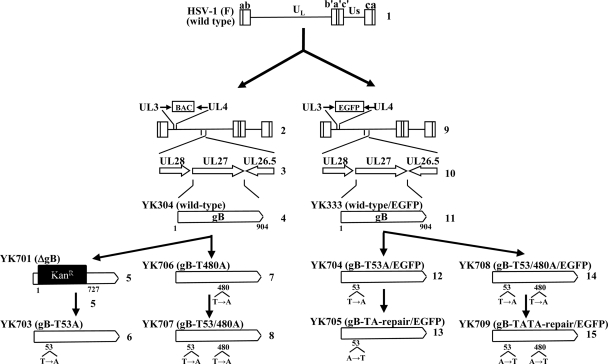
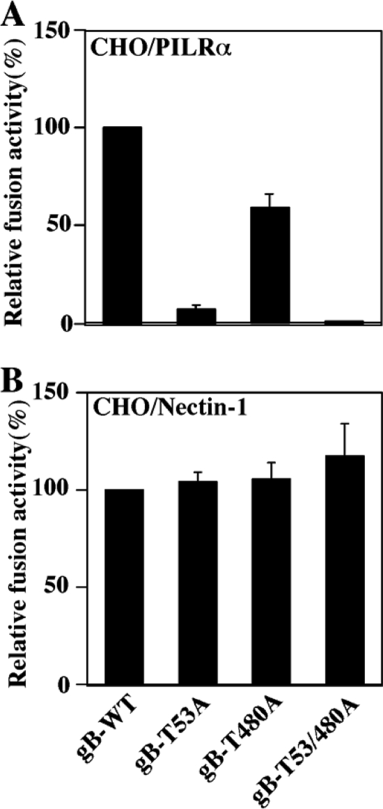
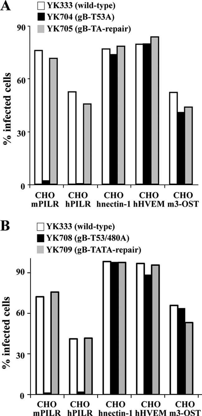
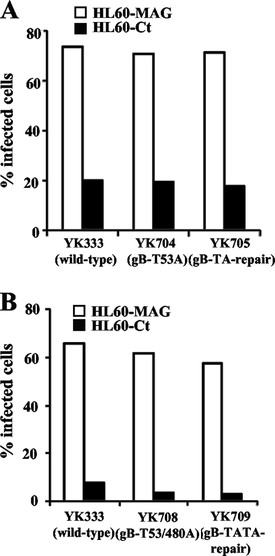
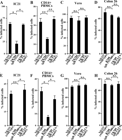
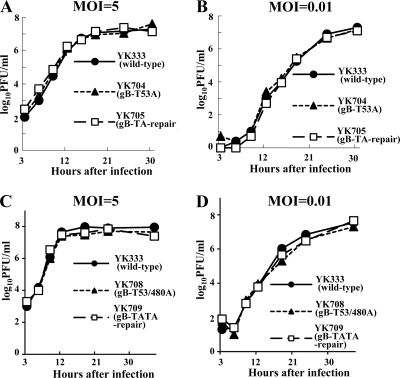
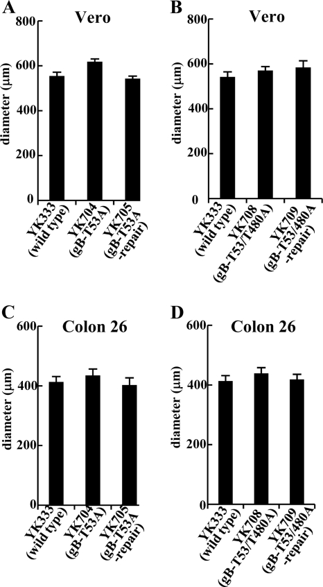
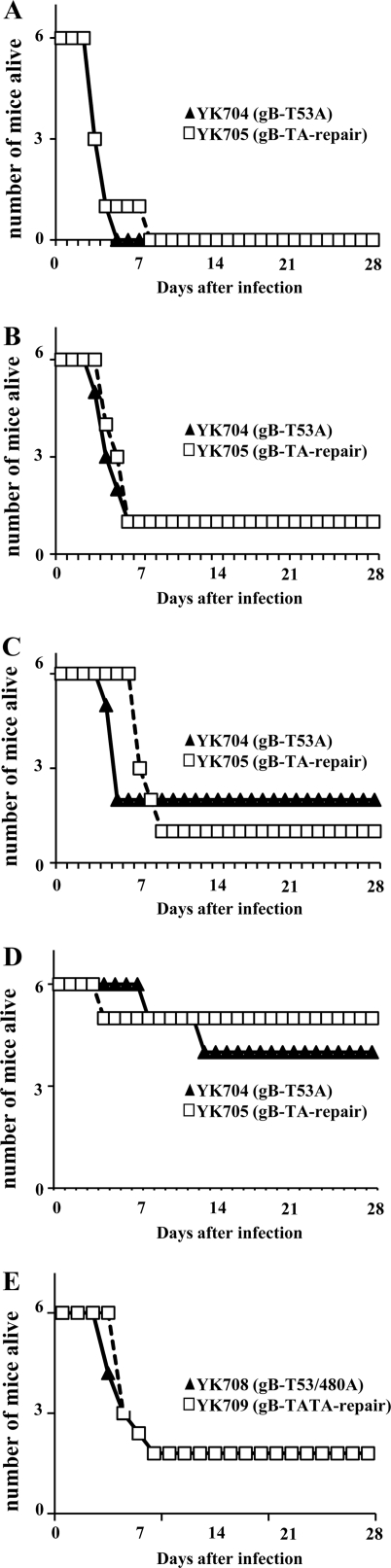
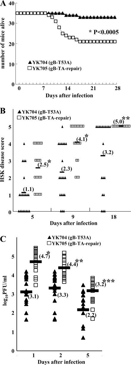
Paired immunoglobulin-like type 2 receptor α (PILRα) is a herpes simplex virus 1 (HSV-1) entry receptor that associates with O-glycans on HSV-1 envelope glycoprotein B (gB). Two threonine residues (Thr-53 and Thr-480) in gB, which are required for the addition of the principal gB O-glycans, are essential for binding to soluble PILRα. However, the role of the two threonines in PILRα-dependent viral entry remains to be elucidated. Therefore, we constructed a recombinant HSV-1 carrying an alanine replacement of gB Thr-53 alone (gB-T53A) or of both gB Thr-53 and Thr-480 (gB-T53/480A) and demonstrated that these mutations abrogated viral entry in CHO cells expressing PILRα. In contrast, the mutations had no effect on viral entry in CHO cells expressing known host cell receptors for HSV-1 gD, viral entry in HL60 cells expressing myelin-associated glycoprotein (MAG) (another HSV-1 gB receptor), viral attachment to heparan sulfate, and viral replication in PILRα-negative cells. These results support the hypothesis that gB Thr-53 and Thr-480 as well as gB O-glycosylation, probably at these sites, are critical for PILRα-dependent viral entry. Interestingly, following corneal inoculation in mice, the gB-T53A and gB-T53/480A mutations significantly reduced viral replication in the cornea, the development of herpes stroma keratitis, and neuroinvasiveness. The abilities of HSV-1 to enter cells in a PILRα-dependent manner and to acquire specific carbohydrates on gB are therefore linked to an increase in viral replication and virulence in the experimental murine model.


Figures

FIG. 1.
FIG. 1.
Construction of recombinant viruses. (Diagram 1) Linear representation of the HSV-1(F) genome. (Diagrams 2 and 9) Linear representations of the genomes of YK304 and YK333, respectively. The YK304 and YK333 genomes contain a bacmid (BAC) or EGFP expression cassette, respectively, in the intergenic region between UL3 and UL4. (Diagrams 3 and 10) Domains encoding the UL26.5, UL27 (gB), and UL28 open reading frames. (Diagrams 4 and 11) The UL27 gene product (gB). (Diagrams 5 to 8 and 12 to 15) Schematic diagrams of the UL27 gene product (gB) in the recombinant viruses YK701, YK703, YK706, YK707, YK704, YK705, YK708, and YK709, respectively.
FIG. 2.
FIG. 2.
Effect of the T53A and/or the T480A mutation in gB on cell-cell fusion. CHO cells were transfected with wild-type gB or mutant gB as well as other HSV-1 glycoproteins (gD, gH, and gL) and luciferase driven by a T7 promoter (effector cells) and were cocultured with CHO cells transfected with PILRα (A) or nectin-1 (B) and T7 polymerase (target cells). After 18 h of incubation, luciferase activity was measured. Cell fusion activities relative to that of wild-type gB are shown. These results are the averages and standard errors of data from three independent experiments.
FIG. 3.
FIG. 3.
Effect of the T53A and T53/480A mutations in gB on viral entry into CHO cells expressing viral receptors. CHO transformants were infected with YK333 (wild-type/EGFP), YK704 (gB-T53A/EGFP), or YK705 (gB-TA-repair/EGFP) (A) or with YK333 (wild-type/EGFP), YK708 (gB-T53/480A/EGFP), or YK709 (gB-TATA-repair/EGFP) (B). At 10 h postinfection, live infected CHO-mPILR, CHO-hPILR, CHO-hNectin-1, and CHO-hHVEM cells were examined by flow cytometry, and infected CHO-m3-OST cells were fixed, permeabilized, stained with anti-ICP4 antibody, and examined by flow cytometry.
FIG. 4.
FIG. 4.
Effect of the T53A and T53/480A mutations in gB on viral entry into HL60 cells expressing MAG. HL60-MAG and HL60-ct cells were infected with YK333 (wild-type/EGFP), YK704 (gB-T53A/EGFP), or YK705 (gB-TA-repair/EGFP) (A) or with YK333 (wild-type/EGFP), YK708 (gB-T53/480A/EGFP), or YK709 (gB-TATA-repair/EGFP) (B) at an MOI of 5. At 10 h postinfection, live infected cells were examined by flow cytometry.
FIG. 5.
FIG. 5.
Effect of the T53A and T53/480A mutations in gB on viral entry in normal cell lines. IC21 cells (A and E), primary CD14-positive PBMCs (B and F), Vero cells (C and G), and Colon 26 cells (D and H) were infected with YK333 (wild-type/EGFP), YK704 (gB-T53A/EGFP), or YK705 (gB-repair/EGFP) (A to D) or with YK333 (wild-type/EGFP), YK708 (gB-T53/480A/EGFP), or YK709 (gB-TATA-repair/EGFP) (E to H), and the fraction of infected cells was determined by flow cytometry. These results are the averages and standard errors of data from three independent experiments. The statistical differences between the proportions of IC21 and PBMCs infected with YK333 (wild-type/EGFP) or YK705 (gB-TA-repair/EGFP) and cells infected with YK704 (gB-T53A/EGFP) were significant in A and B (*, P < 0.05; **, P < 0.001), but the difference was not significant (n.s.) for Vero and Colon 26 cells (C and D), as noted. The statistical differences between the proportions of IC21 and PBMCs infected with YK333 (wild-type/EGFP) or YK708 (gB-TATA-repair/EGFP) and cells infected with YK708 (gB-T53/480A/EGFP) were significant in E and F (*, P < 0.05; **, P < 0.001), but the difference was not significant for Vero and Colon 26 cells (G and H), as noted.
FIG. 6.
FIG. 6.
Effect of the T53A and T53/480A mutations in gB on viral replication in Vero cells. Vero cells were infected with YK333 (wild-type/EGFP), YK704 (gB-T53A/EGFP), or YK705 (gB-TA-repair/EGFP) (A and B) or with YK333 (wild-type/EGFP), YK708 (gB-T53/480A/EGFP), or YK709 (gB-TATA-repair/EGFP) (C and D) at an MOI of 5 (A and C) or 0.01 (B and D). At the times indicated postinfection, total virus from cell culture supernatants and infected cells was assayed on Vero cells.
FIG. 7.
FIG. 7.
Effect of the T53A and T53/480A mutations in gB on plaque formation on Vero and Colon 26 cells. Vero (A and B) or Colon 26 (C and D) cells were infected with YK333 (wild-type/EGFP), YK704 (gB-T53A/EGFP), or YK705 (gB-repair/EGFP) (A and C) or with YK333 (wild-type/EGFP), YK708 (gB-T53/480A/EGFP), or YK709 (gB-TATA-repair/EGFP) (B and D) under plaque assay conditions for 2 days. The results are the average diameters and standard errors of 30 (Vero cells) or 20 (Colon 26 cells) single plaques per each recombinant virus.
FIG. 8.
FIG. 8.
Effect of heparin on recombinant virus adsorption. Subconfluent Vero cells were infected with YK333 (wild-type/EGFP), YK704 (gB-T53A/EGFP), or YK705 (gB-TA-repair/EGFP) (A) or with YK333 (wild-type/EGFP), YK708 (gB-T53/480A/EGFP), or YK709 (gB-TATA-repair/EGFP) (B) at an MOI of 0.1 at 4°C. After 2 h of adsorption at 4°C, cells were washed three times at 4°C with medium 199 supplemented with 1% FCS and 0, 1, 10, or 100 μg heparin/ml. Medium 199 supplemented with 1% FCS, at 37°C, was then added, and the cells were placed into a 37°C incubator. At 10 h postinfection, EGFP-positive cells were determined by using a FACSCalibur instrument. These results are the averages and standard errors of data from three independent experiments, calculated relative to the sample with no heparin.
FIG. 9.
FIG. 9.
Effect of the T53A and T53/480A mutations in gB on mortality of mice following intracerebral infection. Groups of six (A to D) or 10 (E) 3-week-old female ICR mice were infected with 104 PFU (A), 103 PFU (B), 102 PFU (C), or 10 PFU (D) YK704 (gB-T53A/EGFP) or YK705 (gB-TA-repair/EGFP) or with 103 PFU (E) YK708 (gB-T53/480A/EGFP) or YK709 (gB-TATA-repair/EGFP) intracerebrally and monitored for mortality daily for 28 days.
FIG. 10.
FIG. 10.
Effect of the T53 mutation in gB on viral pathogenesis in mice following corneal infection. (A) Groups of 5, 10, or 20 5-week-old female ICR mice were infected with YK704 (gB-T53A/EGFP) or YK705 (gB-TA-repair/EGFP) by corneal scarification and monitored daily, for at least 28 days, for mortality. The results from three independent experiments (one with 5 mice, one with 10 mice, and one with 20 mice) were combined and are shown. The statistical difference between mice infected with YK704 (gB-T53A/EGFP) and those infected with YK705 (gB-TA-repair/EGFP) was significant, as noted (*, P < 0.0005). (B) For the group of 20 mice in the experiment described above (A), each mouse was scored daily for the severity of HSK disease. The HSK scores recorded 5, 9, and 18 days postinfection are shown. Each data point is the HSK score from one mouse. The horizontal bars and numbers in parentheses indicate the averages for each group. The statistical difference between HSK scores for mice infected with YK704 (gB-T53A/EGFP) and those for mice infected with YK705 (gB-TA-repair/EGFP) was significant, as noted (*, P < 0.005; **, P < 0.05). (C) For the mice in B, viral titers in the tear film of infected mice at 1, 2, and 5 days postinfection were determined by standard plaque assays. Each data point represents the titer in the tear film of one mouse. The horizontal bars and numbers in parentheses indicate the averages for each group. The statistical difference between viral titers in mice infected with YK704 (gB-T53A/EGFP) and those in mice infected with YK705 (gB-TA-repair/EGFP) was significant, as noted (*, P < 0.0000001; **, P < 0.00001; ***, P < 0.0005).
FIG. 11.
FIG. 11.
Effect of the T53/480A mutation in gB on viral pathogenesis in mice following corneal infection. (A) Groups of 10 or 20 5-week-old female ICR mice were infected with YK708 (gB-T53/480A/EGFP) or YK709 (gB-TATA-repair/EGFP) by corneal scarification and monitored daily, for at least 28 days, for mortality. The results from two independent experiments (one with 10 mice and two with 20 mice each) were combined and are shown. The statistical difference between mice infected with YK708 (gB-T53/480A/EGFP) and those infected with YK709 (gB-TATA-repair/EGFP) was significant, as noted (*, P < 0.005). (B) For one of the groups of 20 mice in the experiment described above (A), each mouse was scored daily for the severity of HSK disease. The HSK scores recorded 5, 7, and 18 days postinfection are shown. Each data point is the HSK score from one mouse. The horizontal bars and numbers in parentheses indicate the averages for each group. The statistical difference between HSK scores of mice infected with YK708 (gB-T53/480A/EGFP) and those of mice infected with YK709 (gB-TATA-repair/EGFP) was significant, as noted (*, P < 0.0001; **, P < 0.05). (C) For the mice in B, viral titers in the tear film of infected mice at 1, 2, and 5 days postinfection were determined by standard plaque assays. Each data point represents the titer in the tear film of one mouse. The horizontal bars and numbers in parentheses indicate the averages for each group. The statistical difference between viral titers in mice infected with YK708 (gB-T53/480A/EGFP) and those in mice infected with YK709 (gB-TATA-repair/EGFP) was significant, as noted (*, P < 0.005; **, P < 0.01).

Similar articles

Cited by

References

    1. Arii, J., M. Uema, T. Morimoto, H. Sagara, H. Akashi, E. Ono, H. Arase, and Y. Kawaguchi. 2009. Entry of herpes simplex virus 1 and other alphaherpesviruses via the paired immunoglobulin-like type 2 receptor alpha. J. Virol. 83:4520-4527. - PMC - PubMed
    1. Balliet, J. W., and P. A. Schaffer. 2006. Point mutations in herpes simplex virus type 1 oriL, but not in oriS, reduce pathogenesis during acute infection of mice and impair reactivation from latency. J. Virol. 80:440-450. - PMC - PubMed
    1. Bender, F. C., J. C. Whitbeck, H. Lou, G. H. Cohen, and R. J. Eisenberg. 2005. Herpes simplex virus glycoprotein B binds to cell surfaces independently of heparan sulfate and blocks virus entry. J. Virol. 79:11588-11597. - PMC - PubMed
    1. Bosnjak, L., M. Miranda-Saksena, D. M. Koelle, R. A. Boadle, C. A. Jones, and A. L. Cunningham. 2005. Herpes simplex virus infection of human dendritic cells induces apoptosis and allows cross-presentation via uninfected dendritic cells. J. Immunol. 174:2220-2227. - PubMed
    1. Carr, D. J., P. Harle, and B. M. Gebhardt. 2001. The immune response to ocular herpes simplex virus type 1 infection. Exp. Biol. Med. (Maywood) 226:353-366. - PubMed

Publication types

MeSH terms