Crystallographic structure of porcine adenovirus type 4 fiber head and galectin domains
- PMID: 20686025
- PMCID: PMC2950603
- DOI: 10.1128/JVI.00997-10
Crystallographic structure of porcine adenovirus type 4 fiber head and galectin domains
Abstract
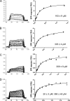
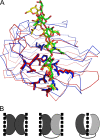
Adenovirus isolate NADC-1, a strain of porcine adenovirus type 4, has a fiber containing an N-terminal virus attachment region, shaft and head domains, and a C-terminal galectin domain connected to the head by an RGD-containing sequence. The crystal structure of the head domain is similar to previously solved adenovirus fiber head domains, but specific residues for binding the coxsackievirus and adenovirus receptor (CAR), CD46, or sialic acid are not conserved. The structure of the galectin domain reveals an interaction interface between its two carbohydrate recognition domains, locating both sugar binding sites face to face. Sequence evidence suggests other tandem-repeat galectins have the same arrangement. We show that the galectin domain binds carbohydrates containing lactose and N-acetyl-lactosamine units, and we present structures of the galectin domain with lactose, N-acetyl-lactosamine, 3-aminopropyl-lacto-N-neotetraose, and 2-aminoethyl-tri(N-acetyl-lactosamine), confirming the domain as a bona fide galectin domain.
Figures


Similar articles
-
Crystallization of the head and galectin-like domains of porcine adenovirus isolate NADC-1 fibre.Acta Crystallogr Sect F Struct Biol Cryst Commun. 2009 Nov 1;65(Pt 11):1149-52. doi: 10.1107/S1744309109038287. Epub 2009 Oct 30. Acta Crystallogr Sect F Struct Biol Cryst Commun. 2009. PMID: 19923738 Free PMC article.
-
X-ray structure of a protease-resistant mutant form of human galectin-8 with two carbohydrate recognition domains.FEBS J. 2012 Oct;279(20):3937-51. doi: 10.1111/j.1742-4658.2012.08753.x. Epub 2012 Sep 11. FEBS J. 2012. PMID: 22913484
-
The N-terminal carbohydrate recognition domain of galectin-8 recognizes specific glycosphingolipids with high affinity.Glycobiology. 2003 Oct;13(10):713-23. doi: 10.1093/glycob/cwg094. Epub 2003 Jul 8. Glycobiology. 2003. PMID: 12851289
-
Full-length galectin-8 and separate carbohydrate recognition domains: the whole is greater than the sum of its parts?Biochem Soc Trans. 2020 Jun 30;48(3):1255-1268. doi: 10.1042/BST20200311. Biochem Soc Trans. 2020. PMID: 32597487 Review.
-
Tropism and transduction of oncolytic adenovirus 5 vectors in cancer therapy: Focus on fiber chimerism and mosaicism, hexon and pIX.Virus Res. 2018 Sep 15;257:40-51. doi: 10.1016/j.virusres.2018.08.012. Epub 2018 Aug 17. Virus Res. 2018. PMID: 30125593 Review.
Cited by
-
Structural basis for norovirus inhibition and fucose mimicry by citrate.J Virol. 2012 Jan;86(1):284-92. doi: 10.1128/JVI.05909-11. Epub 2011 Oct 26. J Virol. 2012. PMID: 22031945 Free PMC article.
-
Virus recognition of glycan receptors.Curr Opin Virol. 2019 Feb;34:117-129. doi: 10.1016/j.coviro.2019.01.004. Epub 2019 Mar 5. Curr Opin Virol. 2019. PMID: 30849709 Free PMC article. Review.
-
Crystal structure of raptor adenovirus 1 fibre head and role of the beta-hairpin in siadenovirus fibre head domains.Virol J. 2016 Jun 22;13:106. doi: 10.1186/s12985-016-0558-7. Virol J. 2016. PMID: 27334597 Free PMC article.
-
An adenovirus vector incorporating carbohydrate binding domains utilizes glycans for gene transfer.PLoS One. 2013;8(2):e55533. doi: 10.1371/journal.pone.0055533. Epub 2013 Feb 1. PLoS One. 2013. PMID: 23383334 Free PMC article.
-
Crystal structure of the fibre head domain of bovine adenovirus 4, a ruminant atadenovirus.Virol J. 2015 May 22;12:81. doi: 10.1186/s12985-015-0309-1. Virol J. 2015. PMID: 25994880 Free PMC article.
References
-
- Arnberg, N. 2009. Adenovirus receptors: implications for tropism, treatment and targeting. Rev. Med. Virol. 19:165-178. - PubMed
-
- Bachtarzi, H., M. Stevenson, and K. Fisher. 2008. Cancer gene therapy with targeted adenoviruses. Expert Opin. Drug Deliv. 5:1231-1240. - PubMed
-
- Barondes, S. H., D. N. Cooper, M. A. Gitt, and H. Leffler. 1994. Galectins. Structure and function of a large family of animal lectins. J. Biol. Chem. 269:20807-20810. - PubMed
-
- Bewley, M. C., K. Springer, Y. B. Zhang, P. Freimuth, and J. M. Flanagan. 1999. Structural analysis of the mechanism of adenovirus binding to its human cellular receptor, CAR. Science 286:1579-1583. - PubMed
-
- Blixt, O., S. Head, T. Mondala, C. Scanlan, M. E. Huflejt, R. Alvarez, M. C. Bryan, F. Fazio, D. Calarese, J. Stevens, N. Razi, D. J. Stevens, J. J. Skehel, I. van Die, D. R. Burton, I. A. Wilson, R. Cummings, N. Bovin, C. H. Wong, and J. C. Paulson. 2004. Printed covalent glycan array for ligand profiling of diverse glycan binding proteins. Proc. Natl. Acad. Sci. U. S. A. 101:17033-17038. - PMC - PubMed
Publication types
MeSH terms
Substances
Grants and funding
LinkOut - more resources
Full Text Sources
Other Literature Sources

