. 2010 Oct;84(19):10209-19.
doi: 10.1128/JVI.00632-10. Epub 2010 Aug 4.

Long-term restriction by APOBEC3F selects human immunodeficiency virus type 1 variants with restored Vif function

Affiliations

Long-term restriction by APOBEC3F selects human immunodeficiency virus type 1 variants with restored Vif function

John S Albin et al. J Virol. 2010 Oct.

Abstract

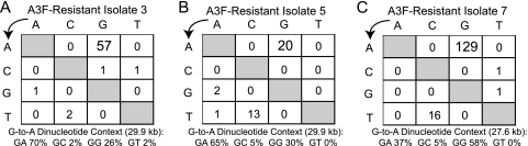
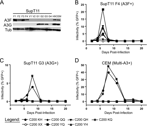
Tandem stop mutations K26X and H27X in human immunodeficiency virus type 1 (HIV-1) vif compromise virus replication in human T-cell lines that stably express APOBEC3F (A3F) or APOBEC3G (A3G). We previously reported that partial resistance to A3G could develop in these Vif-deficient viruses through a nucleotide A200-to-T/C transversion and a vpr null mutation, but these isolates were still susceptible to restriction by A3F. Here, long-term selection experiments were done to determine how these A3G-selected isolates might evolve to spread in the presence of A3F. We found that A3F, like A3G, is capable of potent, long-term restriction that eventually selects for heritable resistance. In all 7 instances, the selected isolates had restored Vif function to cope with A3F activity. In two isolates, Vif Q26-Q27 and Y26-Q27, the resistance phenotype recapitulated in molecular clones, but when the selected vif alleles were analyzed in the context of an otherwise wild-type viral background, a different outcome emerged. Although HIV-1 clones with Vif Q26-Q27 or Y26-Q27 were fully capable of overcoming A3F, they were now susceptible to restriction by A3G. Concordant with prior studies, a lysine at position 26 proved essential for A3G neutralization. These data combine to indicate that A3F and A3G exert at least partly distinct selective pressures and that Vif function may be essential for the virus to replicate in the presence of A3F.


Figures

FIG. 1.
FIG. 1.
Restriction of Vif-deficient HIV-1 by APOBEC3F (A3F) selects resistant virus variants. (A) Western blot showing the expression of A3F and A3G in H9 and CEM cells and the CEM-SS-derived cell lines used in these studies. F1 and F2, A3F-expressing CEM-SS cell lines; G1 and G2, A3G-expressing CEM-SS cell lines; V1 and V2, CEM-SS transfected with a vector control. Tub, tubulin. (B to E) Growth curves in the indicated cells for the following HIV-1 isolates: Vif-proficient (A200 KH), Vif-deficient (A200 XX), A3G-resistant [T200 XX vpr (A3G-R2) and T200 XX vpr (A3G-R3)], and representative A3F-resistant viruses derived from parent A3G-resistant viruses [T200 YQ vpr (A3F-R5) and T200 QQ vpr (A3F-R7) where vif genotypes are retrospectively indicated according to subsequent sequencing (Table 2)]. The starting MOI was approximately 0.02, and similar results were obtained using the second A3F-expressing and vector control CEM-SS cell lines (data not shown). The low peaks observed for some A200 KH growth curves, particularly in panel D, are due to the high cytotoxicity of this virus, which sometimes results in low apparent titers as infected cells are rapidly killed. In addition to the notably low A200 KH CEM-SS V1 curve, a growth curve for A200 KH in another CEM-SS vector line from this experiment, CEM-SS V3, is indicated by a black arrow in panel D to visually demonstrate that the wild-type virus spreads in the absence of restrictive levels of APOBEC3 proteins. The x axis is offset from zero in all curves to permit better visualization of viruses showing little or no growth. Throughout, open symbols indicate viruses lacking Vif expression; closed symbols indicate full-length vif alleles. Similarly, broken lines are used for vpr viruses, while solid black lines are used for vpr+ viruses. A3F+, A3F expressing; A3−, not expressing A3; GFP+, GFP positive; multi-A3+, expresses ≥5 APOBEC3 proteins.
FIG. 2.
FIG. 2.
Hypermutation patterns in selected A3F-resistant isolates. The frequency of each base change is given for the clones described in experiments 1 and 2 of Table 2, as is the predominance of the dinucleotide context in which G-to-A mutations occur. Similar to previous authors, we note a substantial C-to-T transition rate in the presence of A3F in addition to the expected G-to-A hypermutations (4, 20, 26). The viruses selected also contain substantial 5′-GG-3′ to 5′-AG-3′ transitions, particularly in the case of A3F-resistant isolate 7, suggesting ongoing mutation by A3G in addition to A3F.
FIG. 3.
FIG. 3.
Restoration of the vif open reading frame accounts for phenotypic resistance to A3F-mediated restriction. Spreading infections at an MOI of 0.05 were initiated in CEM-SS cells stably transfected with A3F (A) or A3G (B) or a vector control (C) as well as in nonpermissive CEM cells (D) using viruses derived from proviral molecular clones with the indicated genotypes. The mildly enhanced infectivity of A3G-resistant viruses relative to their parent A200 XX viruses in A3F-expressing CEM-SS cells is sometimes observed in experiments such as the one shown that start from a higher MOI (compare the lower MOI in Fig. 1B with the higher MOI in panel A). In contrast, A3F-selected viruses consistently display robust peaks at any MOI in the presence of A3F. The peaks of A200 KH growth are indicated by arrows in panels A and B to differentiate them from the descending T200 YQ vpr (A3F-R5) curve and the superimposed T200 YQ vpr (A3F-R5) peak, respectively. Similar results were obtained using proviral molecular clones corresponding to other selected isolates as well as additional CEM-SS clones stably transfected with A3F, A3G, or a vector control (data not shown).
FIG. 4.
FIG. 4.
The identity of Vif amino acids 26 and/or 27 rather than Vpr status is critical for the ability to replicate on naturally nonpermissive cells. Spreading infection curves are shown for viruses with wild-type (KH), A3F-selected missense (QQ and YQ), and nonsense (XX) codons at positions 26 and 27 of vif in a Vpr-proficient context. Spreading infections were carried out from a starting MOI of 0.01 on CEM and H9 cells (A and B) as well as CEM-SS and SupT11 clones transfected with a vector control (data not shown). Different alleles of vif in Vpr-deficient contexts showed the same growth patterns on CEM and H9 cells as their Vpr-proficient counterparts (Table 1 and data not shown).
FIG. 5.
FIG. 5.
Functional Vif proteins selected by A3F are deficient in their ability to degrade A3G. A titration experiment analyzing the infectivity of particles produced by the cotransfection of constant amounts of A3F-V5 (A) or A3G-V5 (B) in the presence of increasing amounts of Vif-HA. The identities of amino acids 26 and 27 are indicated for each Vif-expressing construct. Infectivity data represent the mean plus standard error of the mean (SEM) (error bar) of three independent experiments where infectivity is determined relative to that of particles produced under the same cotransfection conditions in each experiment with a vector control in place of the APOBEC3 expression construct (not shown). Immunoblots shown are taken from one of these three experiments. While Vif-QQ and Vif-YQ are notably deficient in their ability to neutralize A3G relative to the wild-type Vif-KH, a mild effect is seen at higher levels of Vif expression, which achieves statistical significance by a paired two-tailed t test for Vif-QQ, but not Vif-YQ (panel B and data not shown). V, Vif vector and A3F-V5 or A3G-V5 expression constructs cotransfected; these conditions were tested once in each experiment but are loaded in the immunoblots and plotted in the histograms three times each for direct visual comparison with the addition of each Vif protein.
FIG. 6.
FIG. 6.
A3F-selected vif alleles are nonfunctional for the neutralization of A3G but can be rescued by restoration of the wild-type K26 residue. (A) Western blots showing expression levels of A3F and A3G in the SupT11-derived cell lines used in these experiments as well as in H9 and CEM cells. Spreading infection curves from a starting MOI of 0.01 are shown for the wild type (C200 KH) and for Vif-deficient (C200 XX) and A3F-selected (C200 QQ and YQ) mutants as well as mutants completing the matrix of combinations of wild-type and selected residues at positions 26 and 27 of vif (C200 QH, YH and KQ) on SupT11 cells transfected with A3F (B) or A3G (C) as well as nonpermissive CEM cells (D). Results demonstrate that K26 is critical for the neutralization of A3G and the APOBEC3 repertoire found in CEM cells, but not A3F. Results concordant with results in panels B to D were observed using additional SupT11-derived cell lines expressing A3F or A3G as well as SupT11 clones transfected with a vector control and H9 cells (data not shown). Higher peaks than that shown in panel B are usually observed with the C200 QQ virus (e.g., Fig. 1 and 3 and data not shown); in addition to those curves and many not shown, the ability of C200 QQ to spread in the presence of A3F is indicated by the fact that it efficiently kills the culture in which it replicates (see the legend to Fig. 1).
FIG. 7.
FIG. 7.
Long-term culture of A3F-resistant viruses in CEM cells selects for restoration of a positive charge at Vif residue 26. Passage of CEM-resistant viral isolates in CEM cells demonstrating their functional resistance to the nonpermissive phenotype of CEM. Phenotypic resistance of the selected alleles to the nonpermissive phenotype as encountered in CEM cells was also confirmed using viruses derived from molecular clones (Table 1 and data not shown). A summary of the sequence evolution observed in resistant isolates that were confirmed by second passage and for which sequence was available is given in experiment 5 of Table 2.

References

    1. Abudu, A., A. Takaori-Kondo, T. Izumi, K. Shirakawa, M. Kobayashi, A. Sasada, K. Fukunaga, and T. Uchiyama. 2006. Murine retrovirus escapes from murine APOBEC3 via two distinct novel mechanisms. Curr. Biol. 16:1565-1570. - PubMed
    1. Albin, J. S., and R. S. Harris. 2010. Interactions of host APOBEC3 restriction factors with HIV-1 in vivo: implications for therapeutics. Expert Rev. Mol. Med. 12:1-26. - PMC - PubMed
    1. Bishop, K. N., R. K. Holmes, and M. H. Malim. 2006. Antiviral potency of APOBEC proteins does not correlate with cytidine deamination. J. Virol. 80:8450-8458. - PMC - PubMed
    1. Bishop, K. N., R. K. Holmes, A. M. Sheehy, N. O. Davidson, S. J. Cho, and M. H. Malim. 2004. Cytidine deamination of retroviral DNA by diverse APOBEC proteins. Curr. Biol. 14:1392-1396. - PubMed
    1. Bishop, K. N., M. Verma, E. Y. Kim, S. M. Wolinsky, and M. H. Malim. 2008. APOBEC3G inhibits elongation of HIV-1 reverse transcripts. PLoS Pathog. 4:e1000231. - PMC - PubMed

Publication types

MeSH terms

LinkOut - more resources