. 2010 Oct;84(20):10581-91.
doi: 10.1128/JVI.00925-10. Epub 2010 Aug 4.

Basic residues within the ebolavirus VP35 protein are required for its viral polymerase cofactor function

Affiliations

Basic residues within the ebolavirus VP35 protein are required for its viral polymerase cofactor function

Kathleen C Prins et al. J Virol. 2010 Oct.

Abstract

The ebolavirus (EBOV) VP35 protein binds to double-stranded RNA (dsRNA), inhibits host alpha/beta interferon (IFN-α/β) production, and is an essential component of the viral polymerase complex. Structural studies of the VP35 C-terminal IFN inhibitory domain (IID) identified specific structural features, including a central basic patch and a hydrophobic pocket, that are important for dsRNA binding and IFN inhibition. Several other conserved basic residues bordering the central basic patch and a separate cluster of basic residues, called the first basic patch, were also identified. Functional analysis of alanine substitution mutants indicates that basic residues outside the central basic patch are not required for dsRNA binding or for IFN inhibition. However, minigenome assays, which assess viral RNA polymerase complex function, identified these other basic residues to be critical for viral RNA synthesis. Of these, a subset located within the first basic patch is important for VP35-nucleoprotein (NP) interaction, as evidenced by the inability of alanine substitution mutants to coimmunoprecipitate with NP. Therefore, first basic patch residues are likely critical for replication complex formation through interactions with NP. Coimmunoprecipitation studies further demonstrate that the VP35 IID is sufficient to interact with NP and that dsRNA can modulate VP35 IID interactions with NP. Other basic residue mutations that disrupt the VP35 polymerase cofactor function do not affect interaction with NP or with the amino terminus of the viral polymerase. Collectively, these results highlight the importance of conserved basic residues from the EBOV VP35 C-terminal IID and validate the VP35 IID as a potential therapeutic target.


Figures

FIG. 1.
FIG. 1.
Highly conserved basic residues are located in three regions within VP35 IID. Ribbon representation of VP35 IID highlighting residues from the first basic patch (A) and central basic patch (B) and border basic residues located outside the central basic patch (C). Electrostatic surface representation of the first basic patch (D) and central basic patch and bordering basic residues (E) (scale, −10 kT/e to +10 kT/e).
FIG. 2.
FIG. 2.
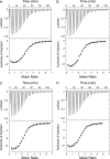
ITC binding isotherms and corresponding raw data for 8-bp dsRNA binding to WT (A) and mutant R225A (B), K248A (C), and K251A (D) VP35 IIDs. The corresponding average KD values for WT, R225A, K248A, and K251A are 0.7, 1.0, 2.4, and 0.8 μM, respectively.
FIG. 3.
FIG. 3.
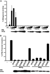
Viral polymerase cofactor function of basic residue-to-alanine first basic patch mutants. Viral polymerase activity was assessed in the minigenome assay. Empty vector (−), WT VP35 (VP35), or the indicated VP35 mutants were assessed. Cells were transfected with empty vector or with increasing amounts of wild-type or mutant VP35 plasmid and expression plasmids for ZEBOV NP, VP30, L, T7 RNA polymerase, and luciferase. A luciferase expression plasmid was cotransfected as a control for transfection efficiency. A plasmid expressing a negative-sense minigenome RNA and reporter construct was also transfected. Minigenome activity was quantified by measuring CAT activity, and this was normalized to the level of expression of luciferase. Error bars represent 1 standard deviation for three experiments. (Lower panel) Western blots (WB) for VP35 proteins. The concentrations of VP35 plasmids used were 63 ng, 125 ng, and 250 ng. WT VP35 at 250 ng was set as 100% minigenome activity.
FIG. 4.
FIG. 4.
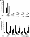
Viral polymerase cofactor function and IFN-antagonist function of basic residue-to-glutamine first basic patch mutants. (A) Empty vector (EV), wild-type VP35 (VP35), or the indicated first basic patch VP35 mutants were assessed in the minigenome assay, as described in the legend to Fig. 3. (B) Inhibition of IFN-β (IFNb) promoter activation by wild-type and mutant VP35 proteins. HEK293T cells were transfected with empty vector (−) or increasing amounts (1, 2, or 4 μg) of WT VP35 (VP35) or mutant VP35 expression plasmid, an IFN-β promoter-firefly luciferase reporter plasmid, and a constitutively expressed Renilla luciferase reporter plasmid. Firefly luciferase activity was normalized to the level of Renilla luciferase activity. Data are presented such that the activity of the empty vector, SeV-infected samples (+), is set equal to 100%. Error bars represent 1 SD, and corresponding Western blots (WB) of cell lysates are shown below the graphs.
FIG. 5.
FIG. 5.
Viral polymerase cofactor function and IFN-antagonist function of border residue mutants. (A) Empty vector (first bar), WT VP35 (VP35), or the indicated border basic residue mutants were assessed for function in the minigenome assay, as described in the legend to Fig. 3. (B) Inhibition of IFN-β promoter activation by WT and mutant VP35 proteins. HEK293T cells were transfected with empty vector (first bar) or increasing amounts (1, 2, or 4 μg) of WT VP35 or mutant VP35 expression plasmid, an IFN-β promoter-firefly luciferase reporter plasmid, and a constitutively expressed Renilla luciferase reporter plasmid. Firefly luciferase activity was normalized to the level of Renilla luciferase activity. Data are presented such that the activity of empty vector, SeV-infected samples, is set equal to 100%. Error bars represent one SD, and corresponding Western blots (WB) of cell lysates are shown below the graphs.
FIG. 6.
FIG. 6.
Viral polymerase cofactor function of arginine-to-lysine and lysine-to-arginine mutations for representative residues from the first basic patch, border residues, and the central basic patch. Empty vector (−), WT VP35 (VP35), or the indicated mutants were assessed for function in the minigenome assay, as described in the legend to Fig. 3. Error bars represent SDs, and corresponding Western blots (WB) of cell lysates are shown below the graphs. The concentrations of VP35 plasmids used were 125 ng, 250 ng, and 500 ng. Data were expressed as the fold induction over that by the negative control (no VP35).
FIG. 7.
FIG. 7.
Full-length WT and mutant VP35 interactions with EBOV NP. Cells were transfected with WT or mutant VP35 and EBOV NP. The left-most lane (−) corresponds to a sample in which NP was expressed in the absence of VP35. Lysates were subjected to IP with anti-VP35 antibody. Proteins were detected by Western blotting with anti-NP and anti-VP35 monoclonal antibodies.
FIG. 8.
FIG. 8.
The VP35 IID is sufficient to interact with NP, and this interaction is impaired by either specific point mutations or the presence of dsRNA. (A) MBP or MBP-VP35 IID was added to NP-transfected cell lysates. MBP was immobilized by amylose resin, and proteins were detected with anti-MBP (α-MBP) or anti-NP (α-NP) antibodies. (B) MBP alone, MBP-VP35 IID WT, or mutants with point mutations were added to lysates containing EBOV NP. MBP was precipitated using amylose resin and was detected with NP and MBP antibodies. (C) MBP, WT MPB-VP35 IID, or MBP-VP35 IID mutants (R312A, K322A, or F239A) were added to NP-transfected lysates in the absence or presence of poly(I:C). The final concentration of each MBP was 2.4 μM, and poly(I:C) was present at either 16 or 98 nM. MBPs were pulled down, and both MBP and NP were detected in the same way as described for panels A and B. The input whole-cell extract lane (WCE) expressing NP represents 4% of the total lysate used for each group, while the MBP pull-down lane represents 15% of the total pull down.
FIG. 9.
FIG. 9.
Full-length WT and mutant VP35 interactions with EBOV L. Cells were transfected with WT or mutant VP35 and EBOV HA-L amino acids 1 to 505 (L1-505). Lysates were immunoprecipitated with anti-VP35 antibody and protein G beads. Proteins were detected with HA or VP35 monoclonal antibodies. WT and mutant full-length VP35 can coimmunoprecipitate with HA-L amino acids 1 to 505. WC, whole-cell extract.

Similar articles

Cited by

References

    1. Basler, C. F., and G. K. Amarasinghe. 2009. Evasion of interferon responses by Ebola and Marburg viruses. J. Interferon Cytokine Res. 29:511-520. - PMC - PubMed
    1. Basler, C. F., A. Mikulasova, L. Martinez-Sobrido, J. Paragas, E. Muhlberger, M. Bray, H. D. Klenk, P. Palese, and A. Garcia-Sastre. 2003. The Ebola virus VP35 protein inhibits activation of interferon regulatory factor 3. J. Virol. 77:7945-7956. - PMC - PubMed
    1. Basler, C. F., X. Wang, E. Muhlberger, V. Volchkov, J. Paragas, H. D. Klenk, A. Garcia-Sastre, and P. Palese. 2000. The Ebola virus VP35 protein functions as a type I IFN antagonist. Proc. Natl. Acad. Sci. U. S. A. 97:12289-12294. - PMC - PubMed
    1. Becker, S., C. Rinne, U. Hofsass, H. D. Klenk, and E. Muhlberger. 1998. Interactions of Marburg virus nucleocapsid proteins. Virology 249:406-417. - PubMed
    1. Boehmann, Y., S. Enterlein, A. Randolf, and E. Muhlberger. 2005. A reconstituted replication and transcription system for Ebola virus Reston and comparison with Ebola virus Zaire. Virology 332:406-417. - PubMed

Publication types

MeSH terms