The viral latency-associated nuclear antigen augments the B-cell response to antigen in vivo
- PMID: 20686032
- PMCID: PMC2950586
- DOI: 10.1128/JVI.00848-10
The viral latency-associated nuclear antigen augments the B-cell response to antigen in vivo
Abstract
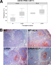
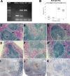
Gammaherpesviruses, including Kaposi sarcoma-associated herpesvirus (KSHV), establish latency in B cells. We hypothesized that the KSHV latency-associated nuclear antigen (LANA/orf73) provides a selective advantage to infected B cells by driving proliferation in response to antigen. To test this, we used LANA B-cell transgenic mice. Eight days after immunization with antigen without adjuvant, LANA mice had significantly more activated germinal center (GC) B cells (CD19(+) PNA(+) CD71(+)) than controls. This was dependent upon B-cell receptor since LANA did not restore the GC defect of CD19 knockout mice. However, LANA was able to restore the marginal zone defect in CD19 knockout mice.
Figures


Similar articles
-
Tissue specificity of the Kaposi's sarcoma-associated herpesvirus latent nuclear antigen (LANA/orf73) promoter in transgenic mice.J Virol. 2002 Nov;76(21):11024-32. doi: 10.1128/jvi.76.21.11024-11032.2002. J Virol. 2002. PMID: 12368345 Free PMC article.
-
Kaposi's Sarcoma-Associated Herpesvirus Latency-Associated Nuclear Antigen Inhibits Major Histocompatibility Complex Class II Expression by Disrupting Enhanceosome Assembly through Binding with the Regulatory Factor X Complex.J Virol. 2015 May;89(10):5536-56. doi: 10.1128/JVI.03713-14. Epub 2015 Mar 4. J Virol. 2015. PMID: 25740990 Free PMC article.
-
Kaposi Sarcoma Herpesvirus (KSHV) Latency-Associated Nuclear Antigen (LANA) recruits components of the MRN (Mre11-Rad50-NBS1) repair complex to modulate an innate immune signaling pathway and viral latency.PLoS Pathog. 2017 Apr 21;13(4):e1006335. doi: 10.1371/journal.ppat.1006335. eCollection 2017 Apr. PLoS Pathog. 2017. PMID: 28430817 Free PMC article.
-
KSHV LANA--the master regulator of KSHV latency.Viruses. 2014 Dec 11;6(12):4961-98. doi: 10.3390/v6124961. Viruses. 2014. PMID: 25514370 Free PMC article. Review.
-
KSHV Genome Replication and Maintenance in Latency.Adv Exp Med Biol. 2018;1045:299-320. doi: 10.1007/978-981-10-7230-7_14. Adv Exp Med Biol. 2018. PMID: 29896673 Review.
Cited by
-
Cancers associated with human gammaherpesviruses.FEBS J. 2022 Dec;289(24):7631-7669. doi: 10.1111/febs.16206. Epub 2021 Oct 2. FEBS J. 2022. PMID: 34536980 Free PMC article. Review.
-
Kaposi sarcoma associated herpesvirus pathogenesis (KSHV)--an update.Curr Opin Virol. 2013 Jun;3(3):238-44. doi: 10.1016/j.coviro.2013.05.012. Epub 2013 Jun 13. Curr Opin Virol. 2013. PMID: 23769237 Free PMC article. Review.
-
Quantitative analysis of the bidirectional viral G-protein-coupled receptor and lytic latency-associated nuclear antigen promoter of Kaposi's sarcoma-associated herpesvirus.J Virol. 2012 Sep;86(18):9683-95. doi: 10.1128/JVI.00881-12. Epub 2012 Jun 27. J Virol. 2012. PMID: 22740392 Free PMC article.
-
Latency locus complements MicroRNA 155 deficiency in vivo.J Virol. 2013 Nov;87(21):11908-11. doi: 10.1128/JVI.01620-13. Epub 2013 Aug 21. J Virol. 2013. PMID: 23966392 Free PMC article.
-
Kaposi's Sarcoma-Associated Herpesvirus Latency Locus Compensates for Interleukin-6 in Initial B Cell Activation.J Virol. 2015 Dec 9;90(4):2150-4. doi: 10.1128/JVI.02456-15. Print 2016 Feb 15. J Virol. 2015. PMID: 26656696 Free PMC article.
References
-
- An, F.-Q., N. Compitello, E. Horwitz, M. Sramkoski, E. S. Knudsen, and R. Renne. 2005. The latency-associated nuclear antigen of Kaposi's sarcoma-associated herpesvirus modulates cellular gene expression and protects lymphoid cells from p16 INK4A-induced cell cycle arrest. J. Biol. Chem. 280:3862-3874. - PubMed
-
- An, J., Y. Sun, and M. B. Rettig. 2004. Transcriptional coactivation of c-Jun by the KSHV-encoded LANA. Blood 103:222-228. - PubMed
-
- Ansari, M. Q., D. B. Dawson, R. Nador, C. Rutherford, N. R. Schneider, M. J. Latimer, L. Picker, D. M. Knowles, and R. W. McKenna. 1996. Primary body cavity-based AIDS-related lymphomas. Am. J. Clin. Pathol. 105:221-229. - PubMed
Publication types
MeSH terms
Substances
Grants and funding
LinkOut - more resources
Full Text Sources
Molecular Biology Databases
Miscellaneous

