. 2010 Nov;84(21):10999-1009.
doi: 10.1128/JVI.00526-10. Epub 2010 Aug 4.

Ultrastructural and biophysical characterization of hepatitis C virus particles produced in cell culture

Affiliations

Ultrastructural and biophysical characterization of hepatitis C virus particles produced in cell culture

Pablo Gastaminza et al. J Virol. 2010 Nov.

Abstract

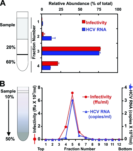
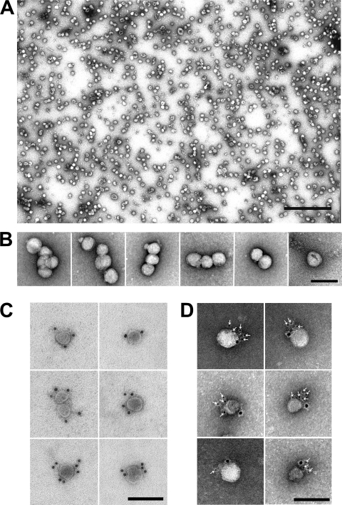
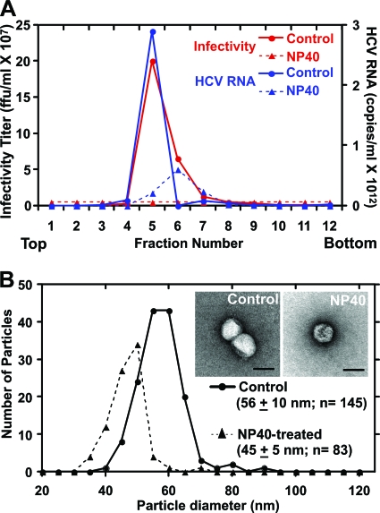
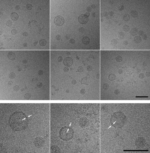
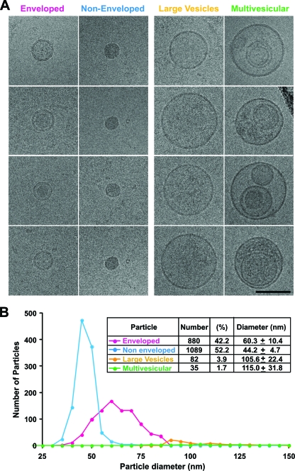
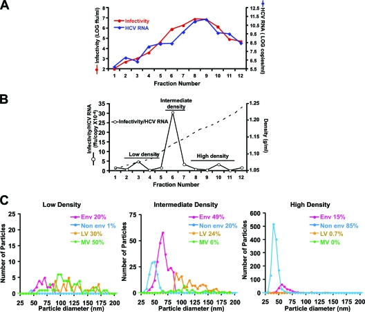
We analyzed the biochemical and ultrastructural properties of hepatitis C virus (HCV) particles produced in cell culture. Negative-stain electron microscopy revealed that the particles were spherical (∼40- to 75-nm diameter) and pleomorphic and that some of them contain HCV E2 protein and apolipoprotein E on their surfaces. Electron cryomicroscopy revealed two major particle populations of ∼60 and ∼45 nm in diameter. The ∼60-nm particles were characterized by a membrane bilayer (presumably an envelope) that is spatially separated from an internal structure (presumably a capsid), and they were enriched in fractions that displayed a high infectivity-to-HCV RNA ratio. The ∼45-nm particles lacked a membrane bilayer and displayed a higher buoyant density and a lower infectivity-to-HCV RNA ratio. We also observed a minor population of very-low-density, >100-nm-diameter vesicular particles that resemble exosomes. This study provides low-resolution ultrastructural information of particle populations displaying differential biophysical properties and specific infectivity. Correlative analysis of the abundance of the different particle populations with infectivity, HCV RNA, and viral antigens suggests that infectious particles are likely to be present in the large ∼60-nm HCV particle populations displaying a visible bilayer. Our study constitutes an initial approach toward understanding the structural characteristics of infectious HCV particles.


Figures

FIG. 1.
FIG. 1.
Infectious HCV particles purified by sucrose gradient centrifugation. (A) Concentrated supernatants isolated from infected cells were subjected to ultracentrifugation on a sucrose step gradient. (B) The particles collected from the 20-60% interface (fraction 3) were further purified in a continuous 10 to 50% sucrose gradient. Infectivity and HCV RNA levels were determined by titration and quantitative PCR, and the results were expressed as the numbers of FFU per ml and HCV RNA genome equivalents (GE) per ml, respectively.
FIG. 2.
FIG. 2.
HCV particles are pleomorphic and display E2 and apolipoprotein E on their surface. (A) Electron micrograph of highly purified, negatively stained HCV particles demonstrates their fairly uniform size but pleomorphic shape. Particles were purified as described in the legend of Fig. 1. Scale bar, 500 nm. (B) Close-up views show the heterogeneous sizes and shapes. (C) Representative images show specific decoration of a subset (5 to 20%) of particles with an antibody directed against the viral envelope protein E2. (D) Representative images show specific decoration of E2-positive particles (6-nm gold) with anti-apoE antibodies (12-nm gold). Scale bar, 100 nm (B, C, and D).
FIG. 3.
FIG. 3.
Particles treated with 0.2% NP-40 are noninfectious and display a decrease in diameter. Detergent-treated and untreated particles were subjected to ultracentrifugation in a continuous 10 to 50% sucrose gradient. (A) Detergent treatment eliminated infectivity and resulted in a substantial loss of HCV RNA. (B) Particle diameter decreased from ∼60 to ∼45 nm with detergent treatment. Inset shows representative negatively stained particles. Scale bar, 50 nm.
FIG. 4.
FIG. 4.
cryoEM showed that purified HCV particles are predominantly spherical and consist of subpopulations with different diameters. Note that the large-diameter particles are surrounded by an envelope (arrowheads), which is not detectable in the small-diameter particles. Scale bar, 100 nm.
FIG. 5.
FIG. 5.
Purified virus preparations contained two major (enveloped and nonenveloped) and two minor (large vesicle and multivesicular) subpopulations. (A) Gallery of representative electron cryomicrographs showing the four particle classes. Scale bar, 100 nm. (B) Histogram and table of 2,086 particles from four independent virus preparations revealed roughly similar numbers of enveloped and nonenveloped particles, with minor populations of large vesicles having a uniform interior density, and a few multivesicular particles that are probably nonviral and may represent exosomes.
FIG. 6.
FIG. 6.
Enrichment of different particle populations by isopycnic sucrose density gradient ultracentrifugations. (A) Representative plot of the gradient fractions showing density, infectivity, and HCV RNA content. (B) The infectivity-HCV RNA ratio calculated from the data in panel A highlights the fractions with increased specific infectivity. (C) Plots of the size distribution and frequency of each population in pooled fractions having low, intermediate, and high densities. Population distributions were obtained from the combined analysis of two independent viral preparations.

Similar articles

Cited by

References

    1. Andre, P., F. Komurian-Pradel, S. Deforges, M. Perret, J. L. Berland, M. Sodoyer, S. Pol, C. Brechot, G. Paranhos-Baccala, and V. Lotteau. 2002. Characterization of low- and very-low-density hepatitis C virus RNA-containing particles. J. Virol. 76:6919-6928. - PMC - PubMed
    1. Appel, N., M. Zayas, S. Miller, J. Krijnse-Locker, T. Schaller, P. Friebe, S. Kallis, U. Engel, and R. Bartenschlager. 2008. Essential role of domain III of nonstructural protein 5A for hepatitis C virus infectious particle assembly. PLoS Pathog. 4:e1000035. - PMC - PubMed
    1. Atshaves, B. P., A. L. McIntosh, H. R. Payne, A. M. Gallegos, K. Landrock, N. Maeda, A. B. Kier, and F. Schroeder. 2007. SCP-2/SCP-x gene ablation alters lipid raft domains in primary cultured mouse hepatocytes. J. Lipid Res. 48:2193-2211. - PubMed
    1. Baumert, T. F., S. Ito, D. T. Wong, and T. J. Liang. 1998. Hepatitis C virus structural proteins assemble into viruslike particles in insect cells. J. Virol. 72:3827-3836. - PMC - PubMed
    1. Baumert, T. F., J. Vergalla, J. Satoi, M. Thomson, M. Lechmann, D. Herion, H. B. Greenberg, S. Ito, and T. J. Liang. 1999. Hepatitis C virus-like particles synthesized in insect cells as a potential vaccine candidate. Gastroenterology 117:1397-1407. - PubMed

Publication types

LinkOut - more resources