. 2010 Oct;84(20):10488-500.
doi: 10.1128/JVI.00894-10. Epub 2010 Aug 4.

Convergence of Kaposi's sarcoma-associated herpesvirus reactivation with Epstein-Barr virus latency and cellular growth mediated by the notch signaling pathway in coinfected cells

Affiliations

Convergence of Kaposi's sarcoma-associated herpesvirus reactivation with Epstein-Barr virus latency and cellular growth mediated by the notch signaling pathway in coinfected cells

Sophia Spadavecchia et al. J Virol. 2010 Oct.

Abstract

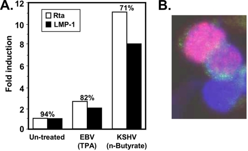
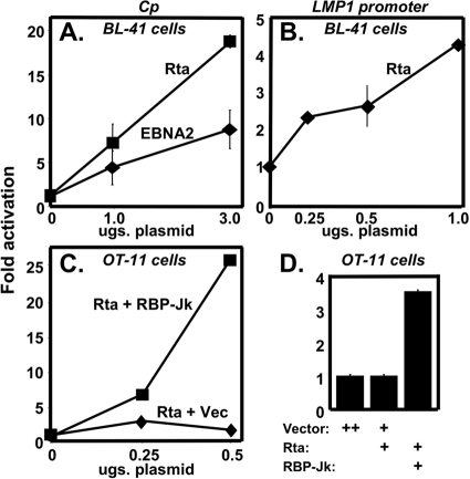
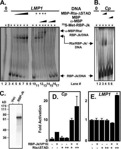
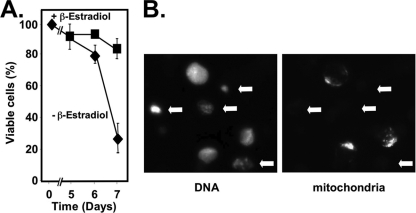
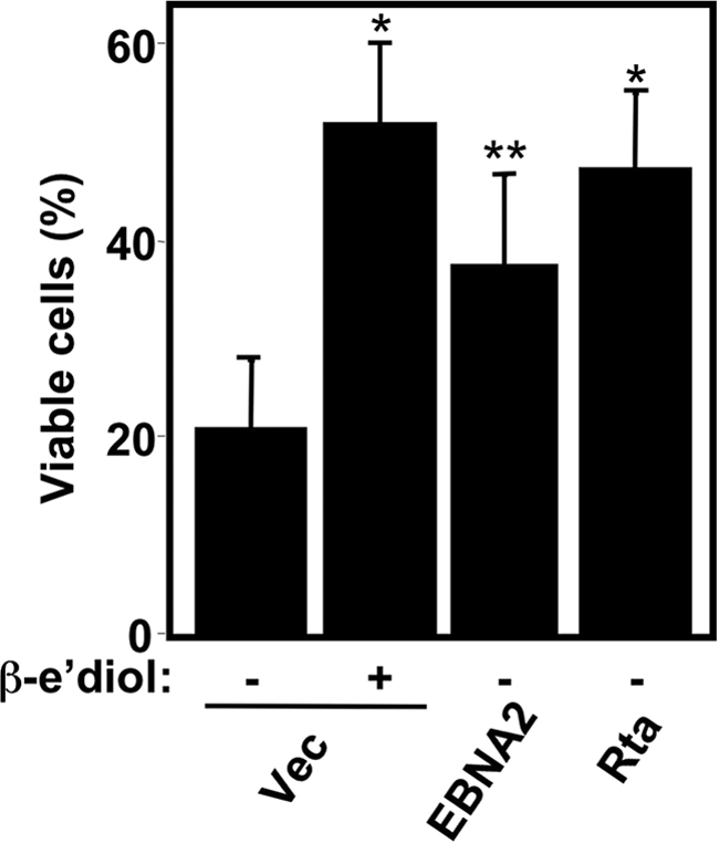
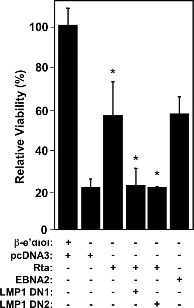
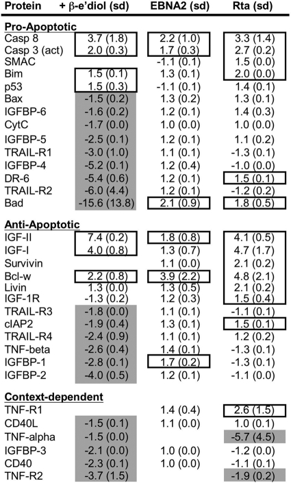
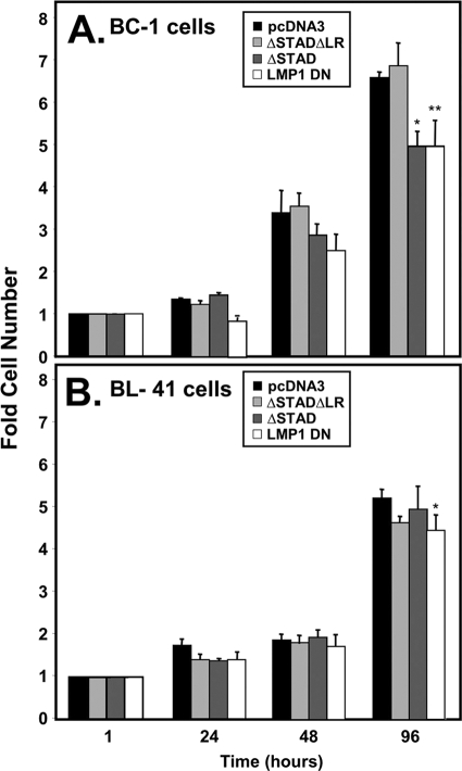
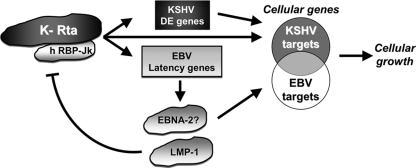
Kaposi's sarcoma-associated herpesvirus (KSHV) is the etiologic agent of primary effusion lymphoma (PEL). All PEL cell lines are infected with KSHV, and 70% are coinfected with Epstein-Barr virus (EBV). KSHV reactivation from latency requires promoter-specific transactivation by the KSHV Rta protein through interactions with RBP-Jk (CSL), the cellular DNA-binding component of the Notch signal transduction pathway. EBV transformation of primary B cells requires EBV nuclear antigen 2 (EBNA-2) to interact with RBP-Jk to direct the latent viral and cellular gene expression program. Although KSHV Rta and EBV EBNA-2 both require RBP-Jk for transactivation, previous studies have suggested that RBP-Jk-dependent transactivators do not function identically. We have found that the EBV latent protein LMP-1 is expressed in less than 5% of KSHV(+)/EBV(+) PEL cells but is induced in an Rta-dependent fashion when KSHV reactivates. KSHV Rta transactivates the EBV latency promoters in an RBP-Jk-dependent fashion and forms a ternary complex with RBP-Jk on the promoters. In B cells that are conditionally transformed by EBV alone, we show that KSHV Rta complements a short-term EBNA-2 growth deficiency in an autocrine/paracrine manner. Complementation of EBNA-2 deficiency by Rta depends on RBP-Jk and LMP-1, and Rta transactivation is required for optimal growth of KSHV(+)/EBV(+) PEL lines. Our data suggest that Rta can contribute to EBV-driven cellular growth by transactivating RBP-Jk-dependent EBV latency genes. However, our data also suggest that EBNA-2 and Rta induce distinct alterations in the cellular proteomes that contribute to the growth of infected cells.


Figures

FIG. 1.
FIG. 1.
KSHV reactivation induces LMP-1 expression in KSHV+/EBV+ cells. (A) BC-1 cells were treated with 20 ng of TPA/ml or 3 mM sodium butyrate, as indicated, to stimulate reactivation of EBV and KSHV, respectively. At 18 h postinduction, LMP1 and Rta were detected by immunofluorescence. A minimum of 500 immunopositive cells were counted, and the fold induction of either protein was calculated by comparison to untreated cells. The numbers above each bar graph represent the percentage of Rta-expressing cells that also expressed LMP1. (B) Representative image demonstrating correspondence between Rta and LMP-1 expression.
FIG. 2.
FIG. 2.
Rta transactivates LMP-1 in KSHV+/EBV+ cells. BC-1 cells were electroporated with 10 μg of V5-Rta plasmid and 10 and 20 μg of either empty expression vector (vector), RtaΔSTAD vector (ΔSTAD), or RtaΔSTADΔLR (ΔSTADΔLR) vector. At 42 h postelectroporation, LMP1 and V5-Rta were detected by immunofluorescence. (A) Graph representing the percentages of V5-Rta-positive (transfected) cells, in which LMP1 was induced. (B) Representative images demonstrating induction of LMP-1 in cells transfected with the V5-Rta expression vector with either empty vector, RtaΔSTAD vector, RtaΔSTADΔLR vector. (C) Total RNA was purified from BC-1 cells transfected with empty vector and treated with sodium butyrate (n-BA) or transfected with the indicated plasmids alone. The fold activation of LMP-1 was quantitated by qRT-PCR measuring LMP-1 and GAPDH RNA and comparing each to cells transfected with empty vector (Vec).
FIG. 3.
FIG. 3.
KSHV Rta transactivates EBV latency III promoters in an RBP-Jk-dependent manner. Uninfected BL-41 cells were coelectroporated with 2 μg of a luciferase reporter construct of the EBV Cp (A) or the LMP-1 promoter (B) alone and increasing amounts of either KSHV Rta or EBNA-2 expression plasmids, as indicated. The fold activation was calculated for each amount of Rta or EBNA-2 plasmid by comparison to the luciferase expressed by the reporter vectors coelectroporated with empty expression vector (“0”). OT-11 (RBP-Jk−/−) cells were transfected with 0.25 μg of the EBV Cp (C) or the LMP-1 promoter (D) reporters alone, or together with Rta expression vector with increasing amounts of either RBP-Jk expression vector or empty vector (Vec) (as indicated). The fold activation was calculated as in panels A and B.
FIG. 4.
FIG. 4.
Rta forms a ternary complex with RBP-Jk and the EBV Cp and LMP1 promoter DNAs. (A) EMSA was performed by incubating [35S]methionine (35S-Met)-labeled RBP-Jk alone or together with LMP-1 promoter DNA, MBP-Rta protein, and/or anti-MBP serum, as indicated. (B) EMSA was performed by incubating 35S-Met-labeled RBP-Jk alone or together with Cp promoter DNA and MBP-Rta or MBP proteins, as indicated. (C) 35S-Met-labeled RBP-Jk as visualized by SDS-PAGE/autoradiogram. The position of migration of molecular mass standards are indicated at left in kilodaltons. Rta stimulates DNA binding of RBP-Jk to the EBV Cp RBP-Jk element (D) and the LMP-1 promoter (E). BL-41 cells were electroporated with the indicated plasmids, as in Fig. 3 and reference , and the reporter plasmid 2× EBV RBP-hsp-luc (D) and pLMP-1-GL3basic (E). In panel E, RtaΔSTAD weakly repressed the LMP-1 promoter alone, so the values for RBP-Jk/VP16+RtaΔSTAD were normalized to RtaΔSTAD alone, which was set to 1-fold. *, P = 0.0011 (compared to the RBP-Jk/VP16, alone, column).
FIG. 5.
FIG. 5.
EBNA-2 is required for mitochondrial function in EBV-transformed B cells. EREB2-5 cells were grown in the presence or absence of β-estradiol and stained with MitoTracker CMX-Ros dye at the indicated time points. Cells were fixed and quantitated by visual inspection using fluorescence microscopy. (A) The percentages of total viable cells are plotted over time with the indicated treatments. (B) Representative image demonstrating healthy and apoptotic cells (arrows).
FIG. 6.
FIG. 6.
Rta complements the short-term growth defect of EBNA-2 deficiency in EBV-infected cells. (A) EREB2-5 cells were electroporated with plasmids expressing the indicated proteins (1, 2.5, 5, 10, and 20 μg of Rta [pcDNA3-FLg50], or 20 μg of EBNA-2 positive control). β-Estradiol (β-e'diol) was either maintained or removed from the growth media at the time of electroporation. Mitochondrial function was determined 7 days postelectroporation by MitoTracker dye. Quantitation is expressed as the fold relative to cells electroporated with empty vector in the absence of β-estradiol. (B) EREB2-5 were electroporated with 20 μg of the indicated plasmids, and β-estradiol (β-e'diol) was either maintained or removed from the growth medium at the time of electroporation. Cell death was measured 7 days posttransfection by fluorogenic cytotoxicity assay. (C) Representative images of cells. Functioning mitochondria are red, and DNA is blue (DAPI). (D) Relationship of Rta-transfected cells to functioning mitochondria. EREB2-5 cells were transfected with plasmids expressing Rta and histone H2b-GFP. Green, transfected cells; red, functioning mitochondria; blue, DNA. Green/Blue/Red overlap appears white in color.
FIG. 7.
FIG. 7.
Medium conditioned by Rta-transfected cells complements the short-term growth defect of EBNA2 deficiency in EBV-infected cells. EREB2-5 cells were grown in the absence of β-estradiol for 7 days, after which their media were replaced with the conditioned medium from EREB2-5 cells that had been electroporated with the indicated plasmids and cultured in the presence or absence of β-estradiol (β-e'diol). After growth for an additional 7 days, the mitochondrial function of the cells was determined by MitoTracker dye. *, P < 0.0001; **, P < 0.0357 (compared to the first column).
FIG. 8.
FIG. 8.
LMP-1 signaling is required for Rta to complement the short-term growth defect of EBNA2 deficiency. EREB2-5 cells were electroporated with plasmids expressing the indicated proteins. Relative viabilities were calculated by dividing MitoTracker-positive (Red) cells by total cells. “LMP1 DN1” is pSG5-LMP1-AAAG, and “LMP1 DN2” is psVHA-LMP1 DN. *, P < 0.0001 (Rta alone compared to the first column, Rta with DNs compared to Rta alone).
FIG. 9.
FIG. 9.
DNA binding of Rta and RBP-Jk are required for Rta to complement the short-term growth defect of EBNA2 deficiency. EREB2-5 cells were electroporated with plasmids expressing the indicated proteins. Mitochondrial function was determined 7 days postelectroporation by MitoTracker dye. Relative viabilities were calculated by dividing MitoTracker-positive cells (Red) by total cells. “RBP-Jk DN” is pEF-BOS Neo/RBP-J R218H-myc. “Rta-ILL” is pV5-ORF50-ILL140AAA. *, P = 0.0264 (Rta compared to the first column, Rta plus DN compared to Rta alone).
FIG. 10.
FIG. 10.
The partial cellular proteomes associated with rescue of the short-term EREB2-5 growth defect by EBNA2 and Rta are distinct. EREB2-5 cells were electroporated with the indicated plasmids and cultured in the absence of β-estradiol (EBNA2 and Rta) or were transfected with empty vector and cultured in the presence of β-estradiol (+ β-e'diol). Equal amounts of protein extracts from each condition were analyzed by using a RayBio human apoptosis antibody array kit (RayBiotech). The figure shows the fold changes and standard deviations (sd) determined by comparison to results from vector-transfected EREB2-5 cells that were cultured in the absence of β-estradiol (data not shown). Fold changes greater than or equal to 1.5 are indicated by boxes, and fold changes less than or equal to −1.5 are indicated by gray shading.
FIG. 11.
FIG. 11.
Rta transactivation is required for optimal growth of KSHV+/EBV+ PEL cells. BC-1 cells (A) and BL-41 cells (B) were electroporated with equal amounts of the indicated expression vectors, and total live cells were counted at the indicated times postelectroporation. The fold cell number equals total live cells at the indicated times, divided by total live cells 1 h postelectroporation. “LMP-1 DN” is pSG5-LMP-1-AAAG. (A) *, P < 0.0231; **, P < 0.0633 (both compared to the pcDNA3 column). (B) *, P = 0.3508 (not significantly different than empty vector [pcDNA3]).
FIG. 12.
FIG. 12.
Model of convergence of KSHV reactivation with EBV latency in KSHV+/EBV+ B cells. Rta transactivates EBV latency promoters as if they were KSHV delayed early promoters. Rta activates EBV latency type III promoters in a background of latency type I. In accordance with previous published data suggesting a negative, reciprocal feedback between KSHV and EBV (see the text), the subsequent expression of LMP1 would inhibit Rta. Sets of cellular proteins induced by Rta and EBNA2 partially overlap.

Similar articles

Cited by

References

    1. Abramoff, M., P. Magelhaes, and S. Ram. 2004. Image processing with ImageJ. Biophotonics Int. 11:36-42.
    1. Adriaenssens, E., A. Mougel, G. Goormachtigh, E. Loing, V. Fafeur, C. Auriault, and J. Coll. 2004. A novel dominant-negative mutant form of Epstein-Barr virus latent membrane protein-1 (LMP1) selectively and differentially impairs LMP1 and TNF signaling pathways. Oncogene 23:2681-2693. - PubMed
    1. Brennan, P., J. E. Floettmann, A. Mehl, M. Jones, and M. Rowe. 2001. Mechanism of action of a novel latent membrane protein-1 dominant negative. J. Biol. Chem. 276:1195-1203. - PubMed
    1. Bu, W., K. D. Carroll, D. Palmeri, and D. M. Lukac. 2007. The Kaposi's sarcoma-associated herpesvirus/human herpesvirus-8 ORF50/Rta lytic switch protein functions as a tetramer. J. Virol. 81:5788-5806. - PMC - PubMed
    1. Bu, W., D. Palmeri, R. Krishnan, R. Marin, V. M. Aris, P. Soteropoulous, and D. M. Lukac. 2008. Identification of direct transcriptional targets of the KSHV Rta lytic switch protein by conditional nuclear localization. J. Virol. 82:10709-10723. - PMC - PubMed

Publication types

MeSH terms

Substances