. 2010 Oct;84(20):10569-80.
doi: 10.1128/JVI.00103-10. Epub 2010 Aug 4.

Infectious Lassa virus, but not filoviruses, is restricted by BST-2/tetherin

Affiliations

Infectious Lassa virus, but not filoviruses, is restricted by BST-2/tetherin

Sheli R Radoshitzky et al. J Virol. 2010 Oct.

Abstract

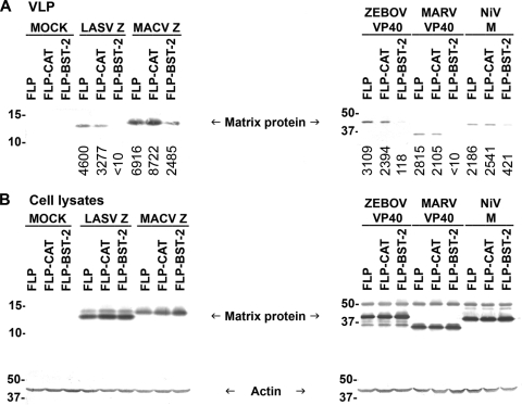
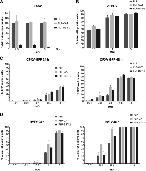
Bone marrow stromal antigen 2 (BST-2/tetherin) is a cellular membrane protein that inhibits the release of HIV-1. We show for the first time, using infectious viruses, that BST-2 also inhibits egress of arenaviruses but has no effect on filovirus replication and spread. Specifically, infectious Lassa virus (LASV) release significantly decreased or increased in human cells in which BST-2 was either stably expressed or knocked down, respectively. In contrast, replication and spread of infectious Zaire ebolavirus (ZEBOV) and Lake Victoria marburgvirus (MARV) were not affected by these conditions. Replication of infectious Rift Valley fever virus (RVFV) and cowpox virus (CPXV) was also not affected by BST-2 expression. Elevated cellular levels of human or murine BST-2 inhibited the release of virus-like particles (VLPs) consisting of the matrix proteins of multiple highly virulent NIAID Priority Pathogens, including arenaviruses (LASV and Machupo virus [MACV]), filoviruses (ZEBOV and MARV), and paramyxoviruses (Nipah virus). Although the glycoproteins of filoviruses counteracted the antiviral activity of BST-2 in the context of VLPs, they could not rescue arenaviral (LASV and MACV) VLP release upon BST-2 overexpression. Furthermore, we did not observe colocalization of filoviral glycoproteins with BST-2 during infection with authentic viruses. None of the arenavirus-encoded proteins rescued budding of VLPs in the presence of BST-2. Our results demonstrate that BST-2 might be a broad antiviral factor with the ability to restrict release of a wide variety of human pathogens. However, at least filoviruses, RVFV, and CPXV are immune to its inhibitory effect.


Figures

FIG. 1.
FIG. 1.
BST-2 inhibits budding of arenaviral, filoviral, and paramyxoviral VLPs. (A) Human 293T cells were cotransfected with plasmids encoding hemagglutinin (HA)-tagged matrix proteins of LASV, MACV, ZEBOV, MARV, or NiV, together with an empty plasmid, or plasmid encoding VPS4A-E228Q, human BST-2 (hBST-2), or murine BST-2 (mBST-2). Cell lysates and supernatants were harvested 48 h after transfection. VLPs in clarified medium were pelleted through a sucrose cushion, and HA-tagged matrix proteins in VLPs were analyzed by Western blotting. Numbers below each lane indicate values obtained with densitometric scanning using the ImageJ program (NIH). (B) Expression of the HA-tagged matrix proteins, actin, VPS4A-E228Q, hBST-2, or mBST-2 in cell lysates was determined by Western blotting. Shown is a representative Western blot from three independent experiments.
FIG. 2.
FIG. 2.
Release of arenaviral, filoviral, and paramyxoviral VLPs is reduced in cells stably expressing human BST-2. (A) Human 293 cells stably expressing human BST-2 (FLP-BST-2), CAT (FLP-CAT), or an empty plasmid (pCDNA5/FRT, FLP) were transfected with plasmids encoding HA-tagged matrix proteins of LASV, MACV, ZEBOV, MARV, or NiV. Cell lysates and supernatants were treated as in Fig. 1, and HA-tagged matrix proteins in VLPs were analyzed by Western blotting. Numbers below each lane indicate values obtained with densitometric scanning using the ImageJ program (NIH). (B) Expression of the HA-tagged matrix proteins and actin in cell lysates was determined by Western blot analysis. Shown is a representative Western blot from three independent experiments.
FIG. 3.
FIG. 3.
Filoviral GP1,2, but none of the arenavirus-encoded proteins, counteract human BST-2. (A) Human 293 cells stably expressing human BST-2 were cotransfected with ZEBOV HA-VP40 DNA and plasmids encoding ZEBOV VP30-V5, VP35-V5, V5-VP24, GP1,2, GP1,2ΔMLD-V5, sGP-V5, ssGP-V5, Δ-peptide-V5, or NP-V5 or HIV-1 Vpu-V5. Alternatively, cells were transfected with MARV HA-VP40 DNA, together with plasmids encoding MARV GP1,2 or HIV-1 Vpu-V5. Cell lysates and supernatants were treated as in Fig. 1, and HA-tagged VP40 in VLPs were analyzed by Western blotting. Numbers below each lane indicate values obtained with densitometric scanning using the ImageJ program (NIH). (B) Expression of HA-tagged VP40, actin, and other filovirus-encoded proteins in cell lysates was determined by Western blotting. Expression of HIV-1 Vpu (∼16 kDa), ZEBOV VP30 (∼30 kDa), VP35 (∼35 kDa), VP24 (∼24 kDa), GP1,2 ΔMLD (∼75 kDa), sGP (∼50 kDa), ssGP (∼47 kDa), Δ-peptide (∼10 kDa), and NP (∼104 kDa) was detected using anti-V5 antibody. Expression of ZEBOV (∼140 kDa) and MARV (∼170 kDa) GP1,2 was detected using anti-GP antibodies (6D8 and 5D7, respectively). (C) Human 293 cells stably expressing human BST-2 were cotransfected with LASV Z-HA DNA and plasmids encoding LASV NP-V5, SSP-V5, or GPC or HIV-1 Vpu-V5. Alternatively, cells were transfected with MACV Z-HA and MACV NP-V5, GPC, L-FLAG, or HIV-1 Vpu-V5. Cell lysates and supernatants were treated as in Fig. 1, and HA-tagged Z proteins in VLPs were analyzed by Western blotting. Numbers below each lane indicate values obtained with densitometric scanning using the ImageJ program (NIH). (D) Expression of HA-tagged Z, actin, and other arenavirus-encoded proteins in cell lysates was determined by Western blotting. Expression of HIV-1 Vpu (∼16 kDa), arenaviral NP (∼63 kDa), and LASV SSP (∼10 kDa) was detected by using anti-V5 antibody. Expression of MACV L-FLAG (∼200 kDa) was detected using anti-FLAG antibody. Expression of LASV GPC (∼75 kDa) and GP1 (∼42 kDa) was detected with an antibody against GP1 (161-6). Shown is a representative Western blot from three independent experiments.
FIG. 4.
FIG. 4.
The increase in ZEBOV VLP release when GP1,2 is expressed in the presence of BST-2 is significantly higher than that observed in the absence of BST-2. (A) Human 293 cells stably expressing human BST-2 (FLP-BST-2) or an empty plasmid (FLP) were transfected with ZEBOV VP40, together with an empty plasmid or a plasmid encoding GP1,2, or HIV-1 Vpu. Cell lysates and supernatants were treated as in Fig. 1, and HA-tagged VP40 in VLPs was analyzed by Western blotting. Numbers below each lane indicate values obtained with densitometric scanning using the ImageJ program (NIH). (B) Expression of the HA-tagged VP40, actin, GP1,2, and HIV-1 Vpu-V5 in cell lysates was determined by Western blot analysis. Shown is a representative Western blot from three independent experiments.
FIG. 5.
FIG. 5.
Expression of filoviral GP1,2 has little or no effect on release of arenaviral VLPs in BST-2-expressing cells. (A) Human 293 cells stably expressing human BST-2 were cotransfected with arenaviral Z-HA DNA and an empty plasmid or plasmids encoding filoviral GP1,2 or HIV-1 Vpu-V5. Cell lysates and supernatants were treated as in Fig. 1, and HA-tagged Z proteins in VLPs were analyzed by Western blotting. (B) Expression of HA-tagged Z, actin, HIV Vpu-V5, and filoviral GP1,2 in cell lysates was determined by Western blotting. Shown is a representative Western blot from three independent experiments.
FIG. 6.
FIG. 6.
Human BST-2 overexpression inhibits infectious LASV, but not ZEBOV, release and spread. (A) Human 293 cells stably expressing human BST-2 (FLP-BST-2), CAT (FLP-CAT), or an empty plasmid (FLP) were infected with LASV at the indicated MOIs. After 72 h, viral RNA was extracted from the media, and the viral copy number was determined by qRT-PCR. (B to D) Alternatively, cells were infected with ZEBOV (B), CPXV-GFP (C), or RVFV (D), fixed in formalin and stained for high-content quantitative image-based analysis with virus-specific antibodies 24 or 48 h (C and D) or 72 h (B) after infection. Error bars indicate standard deviations.
FIG. 7.
FIG. 7.
Knockdown of human BST-2 expression enhances infectious LASV release, but not ZEBOV or MARV spread. (A) HeLa cells were transfected with siRNA targeting BST-2 or with a control siRNA. After 24, 48, or 72 h, cellular RNA was extracted, and the relative BST-2 expression levels were determined by qRT-PCR. BST-2 protein expression levels in cell lysates were also determined by Western blotting. (B) Cells were infected with LASV (MOI = 0.1) 24 h after transfection. Viral RNA was extracted from the medium 72 h later, and the viral copy number was determined by qRT-PCR. (C) Alternatively, cells were infected with ZEBOV-GFP (MOI = 10) or MARV (MOI = 3). Cells were fixed in formalin 72 h later and stained for high-content quantitative image-based analysis with virus-specific antibodies. Error bars indicate standard deviations.
FIG. 8.
FIG. 8.
BST-2 localization in relation to filoviral GP1,2. (A) Human 293 cells stably expressing human BST-2 were infected with ZEBOV. Cells were fixed in formalin 24 h later and stained with antibodies against BST-2 and ZEBOV GP1,2. (B and C) Alternatively, cells were infected with MARV for 24 h (B) or 48 h (C) and stained with antibodies against BST-2 and MARV GP1,2. Immunofluorescence of anti-filoviral GP1,2 (green) and anti-BST-2 (red) in a single confocal plain is shown as indicated. Right columns show the merged images for triple staining of BST-2 (red), filoviral GP1,2 (green), and the nucleus (blue), as well as bright-field images.

Similar articles

Cited by

References

    1. Agnihothram, S. S., J. York, and J. H. Nunberg. 2006. Role of the stable signal peptide and cytoplasmic domain of G2 in regulating intracellular transport of the Junin virus envelope glycoprotein complex. J. Virol. 80:5189-5198. - PMC - PubMed
    1. Baize, S., J. Kaplon, C. Faure, D. Pannetier, M. C. Georges-Courbot, and V. Deubel. 2004. Lassa virus infection of human dendritic cells and macrophages is productive but fails to activate cells. J. Immunol. 172:2861-2869. - PubMed
    1. Baize, S., D. Pannetier, C. Faure, P. Marianneau, I. Marendat, M. C. Georges-Courbot, and V. Deubel. 2006. Role of interferons in the control of Lassa virus replication in human dendritic cells and macrophages. Microbes Infect. 8:1194-1202. - PubMed
    1. Bartee, E., A. McCormack, and K. Fruh. 2006. Quantitative membrane proteomics reveals new cellular targets of viral immune modulators. PLoS Pathog. 2:e107. - PMC - PubMed
    1. Battles, J. K., and J. M. Dalrymple. 1988. Genetic variation among geographic isolates of Rift Valley fever virus. Am. J. Trop. Med. Hyg. 39:617-631. - PubMed

Publication types

MeSH terms