. 2010 Oct;84(20):10501-9.
doi: 10.1128/JVI.00963-10. Epub 2010 Aug 4.

Epitope-specific regulatory CD4 T cells reduce virus-induced illness while preserving CD8 T-cell effector function at the site of infection

Affiliations

Epitope-specific regulatory CD4 T cells reduce virus-induced illness while preserving CD8 T-cell effector function at the site of infection

Jie Liu et al. J Virol. 2010 Oct.

Abstract

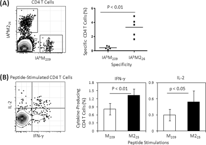
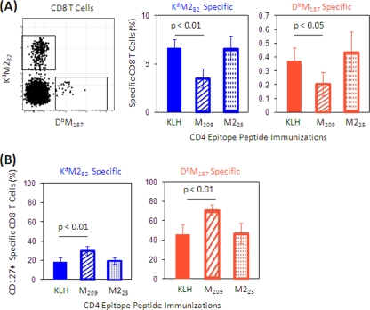
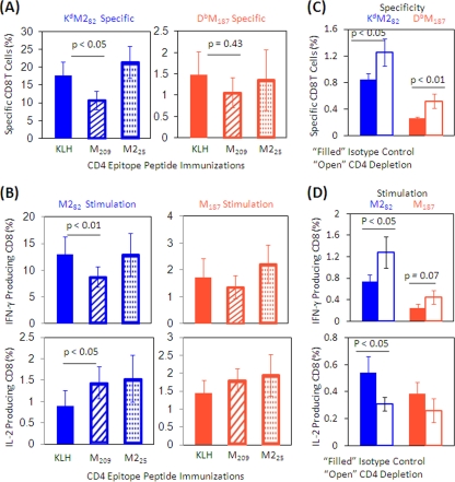
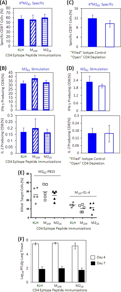
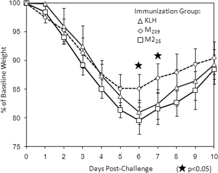
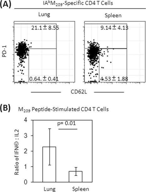
The role of epitope-specific regulatory CD4 T cells in modulating CD8 T-cell-mediated immunopathology during acute viral infection has not been well defined. In the murine model of respiratory syncytial virus (RSV) infection, CD8 T cells play an important role in both viral clearance and immunopathology. We have previously characterized two RSV epitope-specific CD4 T-cell responses with distinct phenotypic properties. One of them, the IA(b)M(209)-specific subset, constitutively expresses FoxP3 and modulates CD8 T-cell function in vitro. We show here that the IA(b)M(209)-specific CD4 T-cell response regulates CD8 T-cell function in vivo and is associated with diminished RSV-induced illness without affecting viral clearance at the site of infection. Achieving the optimal balance of regulatory and effector T-cell function is an important consideration for designing future vaccines.


Figures

FIG. 1.
FIG. 1.
CD4 T-cell responses to RSV M and M2. Lung lymphocytes were isolated at day 7 postinfection and stained with tetramers and phenotyping antibodies to identify specific CD4 T cells (A) or stimulated with MHC class II-restricted CD4 T-cell epitope-containing peptides (M209 or M225) in the presence of costimulators and brefeldin A for 5 h and then stained for lineage phenotype and cytokines intracellularly (B). Representative flow plots are gated on live CD4+ CD8 T cells. Frequencies of specific or cytokine-producing T cells refer to the total CD4 T-cell population. The cytokine-producing cell frequency is background subtracted, which is based on irrelevant OVA peptide stimulation. The data (mean ± the standard deviation [SD], n = 5 to 6/group) represent three independent experiments and are compared by using the Student t test.
FIG. 2.
FIG. 2.
Frequency and CD127 expression of RSV-specific CD8 cells in peripheral blood after administration of rAd5-M/M2. Mice were immunized with KLH or KLH-conjugated peptides M209 or M225 and then immunized with rAd5-M/M2 intramuscularly. (A) Peripheral blood samples were collected 10 days after rAd5-M/M2 immunization and stained with tetramers and phenotyping antibodies to assess the frequency of RSV tetramer-specific CD8 T cells. M209 peptide-immunized mice had a lower frequency of CD8 T-cell responses after rAd5-M/M2 than M25 peptide- or KLH-immunized mice. A representative plot is gated on CD4 CD8+ T cells. The percentages of specific CD8 T cells refer to total CD8 T-cell population. (B) We also assessed the frequency of CD127-expressing specific CD8 T cells in mice immunized with M209 or M25 peptides. The percentages refer to the specific CD8 T cells. The data (mean ± the SD, n = 5/group) are representative of three independent experiments and were compared by using the Student t test. Filled bars indicate the KLH-only-immunized control groups, hatched bars indicate M209-KLH-immunized mice, and dotted bars indicate M225-KLH-immunized mice.
FIG. 3.
FIG. 3.
Frequency and cytokine production of RSV-specific CD8 T cells from the spleen after RSV challenge. Mice were immunized with KLH or KLH-conjugated peptides, followed by rAd5-M/M2 administration intramuscularly, and then challenged with live RSV intranasally. Spleen lymphocytes were isolated at 7 day postchallenge and stained with tetramers and phenotyping antibodies to identify specific CD8 T cells by flow cytometry (A) or stimulated with CD8 epitope-containing peptides in the presence of costimulators and brefeldin A for 5 h and then stained for lineage phenotype and cytokine expressions intracellularly (B). The frequencies of specific CD8 T cells or cytokine-producing cells refer to CD8 T-cell population. The cytokine-producing cell frequency is the background (ranging from 0.01 to 0.08%) subtracted based on irrelevant OVA peptide stimulation. The data (mean ± the SD, n = 5/group) are representative of three independent experiments and compared by using the Student t test. Filled bars indicate the KLH-only-immunized control groups, hatched bars indicate M209-KLH-immunized mice, and dotted bars indicate M225-KLH-immunized mice. We also assessed the CD8 T-cell responses in the spleen in unimmunized mice depleted of CD4 T cells just prior to RSV challenge to remove the impact of CD4 T-cell modulation on the CD8 T-cell responses. Naive mice were treated with anti-CD4 antibody (□) or isotype control (▪) and then challenged with RSV. Spleen lymphocytes were isolated at day 7 postchallenge. Frequencies of specific CD8 T cells (C) and cytokine-producing CD8 T cells (D) from CD4-depleted or control mice stimulated with epitope-containing peptides were assessed as described in panels A and B.
FIG. 4.
FIG. 4.
Frequency and cytokine production of specific CD8 cells from the lung after RSV challenge. Mice were immunized with KLH or KLH-conjugated peptides, followed by rAd5-M/M2 administration intramuscularly, and then challenged with RSV intranasally. Lung lymphocytes were isolated at day 7 postinfection and stained with tetramers and phenotyping antibodies to identify specific CD8 T cells by flow cytometry (A) or stimulated with CD8 epitope-containing peptides in the presence of costimulators and brefeldin A for 5 h and then stained for lineage and cytokine expression intracellularly (B). Frequencies of specific CD8 T cells or cytokine-producing cells refer to the total CD8 T-cell population. The cytokine-producing cell frequency is the background (ranging from 0.03 to 0.2%) subtracted based on irrelevant OVA peptide stimulation. The data (mean ± the SD, n = 5/group) are representative of three independent experiments. Filled bars indicate the KLH-only-immunized control groups, hatched bars indicate M209-KLH-immunized mice, and dotted bars indicate M225-KLH-immunized mice. We then assessed CD8 T-cell responses in the lungs in unimmunized mice depleted of CD4 T cells just prior to RSV challenge to remove the impact of CD4 T-cell modulation on the CD8 T-cell responses. Naive mice were treated with anti-CD4 antibody (□) or isotype control (▪) and then infected with RSV. Lung lymphocytes were isolated at day 7 postinfection. Frequencies of specific (C) or cytokine-producing (D) CD8 T cells from CD4-depleted or control mice stimulated with epitope-containing peptides were assessed as described in panels A and B. (E) Isolated lung lymphocytes were cultured with CD8 epitope peptide M282- and M187-loaded P815 and EL-4 cells, respectively, for 3 h. Dead P815 and EL-4 cells after culturing were assessed with flow cytometry by annexin V staining. The percentages of specific killings refer to total P815 and EL-4 cell populations, with nonspecific cell death subtracted based on irrelevant OVA peptide-loaded P815 and EL-4 cell cultures. The data (mean ± the SD, n = 5/group) are representative of three independent experiments. (F) Supernatant of ground lung tissue isolated at days 4 and 7 postinfection were plated on HEp-2 cell layer and cultured for 4 days. The virus titers are expressed as PFU per gram of lung tissue. The limit of detection is 1.8 log10 PFU/gram of tissue. The data (mean ± the SD, n = 5/group) are representative of two independent experiments. All data were statistically compared by using the Student t test.
FIG. 5.
FIG. 5.
Weight loss after RSV challenge following immunization and boost. Mice were weighed daily after virus challenge (challenge day = day 0). Shown are the percentages of baseline weight. The data (mean ± the SD, n = 5/group) are representative of three independent experiments.
FIG. 6.
FIG. 6.
Phenotype and cytokine expressions of CD4 and CD8 T cells in the spleen and lung. Mice were primed with KLH or KLH-conjugated peptides, followed by rAd5-M/M2 administration intramuscularly, and then challenged with RSV intranasally. (A) Lung and spleen lymphocytes were isolated at day 7 postinfection and stained with tetramers and phenotyping antibodies to identify specific CD8 T cells and their phenotypes by flow cytometry. Representative plots are gated on IAbM209-specific CD4 T cells. (B) Frequencies of PD-1 high or PD-1 low with CD62L expression cells refer to the gated population or stimulated with CD4 epitope peptide M209 in the presence of costimulators and brefeldin A for 5 h and then stained for lineage phenotype and intracellular cytokines. The ratio of CD4 T cells producing IFN-γ and IL-2 are the background subtracted based on irrelevant OVA peptide stimulation. The data (mean ± the SD, n = 5/group) are representative of three independent experiments and compared by using the Student t test.

Similar articles

Cited by

References

    1. Anz, D., V. H. Koelzer, S. Moder, R. Thaler, T. Schwerd, K. Lahl, T. Sparwasser, R. Besch, H. Poeck, V. Hornung, G. Hartmann, S. Rothenfusser, C. Bourquin, and S. Endres. Immunostimulatory RNA blocks suppression by regulatory T cells. J. Immunol. 184:939-946. - PubMed
    1. Aung, S., J. A. Rutigliano, and B. S. Graham. 2001. Alternative mechanisms of respiratory syncytial virus clearance in perforin knockout mice lead to enhanced disease. J. Virol. 75:9918-9924. - PMC - PubMed
    1. Barber, D. L., E. J. Wherry, D. Masopust, B. Zhu, J. P. Allison, A. H. Sharpe, G. J. Freeman, and R. Ahmed. 2006. Restoring function in exhausted CD8 T cells during chronic viral infection. Nature 439:682-687. - PubMed
    1. Belkaid, Y. 2007. Regulatory T cells and infection: a dangerous necessity. Nat. Rev. Immunol. 7:875-888. - PubMed
    1. Biagi, E., I. Di Biaso, V. Leoni, G. Gaipa, V. Rossi, C. Bugarin, G. Renoldi, M. Parma, A. Balduzzi, P. Perseghin, and A. Biondi. 2007. Extracorporeal photochemotherapy is accompanied by increasing levels of circulating CD4+ CD25+ GITR+ Foxp3+ CD62L+ functional regulatory T cells in patients with graft-versus-host disease. Transplantation 84:31-39. - PubMed

MeSH terms