

Identification of alpha interferon-induced genes associated with antiviral activity in Daudi cells and characterization of IFIT3 as a novel antiviral gene
- PMID: 20686046
- PMCID: PMC2950578
- DOI: 10.1128/JVI.00818-10
Identification of alpha interferon-induced genes associated with antiviral activity in Daudi cells and characterization of IFIT3 as a novel antiviral gene
Abstract
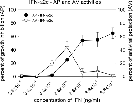
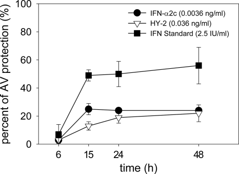
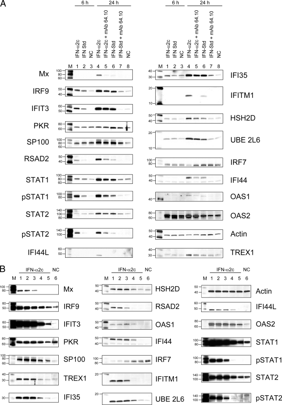
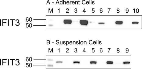
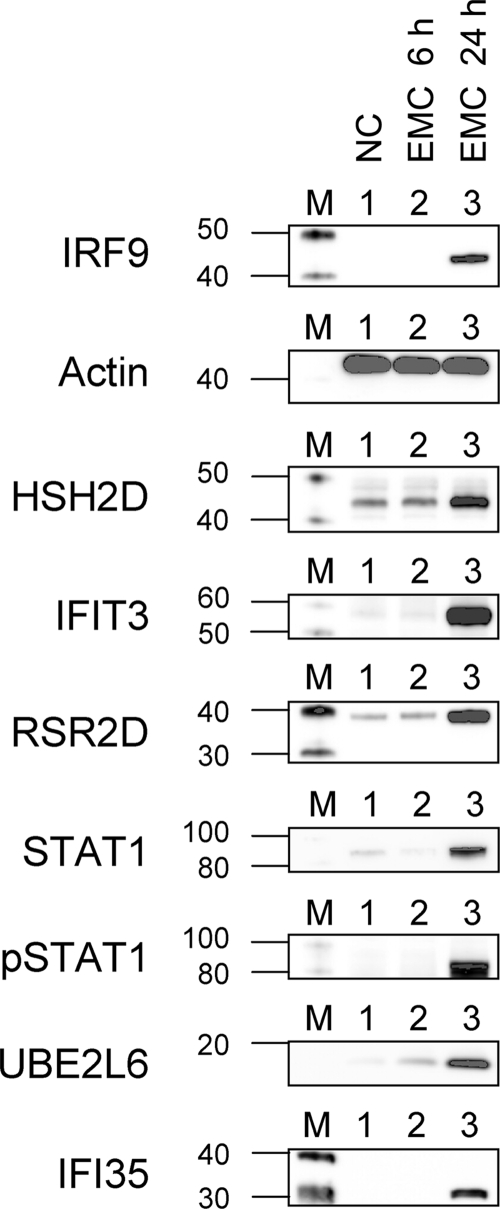
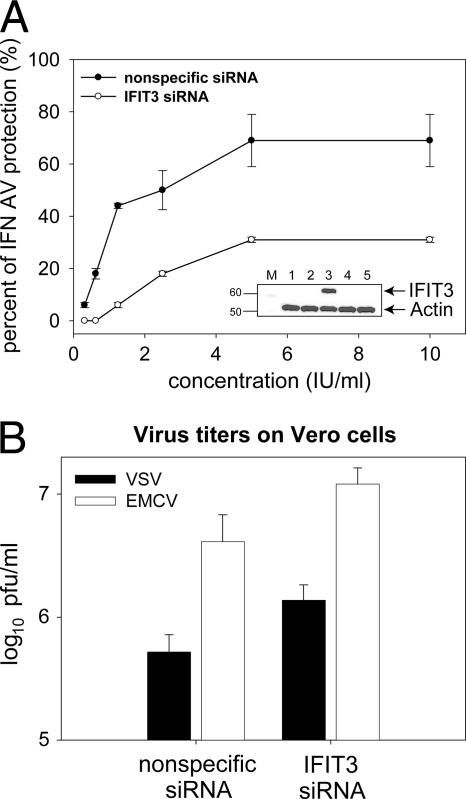
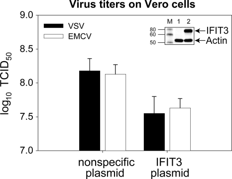
A novel assay was developed for Daudi cells in which the antiviral (AV) and antiproliferative (AP) activities of interferon (IFN) can be measured simultaneously. Using this novel assay, conditions allowing IFN AV protection but no growth inhibition were identified and selected. Daudi cells were treated under these conditions, and gene expression microarray analyses were performed. The results of the analysis identified 25 genes associated with IFN-α AV activity. Upregulation of 23 IFN-induced genes was confirmed by using reverse transcription-PCR. Of 25 gene products, 17 were detected by Western blotting at 24 h. Of the 25 genes, 10 have not been previously linked to AV activity of IFN-α. The most upregulated gene was IFIT3 (for IFN-induced protein with tetratricopeptide repeats 3). The results from antibody neutralizing experiments suggested an association of the identified genes with IFN-α AV activity. This association was strengthened by results from IFIT3-small interfering RNA transfection experiments showing decreased expression of IFIT3 and a reduction in the AV activity induced by IFN-α. Overexpression of IFIT3 resulted in a decrease of virus titer. Transcription of AV genes after the treatment of cells with higher concentrations of IFN having an AP effect on Daudi cells suggested pleiotropic functions of identified gene products.
Figures
Similar articles
-
IFIT3 Is Increased in Serum from Patients with Chronic Hepatitis B Virus (HBV) Infection and Promotes the Anti-HBV Effect of Interferon Alpha via JAK-STAT2 In Vitro.Microbiol Spectr. 2022 Dec 21;10(6):e0155722. doi: 10.1128/spectrum.01557-22. Epub 2022 Oct 31. Microbiol Spectr. 2022. PMID: 36314949 Free PMC article.
-
Human IFIT3 Protein Induces Interferon Signaling and Inhibits Adenovirus Immediate Early Gene Expression.mBio. 2021 Dec 21;12(6):e0282921. doi: 10.1128/mBio.02829-21. Epub 2021 Nov 2. mBio. 2021. PMID: 34724821 Free PMC article.
-
Protective roles of interferon-induced protein with tetratricopeptide repeats 3 (IFIT3) in dengue virus infection of human lung epithelial cells.PLoS One. 2013 Nov 4;8(11):e79518. doi: 10.1371/journal.pone.0079518. eCollection 2013. PLoS One. 2013. PMID: 24223959 Free PMC article.
-
The antiproliferative and antiviral activities of IFN-tau variants in human cells.J Interferon Cytokine Res. 1997 Dec;17(12):769-79. doi: 10.1089/jir.1997.17.769. J Interferon Cytokine Res. 1997. PMID: 9452365
-
Antiviral and antiproliferative activities of human leukocyte interferon potentiated by cimetidine in vitro.J Interferon Res. 1985 Summer;5(3):375-82. doi: 10.1089/jir.1985.5.375. J Interferon Res. 1985. PMID: 2997335
Cited by
-
Defense genes missing from the flight division.Dev Comp Immunol. 2013 Nov;41(3):377-88. doi: 10.1016/j.dci.2013.04.010. Epub 2013 Apr 24. Dev Comp Immunol. 2013. PMID: 23624185 Free PMC article. Review.
-
DNA methylation mediates the effect of cocaine use on HIV severity.Clin Epigenetics. 2020 Sep 14;12(1):140. doi: 10.1186/s13148-020-00934-1. Clin Epigenetics. 2020. PMID: 32928285 Free PMC article.
-
Identification of bovine leukemia virus tax function associated with host cell transcription, signaling, stress response and immune response pathway by microarray-based gene expression analysis.BMC Genomics. 2012 Mar 28;13:121. doi: 10.1186/1471-2164-13-121. BMC Genomics. 2012. PMID: 22455445 Free PMC article.
-
Doxorubicin resistance involves modulation of interferon signaling, transcriptional bursting, and gene co-expression patterns of U-ISGF3-related genes.Neoplasia. 2024 Dec;58:101071. doi: 10.1016/j.neo.2024.101071. Epub 2024 Oct 13. Neoplasia. 2024. PMID: 39405604 Free PMC article.
-
Cutting Edge: Expression of IRF8 in Gastric Epithelial Cells Confers Protective Innate Immunity against Helicobacter pylori Infection.J Immunol. 2016 Mar 1;196(5):1999-2003. doi: 10.4049/jimmunol.1500766. Epub 2016 Feb 3. J Immunol. 2016. PMID: 26843324 Free PMC article.
References
-
- Armstrong, J. A. 1981. Cytopathic effect inhibition assay for interferon: microculture plate assay. Methods Enzymol. 78:381-387. - PubMed
-
- Brass, A. L., I.-C. Huang, Y. Benita, S. P. John, M. N. Krishnan, E. M. Feeley, B. J. Ryan, J. L. Weyer, L. van der Weyden, E. Fikrig, D. J. Adams, R. J. Xavier, M. Farzan, and S. J. Elledge. 2009. The IFITM proteins mediate cellular resistance to influenza A H1N1 virus, West Nile virus, and dengue virus. Cell 139:1243-1254. - PMC - PubMed
-
- Chieux, V., W. Chehadeh, P. Hautecoeur, J. Harvey, P. Wattre, and D. Hober. 2001. Increased levels of antiviral MxA protein in peripheral blood of patients with a chronic disease of unknown etiology. J. Med. Virol. 65:301-308. - PubMed
-
- Deblandre, G. A., O. P. Marinx, S. S. Evans, S. Majjaj, O. Leo, D. Caput, G. A. Huez, and M. G. Wathelet. 1995. Expression cloning of an interferon inducible 17-kDa membrane protein implicated in the control of cell growth. J. Biol. Chem. 270:23860-23866. - PubMed
Publication types
MeSH terms
Substances
Grants and funding
LinkOut - more resources
Full Text Sources
Other Literature Sources
Molecular Biology Databases
