. 2010 Oct;84(20):10877-87.
doi: 10.1128/JVI.00949-10. Epub 2010 Aug 4.

Chikungunya virus nonstructural protein 2 inhibits type I/II interferon-stimulated JAK-STAT signaling

Affiliations

Chikungunya virus nonstructural protein 2 inhibits type I/II interferon-stimulated JAK-STAT signaling

Jelke J Fros et al. J Virol. 2010 Oct.

Abstract

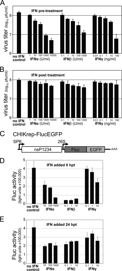
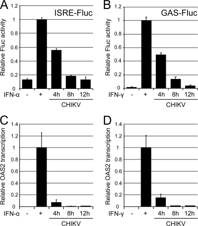
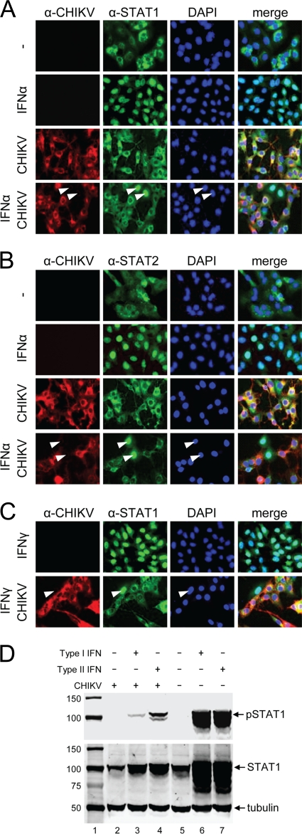
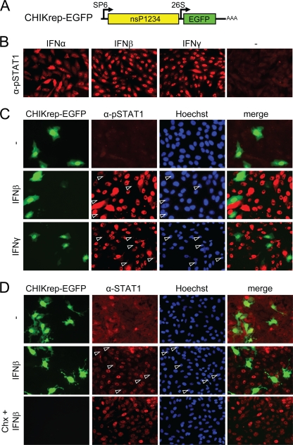
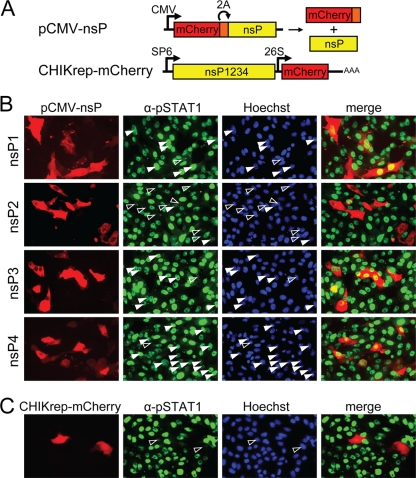
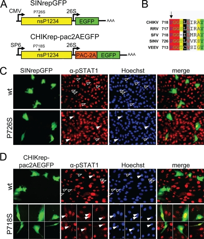
Chikungunya virus (CHIKV) is an emerging human pathogen transmitted by mosquitoes. Like that of other alphaviruses, CHIKV replication causes general host shutoff, leading to severe cytopathicity in mammalian cells, and inhibits the ability of infected cells to respond to interferon (IFN). Recent research, however, suggests that alphaviruses may have additional mechanisms to circumvent the host's antiviral IFN response. Here we show that CHIKV replication is resistant to inhibition by interferon once RNA replication has been established and that CHIKV actively suppresses the antiviral IFN response by preventing IFN-induced gene expression. Both CHIKV infection and CHIKV replicon RNA replication efficiently blocked STAT1 phosphorylation and/or nuclear translocation in mammalian cells induced by either type I or type II IFN. Expression of individual CHIKV nonstructural proteins (nsPs) showed that nsP2 was a potent inhibitor of IFN-induced JAK-STAT signaling. In addition, mutations in CHIKV-nsP2 (P718S) and Sindbis virus (SINV)-nsP2 (P726S) that render alphavirus replicons noncytopathic significantly reduced JAK-STAT inhibition. This host shutoff-independent inhibition of IFN signaling by CHIKV is likely to have an important role in viral pathogenesis.


Figures

FIG. 1.
FIG. 1.
Resistance of CHIKV to type I/II IFN treatment. (A and B) Sensitivity of CHIKV infection to IFN treatment. IFNs were added as indicated to CHIKV-infected Vero cells 6 h prior to infection (A) or 4 h p.i. (B). Supernatants were collected 24 h p.i., and virus titers were determined by plaque assays. Error bars represent standard deviations of duplicates. (C) Schematic representation of the CHIKrep-FlucEGFP replicon expressing an Fluc-EGFP fusion protein. (D and E) Sensitivity of the replication of CHIKV replicon RNA to IFN treatment. Different concentrations of IFNs were added to CHIKV replicon-transfected Vero cells in 96-well plates directly posttransfection (0 h p.t.) (D) or 24 h p.t. (E), and Fluc activity was measured 48 h p.t. Concentrations of IFN-α are expressed in international units (IU) per ml, and IFN-β/γ concentrations are expressed in ng per ml. Error bars represent standard deviations.
FIG. 2.
FIG. 2.
Inhibition of type I/II IFN signaling and ISG induction by CHIKV infection. (A and B) Vero cells were transfected with a pRL-TK plasmid expressing Rluc and either a type I IFN-responsive (ISRE) or a type II IFN-responsive (GAS) Fluc reporter plasmid. At 24 h p.t., cells were infected with CHIKV at an MOI of 5 PFU/ml. At 4, 8, and 12 h p.i., cells were treated with IFN-α at 1,000 IU/ml (A) or with IFN-γ at 100 ng/ml (B) for 6 h; then they were assayed for Fluc and Rluc activities. Activities in mock-infected (uninfected) cells with/without IFN induction were also measured. Fluc values were divided by Rluc readings to compensate for virus-induced downregulation of transcription/translation and were expressed relative to values for mock-infected, IFN-treated samples. Average values from triplicate samples are shown. Error bars represent standard deviations. (C and D) Vero cells, either healthy or infected with CHIKV for 4, 8, or 12 h, were incubated with 1,000 IU of IFN-α (C) or 100 ng of IFN-γ (D) per ml for 10 h. Real-time RT-PCR values for the IFN-stimulated gene OAS2 were normalized to those for the housekeeping gene RPL13A. OAS2 mRNA transcription levels were expressed relative to those of mock-infected, IFN-treated samples. Average values from duplicate samples are shown. Error bars represent standard deviations.
FIG. 3.
FIG. 3.
(A to C) CHIKV infection blocks STAT1/STAT2 nuclear translocation without depleting endogenous STAT1 levels. Vero cells were infected by CHIKV and were treated with IFN-α (A and B) or IFN-γ (C) for 30 min. Cells were fixed and stained with monoclonal antibodies specific for CHIKV envelope protein and STAT1 (A and C) or STAT2 (B). (C) Block in nuclear translocation of STAT1 in CHIKV infection in response to treatment with IFN-γ. Arrowheads indicate cells negatively infected with CHIKV but with nuclear STAT1/2. (D) CHIKV infection blocks STAT1 phosphorylation in Vero cells in response to IFN treatment. pSTAT1, STAT1, and tubulin were detected by Western blotting in CHIKV-infected or mock-infected Vero cells that were either left untreated or induced with type I or type II IFNs. Lane 1, protein size marker (in kilodaltons).
FIG. 4.
FIG. 4.
A CHIKV replicon efficiently inhibits type I/II IFN-induced JAK-STAT signaling independently of host shutoff. (A) Schematic representation of CHIKrepEGFP, expressing EGFP. (B) pSTAT1 nuclear translocation in Vero cells upon induction with type I and type II IFNs. (C) A CHIKV replicon blocks pSTAT1 nuclear translocation upon type I/II IFN induction. Vero cells were immunostained with an anti-pSTAT1 antibody 24 h p.t. (D) CHIKV RNA replication, but not translational shutoff, blocks STAT1 nuclear translocation. Vero cells were transfected with CHIKrep-EGFP replicon RNA in the absence or presence of cycloheximide (Chx). Cells were induced for 30 min with IFN-β at 12 h p.t. and were stained with an anti-STAT1 antibody. Open arrowheads indicate CHIKV replicon-positive cells lacking nuclear STAT1.
FIG. 5.
FIG. 5.
Inhibition of IFN-β-induced STAT1 nuclear translocation by individual CHIKV nsPs. (A) Schematic representation of the pCMV-nsP1, -2, -3, and -4 expression plasmids and the CHIKrep-mCherry replicon, expressing mCherry. CMV, cytomegalovirus immediate-early promoter; 2A, foot-and-mouth disease virus 2A autoprotease. The bacteriophage SP6 and CHIKV 26S promoters are indicated. (B) pSTAT1 nuclear translocation upon IFN-β induction in Vero cells transfected with pCMV-nsP1, -2, -3, or -4. Cells were immunostained with an anti-pSTAT1 antibody. (C) pSTAT1 nuclear translocation upon IFN-β induction in CHIKrep-mCherry-transfected Vero cells. Open arrowheads indicate cells positive for nsP1, -2, -3, or -4- or for the CHIKV replicon that lack nuclear pSTAT1; solid arrowheads indicate nsP1- to nsP4-positive cells with nuclear pSTAT1.
FIG. 6.
FIG. 6.
Mutation of a conserved proline in nsP2 abolishes the inhibitory effect of CHIKV and SINV replicons on JAK-STAT signaling. (A) Schematic representation of the CHIKrep-pac2AEGFP and SINrepLuc replicons. nsP2 mutations P718S and P726S are indicated with asterisks; pac, puromycin acetyltransferase. (B) Partial amino acid alignment of alphavirus nsP2s. RRV, Ross River virus; VEEV, Venezuelan equine encephalitis virus. The conserved proline and amino acid numbers within nsP2 proteins are indicated. (C) pSTAT1 nuclear translocation upon IFN-β induction in SINrepGFP (wild type and mutant nsP2-P726S)-transfected Vero cells. Cells were immunostained with an anti-pSTAT1 antibody. Open arrowheads indicate replicon-positive cells lacking nuclear pSTAT1; solid arrowheads indicate replicon-positive cells with nuclear pSTAT1. (D) Nuclear translocation of phospho-STAT1 upon IFN-β induction in CHIKrep-pac2AEGFP (wild type and mutant nsP2-P718S)-transfected Vero cells. Cells were immunostained with an anti-pSTAT1 antibody.

Similar articles

Cited by

References

    1. Aguilar, P. V., S. C. Weaver, and C. F. Basler. 2007. Capsid protein of Eastern equine encephalitis virus inhibits host cell gene expression. J. Virol. 81:3866-3876. - PMC - PubMed
    1. Ashour, J., M. Laurent-Rolle, P. Y. Shi, and A. Garcia-Sastre. 2009. NS5 of dengue virus mediates STAT2 binding and degradation. J. Virol. 83:5408-5418. - PMC - PubMed
    1. Breakwell, L., P. Dosenovic, G. B. Karlsson Hedestam, M. D'Amato, P. Liljestrom, J. Fazakerley, and G. M. McInerney. 2007. Semliki Forest virus nonstructural protein 2 is involved in suppression of the type I interferon response. J. Virol. 81:8677-8684. - PMC - PubMed
    1. Burke, C. W., C. L. Gardner, J. J. Steffan, K. D. Ryman, and W. B. Klimstra. 2009. Characteristics of alpha/beta interferon induction after infection of murine fibroblasts with wild-type and mutant alphaviruses. Virology 395:121-132. - PMC - PubMed
    1. Couderc, T., F. Chretien, C. Schilte, O. Disson, M. Brigitte, F. Guivel-Benhassine, Y. Touret, G. Barau, N. Cayet, I. Schuffenecker, P. Despres, F. Arenzana-Seisdedos, A. Michault, M. L. Albert, and M. Lecuit. 2008. A mouse model for Chikungunya: young age and inefficient type-I interferon signaling are risk factors for severe disease. PLoS Pathog. 4:e29. - PMC - PubMed

MeSH terms