. 2010 Nov;299(5):C912-21.
doi: 10.1152/ajpcell.00215.2010. Epub 2010 Aug 4.

Cystic fibrosis transmembrane conductance regulator is involved in airway epithelial wound repair

Affiliations

Cystic fibrosis transmembrane conductance regulator is involved in airway epithelial wound repair

Katherine R Schiller et al. Am J Physiol Cell Physiol. 2010 Nov.

Abstract

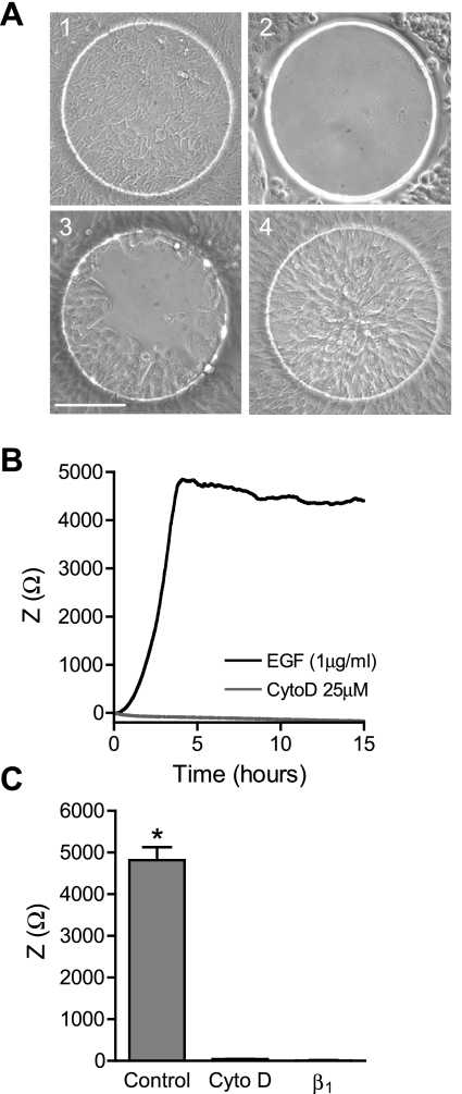
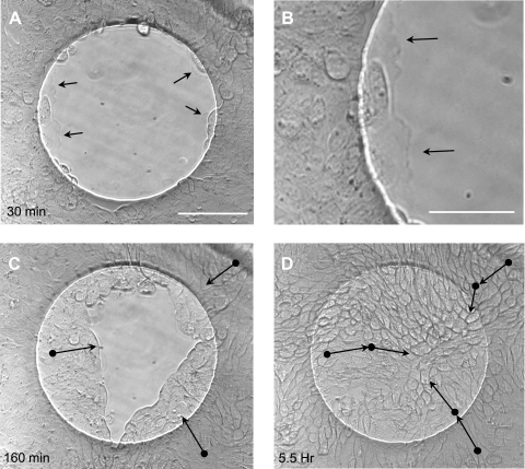
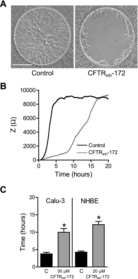
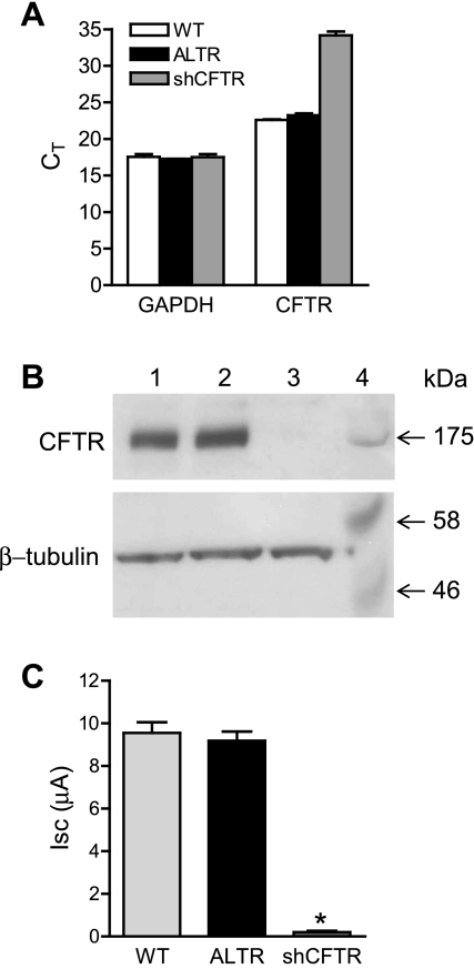
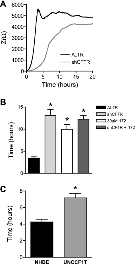
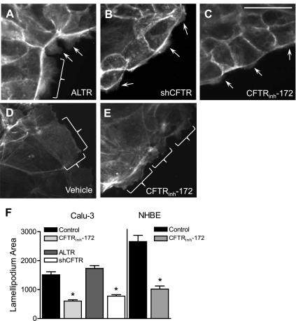
The role of cystic fibrosis (CF) transmembrane conductance regulator (CFTR) in airway epithelial wound repair was investigated using normal human bronchial epithelial (NHBE) cells and a human airway epithelial cell line (Calu-3) of serous gland origin. Measurements of wound repair were performed using continuous impedance sensing to determine the time course for wound closure. Control experiments showed that the increase in impedance corresponding to cell migration into the wound was blocked by treatment with the actin polymerization inhibitor, cytochalasin D. Time lapse imaging revealed that NHBE and Calu-3 cell wound closure was dependent on cell migration, and that movement occurred as a collective sheet of cells. Selective inhibition of CFTR activity with CFTR(inh)-172 or short hairpin RNA silencing of CFTR expression produced a significant delay in wound repair. The CF cell line UNCCF1T also exhibited significantly slower migration than comparable normal airway epithelial cells. Inhibition of CFTR-dependent anion transport by treatment with CFTR(inh)-172 slowed wound closure to the same extent as silencing CFTR protein expression, indicating that ion transport by CFTR plays a critical role in migration. Moreover, morphologic analysis of migrating cells revealed that CFTR inhibition or silencing significantly reduced lamellipodia protrusion. These findings support the conclusion that CFTR participates in airway epithelial wound repair by a mechanism involving anion transport that is coupled to the regulation of lamellipodia protrusion at the leading edge of the cell.


Figures

Fig. 1.
Fig. 1.
Measurement of wound closure by continuous impedance sensing (CIS). CIS was used to measure wound closure in Calu-3 cells. A: images of electrodes: 1) before wounding, 2) immediately after wounding, 3) 1 h after wounding, and 4) at wound closure (3 h). Scale bar = 100 μm. B: averaged impedance (Z) time course during wound closure from a representative experiment of Calu-3 cells treated with 1 μg/ml EGF and vehicle (DMSO; black) or 25 μM cytochalasin D (CtyoD; gray). C: Calu-3 cell impedance at the time of wound closure with control media (1 μg/ml EGF) and at the same time point with cytochalasin D treatment, and final impedance after 24-h treatment of Calu-3 cell suspension with anti-β1-integrin (β1). *Significant differences between means.
Fig. 2.
Fig. 2.
Time lapse images of wound closure in Calu-3 cells. Brightfield phase-contrast images are shown from a time lapse wound closure sequence of Calu-3 cells with control media (serum free + 1 μg/ml EGF). A: image captured 30 min after wounding. Scale bar, 100 μm. B: enlarged image of lamellipodia in A at the leading edge of the wound 30 min after wounding. Scale bar, 50 μm. Arrows in A and B indicate lamellipodia. C: image captured 160 min after wounding. D: image captured 5.5 h after wounding. Movement of individual cells shown in C and D with arrows indicating approximate direction of movement, and starting point of movement indicated by solid black circles. Cells were identified on the basis of their morphology and tracked at 1-min intervals until reaching the positions indicated in the figure.
Fig. 3.
Fig. 3.
Effect of CFTR inhibition on wound closure. A: images of vehicle (DMSO)-treated Calu-3 cells after complete wound closure (3 h; left) or CFTRinh-172-treated cells at the same time point (3 h; right). Scale bar, 100 μm. B: averaged impedance time course during wound closure from a representative experiment of normal human bronchial epithelial (NHBE) cells treated with vehicle (DMSO; black line) or CFTRinh-172 (gray line). C: time required for complete wound closure in Calu-3 and NHBE cells treated with vehicle (black bars) or CFTRinh-172 (gray bars). *Significant differences between means.
Fig. 4.
Fig. 4.
Silencing CFTR in Calu-3 cells. A: cycle threshold (CT) values from quantitative real-time PCR (qRT-PCR) of CFTR and GAPDH expression in wild-type Calu-3 cells (WT, white bar), control altered short hairpin (sh)RNA (ALTR)-expressing Calu-3 cells (black bars), and CFTR-specific shRNA (shCFTR)-expressing Calu-3 cells (gray bars). B: Western blot of CFTR (top) and β-tubulin (bottom) protein levels in ALTR-expressing Calu-3 cells (lane 1), wild-type Calu-3 cells (lane 2), shCFTR-expressing Calu-3 cells (lane 3), and molecular mass markers in kDa (lane 4). C: measurements of 8-cpt-cAMP (2.5 μM)-stimulated short-circuit current (Isc) in monolayers of wild-type Calu-3 cells (light gray bar), ALTR Calu-3 cells (black bar), and CFTR-silenced Calu-3 cells (dark gray bar). *Significant difference between means.
Fig. 5.
Fig. 5.
Wound closure of CFTR-silenced cells. CIS was used to measure wound closure in CFTR-silenced (shCFTR) and control shRNA-expressing (ALTR) Calu-3 cells. A: averaged impedance time course during wound closure from a representative experiment of shCFTR (gray line) and ALTR (black line) Calu-3 cells. B: time required for complete wound closure of ALTR-expressing Calu-3 cells (black bar), shCFTR-expressing cells (light gray bar), wild-type Calu-3 cells treated with CFTRinh-172 (white bar), and shCFTR-expressing cells treated with 30 μM CFTRinh-172 (dark gray bar). C: time required for wound closure of NHBE cells (black bar) and UNCCF1T cells (gray bar). *Significant differences between means.
Fig. 6.
Fig. 6.
Effects of CFTR inhibition on lamellipodia area. Lamellipodia areas were measured at the time of maximum protrusion following wounding of Calu-3 and NHBE cells. A–E: images (taken at ×40 magnification) of Calu-3 cells (A–C) stained with fluorescent phalloidin 1 h after wounding; protruding lamellipodia indicated by brackets and arrows. Shown are ALTR-expressing Calu-3 cells treated with vehicle (DMSO) (A); shCFTR-expressing Calu-3 cells (B); and Calu-3 cells treated with 30 μM CFTRinh-172 (C); scale bar, 20 μm. D and E: NHBE cells stained with fluorescent phalloidin 45 min after wounding; protruding lamellipodia indicated by brackets. Shown are NHBE cells treated with vehicle (DMSO) (D) and NHBE cells treated with 20 μM CFTRinh-172 (E). F: average lamellipodium area (pixels) per cell of Calu-3 and NHBE cells described in A–E. *Significant differences between means.

Comment in

Similar articles

Cited by

References

    1. Alexander RT, Grinstein S. Na+/H+ exchangers and the regulation of volume. Acta Physiol (Oxf) 187: 159–167, 2006 - PubMed
    1. Anderson MP, Gregory RJ, Thompson S, Souza DW, Paul S, Mulligan RC, Smith AE, Welsh MJ. Demonstration that CFTR is a chloride channel by alteration of its anion selectivity. Science 253: 202–205, 1991 - PubMed
    1. Bankers-Fulbright JL, Bartemes KR, Kephart GM, Kita H, O'Grady SM. Beta2-integrin-mediated adhesion and intracellular Ca2+ release in human eosinophils. J Membr Biol 228: 99–109, 2009 - PMC - PubMed
    1. Bilton D. What did we learn from the North American Cystic Fibrosis Conference? J R Soc Med 101, Suppl 1: S6–S9, 2008 - PMC - PubMed
    1. Boucher RC. New concepts of the pathogenesis of cystic fibrosis lung disease. Eur Respir J 23: 146–158, 2004 - PubMed

Publication types

MeSH terms

LinkOut - more resources