. 2010 Nov;299(5):C948-59.
doi: 10.1152/ajpcell.00018.2010. Epub 2010 Aug 4.

Expression profile and protein translation of TMEM16A in murine smooth muscle

Affiliations

Expression profile and protein translation of TMEM16A in murine smooth muscle

Alison J Davis et al. Am J Physiol Cell Physiol. 2010 Nov.

Abstract

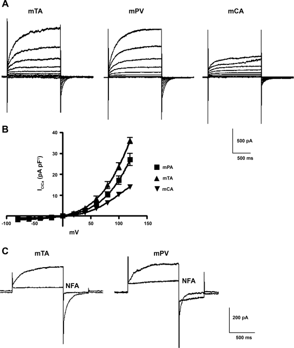
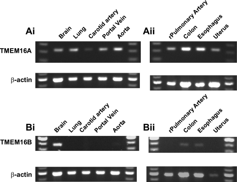
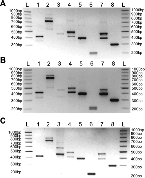
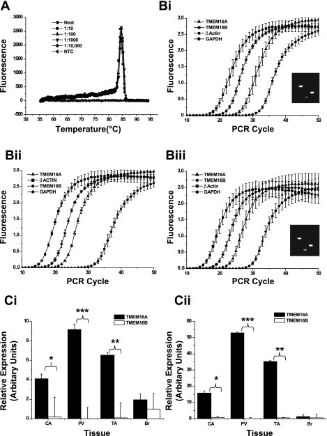
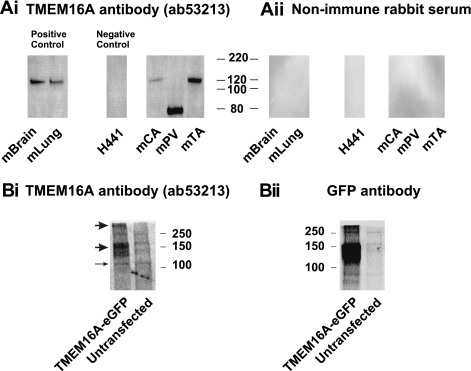
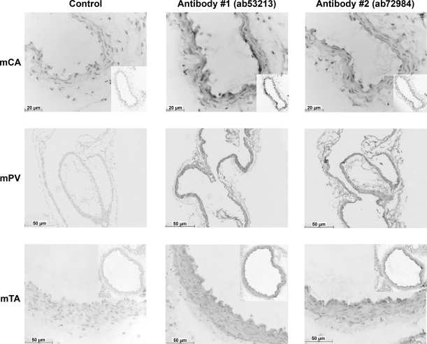
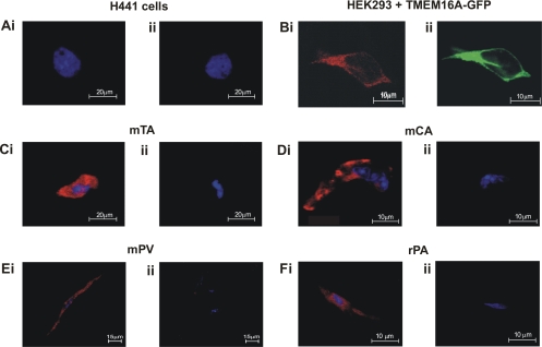
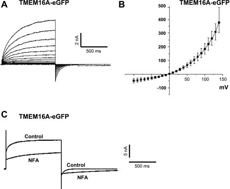
Recently, overexpression of the genes TMEM16A and TMEM16B has been shown to produce currents qualitatively similar to native Ca(2+)-activated Cl(-) currents (I(ClCa)) in vascular smooth muscle. However, there is no information about this new gene family in vascular smooth muscle, where Cl(-) channels are a major depolarizing mechanism. Qualitatively similar Cl(-) currents were evoked by a pipette solution containing 500 nM Ca(2+) in smooth muscle cells isolated from BALB/c mouse portal vein, thoracic aorta, and carotid artery. Quantitative PCR using SYBR Green chemistry and primers specific for transmembrane protein (TMEM) 16A or the closely related TMEM16B showed TMEM16A expression as follows: portal vein > thoracic aorta > carotid artery > brain. In addition, several alternatively spliced variant transcripts of TMEM16A were detected. In contrast, TMEM16B expression was very low in smooth muscle. Western blot analysis with different antibodies directed against TMEM16A revealed a number of products with a consistent band at ∼120 kDa, except portal vein, where an 80-kDa band predominated. TMEM16A protein was identified in the smooth muscle layers of 4-μm-thick slices of portal vein, thoracic aorta, and carotid artery. In isolated myocytes, fluorescence specific to a TMEM16A antibody was detected diffusely throughout the cytoplasm, as well as near the membrane. The same antibody used in Western blot analysis of lysates from vascular tissues also recognized an ∼147-kDa mouse TMEM16A-green fluorescent protein (GFP) fusion protein expressed in HEK 293 cells, which correlated to a similar band detected by a GFP antibody. Patch-clamp experiments revealed that I(ClCa) generated by transfection of TMEM16A-GFP in HEK 293 cells displayed remarkable similarities to I(ClCa) recorded in vascular myocytes, including slow kinetics, steep outward rectification, and a response similar to the pharmacological agent niflumic acid. This study shows that TMEM16A expression is robust in murine vascular smooth muscle cells, consolidating the view that this gene is a viable candidate for the native Ca(2+)-activated Cl(-) channel in this cell type.


Figures

Fig. 1.
Fig. 1.
Characteristics of Ca2+-dependent Cl channels (IClCa) in murine vascular smooth muscle cells. A: representative families of IClCa recorded at test potentials from −100 to +120 mV from a holding potential (HP) of −50 mV in myocytes isolated from mouse thoracic aorta (mTA), portal vein (mPV), and carotid artery (mCA). IClCa was evoked by a pipette solution containing 500 nM free Ca2+. B: mean current-voltage (I-V) relationships for amplitude of IClCa recorded at the end of each test pulse normalized to cell capacitance in each of the 3 different vascular myocytes. Each point is mean ± SE of 6–15 cells from ≥3 different animals. C: typical superimposed IClCa recordings from a murine thoracic aorta (left) and a murine portal vein (right) myocyte obtained in the absence or presence of 100 μM niflumic acid (NFA). Voltage-clamp protocol consisted of 1-s test steps to +70 mV followed by a 500-ms return step to −80 mV (HP = −50 mV). Traces represent results from >4 cells from ≥3 animals.
Fig. 2.
Fig. 2.
RT-PCR expression analysis of transmembrane proteins 16A and 16B (TMEM16A and TMEM16B) in smooth muscle tissues. mRNA was extracted from whole tissues and subjected to reverse transcription, and semiquantitative PCR was performed. After a standard 35 cycles, sequence-verified TMEM16A product (Ai and Aii) was amplified in a range of tissues, including those previously shown to express this gene. In contrast, amplification of TMEM16B (Bi and Bii) product showed a more restricted expression profile. The housekeeping gene β-actin acted as internal positive control for all PCRs. Images represent results from >3 animals.
Fig. 3.
Fig. 3.
TMEM16A splice variant expression in murine vascular tissues. A–C: end-point PCR gels for thoracic aorta (A), carotid artery (B), and portal vein (C). Primers are listed in Table 1. Lane markers are as follows: ladder (L), exon a annealing (lane 1), exon b spanning (lane 2), exon b annealing (lane 3), exon c annealing (lane 4), exon c spanning (lanes 5 and 6), exon d spanning (lane 7), and exon d annealing (lane 8). Double bands in lanes 2 and 7 represent presence and absence of that exon product.
Fig. 4.
Fig. 4.
Quantitative PCR (qPCR) expression analysis of TMEM16A and TMEM16B. A: representative dissociation profile for TMEM16A primers in serially diluted concentrations of murine brain cDNA. B: amplification plots of each PCR primer set tested in murine carotid artery (i), portal vein (ii), and thoracic aorta (iii), including analysis of end-point PCR products on agarose gels for representative qPCR experiment (see insets). In insets, lane 1 is 25-bp ladder, lane 2 is β-actin RT+, lane 3 is GAPDH RT+, lane 4 is TMEM16A RT+, and lane 5 is TMEM16A RT−. C: relative expression of primer sets, normalized to β-actin (Ci) and GAPDH (Cii). Significance was determined by Student's 1-tailed paired t-test: *P = 0.01–0.05; **P = 0.01–0.001; ***P < 0.001. Values are means ± SE of tissues from 3 different animals, each with duplicate PCRs.
Fig. 5.
Fig. 5.
Western blot analysis of murine protein lysates. TMEM16A protein expression in various murine tissues was determined by SDS-PAGE. H441 cells acted as a negative control for TMEM16A protein expression. Aii: no band was detected by Western blot analysis of the same protein lysates samples in Ai when rabbit nonimmune serum was used in place of primary antibody. B: immunodetection of TMEM16A and green fluorescent protein (GFP) in HEK 293 cells transfected with enhanced GFP (eGFP)-tagged TMEM16A. Blot performed using the anti-TMEM16A antibody (Ab 53213) shows molecular weight of the principal product, most likely corresponding to monomeric protein, is consistent with that expected for a TMEM16A-GFP construct (∼138 kDa). There also appears to be a band at approximately the predicted size for endogenous human TMEM16A (∼114 kDa; full-length would be predicted to lie near 117 kDa) in protein obtained from control and TMEM16A-GFP-transfected cells (thin arrow). A similar display of immunodetection is seen in protein samples processed from the same cells, but stained with an anti-GFP antibody (600-101-215, Rockland; 1:1,000 dilution). GFP staining confirms that the band observed at 147 kDa after staining with the anti-TMEM16A antibody is specific to this protein. Images are representative of results from >3 experiments.
Fig. 6.
Fig. 6.
TMEM16A is expressed in the smooth muscle layer of murine vessels. Immunohistochemistry was performed on 4-μm-thick transverse (carotid artery and thoracic aorta) and longitudinal (portal vein) sections. Representative time-matched sections incubated in control serum (without primary antibody) reveal only very faint false-positive staining. Sections treated with the primary antibodies ab53213 and ab72984 show obvious diaminobenzidine staining compared with control. All sections were counterstained for nuclei. Images are representative of results from 3 experiments.
Fig. 7.
Fig. 7.
TMEM16A protein localization in enzymatically isolated smooth muscle cells. A: lack of staining in H441 cells incubated with TMEM16A antibody (ab53213, Ai) compared with cells incubated with secondary antibody only (Aii). Experiments were run in parallel with thoracic aorta myocytes (represented in C). Bi: colocalization of TMEM16A staining (ab53213, 1:50 dilution) with GFP tagging visible. Pattern of TMEM16A immunostaining and GFP location show a clear correlation, indicating that the TMEM16A antibody specifically stains TMEM16A in these cells. Fluorescence immunocytochemistry revealed specific expression of TMEM16A protein in murine carotid artery, portal vein, and thoracic aorta cells (Ci, Di, Ei,) and rat pulmonary artery cells (Fi). Control (no primary antibody) cells showed minimal fluorescence, confirming specificity of the signals (Cii, Dii, Eii, and Fii).
Fig. 8.
Fig. 8.
IClCa in HEK 293 cells transiently transfected with TMEM16A tagged with enhanced GFP. A: representative family of currents from a TMEM16A-transfected HEK 293 cell dialyzed with 500 nM Ca2+. Currents were evoked by stepping in 10-mV increments from HP of −50 mV to 1-s test potentials ranging from −100 to +140 mV following 10 min of dialysis. B: mean current-voltage relationship generated from families of currents similar to those described in A (n = 9). C: superimposed IClCa traces recorded before and after application of 100 μM niflumic acid (NFA). Current recorded in drug-free solution (control) was generated by stepping from HP of −50 mV to +140 mV for 1 s, then repolarizing to −80 mV for 1 s, following 10 min of dialysis with 5 mM ATP and 500 nM Ca2+. NFA (100 μM) was then applied for 10 min, and current trace labeled “NFA” was recorded in a manner similar to control current. Traces represent results from 6 experiments.

Similar articles

Cited by

References

    1. Almaca J, Tian Y, Aldehni F, Ousingsawat J, Kongsuphol P, Rock JR, Harfe BD, Schreiber R, Kunzelmann K. TMEM16 proteins produce volume regulated chloride currents that are reduced in mice lacking TMEM16A. J Biol Chem 284: 28571–28578, 2009 - PMC - PubMed
    1. Angermann JE, Sanguinetti AR, Kenyon JL, Leblanc N, Greenwood IA. Mechanism of the inhibition of Ca2+-activated Cl− currents by phosphorylation in pulmonary arterial smooth muscle cells. J Gen Physiol 128: 73–87, 2006 - PMC - PubMed
    1. Ayon R, Sones W, Forrest AS, Wiwchar M, Valencik ML, Sanguinetti AR, Perrino BA, Greenwood IA, Leblanc N. Complex phosphatase regulation of Ca2+-activated Cl− currents in pulmonary arterial smooth muscle cells. J Biol Chem 284: 32507–32521, 2009 - PMC - PubMed
    1. Britton FC, Ohya S, Horowitz B, Greenwood IA. Comparison of the properties of CLCA1 generated currents and ICl(Ca) in murine portal vein smooth muscle cells. J Physiol 539: 107–117, 2002 - PMC - PubMed
    1. Burg ED, Platoshyn O, Tsigelny IF, Lozano-Ruizn B, Rana BK, Yuan JXJ. Tetramerization domain mutations in KCNA5 affect channel kinetics and cause abnormal trafficking patterns. Am J Physiol Cell Physiol 298: C496–C509, 2010 - PMC - PubMed

Publication types

MeSH terms

LinkOut - more resources