

Multiplex analysis of age-related protein and lipid modifications in human Bruch's membrane
- PMID: 20686107
- PMCID: PMC2992374
- DOI: 10.1096/fj.10-166090
Multiplex analysis of age-related protein and lipid modifications in human Bruch's membrane
Abstract
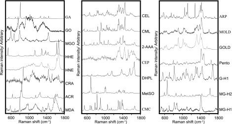
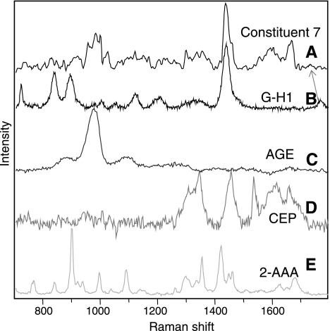
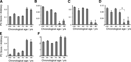
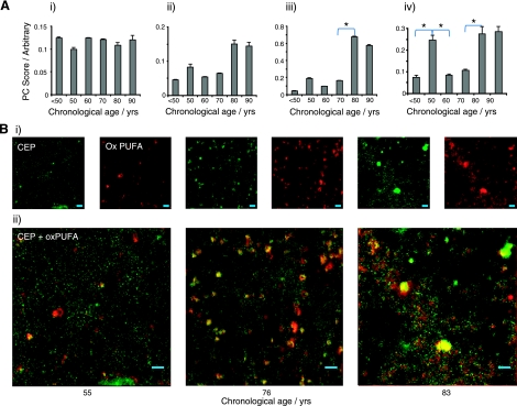
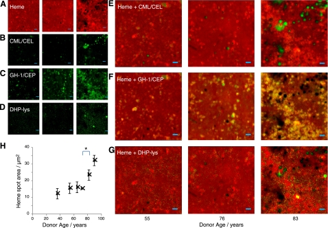
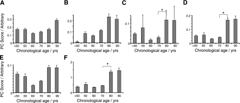
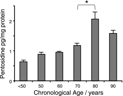
Aging of the human retina is characterized by progressive pathology, which can lead to vision loss. This progression is believed to involve reactive metabolic intermediates reacting with constituents of Bruch's membrane, significantly altering its physiochemical nature and function. We aimed to replace a myriad of techniques following these changes with one, Raman spectroscopy. We used multiplexed Raman spectroscopy to analyze the age-related changes in 7 proteins, 3 lipids, and 8 advanced glycation/lipoxidation endproducts (AGEs/ALEs) in 63 postmortem human donors. We provided an important database for Raman spectra from a broad range of AGEs and ALEs, each with a characteristic fingerprint. Many of these adducts were shown for the first time in human Bruch's membrane and are significantly associated with aging. The study also introduced the previously unreported up-regulation of heme during aging of Bruch's membrane, which is associated with AGE/ALE formation. Selection of donors ranged from ages 32 to 92 yr. We demonstrated that Raman spectroscopy can identify and quantify age-related changes in a single nondestructive measurement, with potential to measure age-related changes in vivo. We present the first directly recorded evidence of the key role of heme in AGE/ALE formation.
Figures
Similar articles
-
Sclera as a surrogate marker for determining AGE-modifications in Bruch's membrane using a Raman spectroscopy-based index of aging.Invest Ophthalmol Vis Sci. 2011 Mar 1;52(3):1593-8. doi: 10.1167/iovs.10-6554. Print 2011 Mar. Invest Ophthalmol Vis Sci. 2011. PMID: 21398274 Free PMC article.
-
Confocal Raman microscopy can quantify advanced glycation end product (AGE) modifications in Bruch's membrane leading to accurate, nondestructive prediction of ocular aging.FASEB J. 2007 Nov;21(13):3542-52. doi: 10.1096/fj.06-7896com. Epub 2007 Jun 12. FASEB J. 2007. PMID: 17567569
-
Advanced glycation end product (AGE) accumulation on Bruch's membrane: links to age-related RPE dysfunction.Invest Ophthalmol Vis Sci. 2009 Jan;50(1):441-51. doi: 10.1167/iovs.08-1724. Epub 2008 Aug 1. Invest Ophthalmol Vis Sci. 2009. PMID: 18676633
-
[Drusen in Bruch's membrane. Their significance for the pathogenesis and therapy of age-associated macular degeneration].Ophthalmologe. 1992 Oct;89(5):363-86. Ophthalmologe. 1992. PMID: 1304217 Review. German.
-
Advanced glycation and advanced lipoxidation: possible role in initiation and progression of diabetic retinopathy.Curr Pharm Des. 2004;10(27):3349-60. doi: 10.2174/1381612043383124. Curr Pharm Des. 2004. PMID: 15544520 Review.
Cited by
-
RNA-seq analysis of ageing human retinal pigment epithelium: Unexpected up-regulation of visual cycle gene transcription.J Cell Mol Med. 2021 Jun;25(12):5572-5585. doi: 10.1111/jcmm.16569. Epub 2021 May 1. J Cell Mol Med. 2021. PMID: 33934486 Free PMC article.
-
Vitamin A cycle byproducts explain retinal damage and molecular changes thought to initiate retinal degeneration.Biol Open. 2021 Nov 15;10(11):bio058600. doi: 10.1242/bio.058600. Epub 2021 Nov 29. Biol Open. 2021. PMID: 34842275 Free PMC article.
-
Nail Properties and Bone Health: A Review.J Funct Biomater. 2018 Apr 23;9(2):31. doi: 10.3390/jfb9020031. J Funct Biomater. 2018. PMID: 29690604 Free PMC article. Review.
-
Proteomic profiling of human retinal pigment epithelium exposed to an advanced glycation-modified substrate.Graefes Arch Clin Exp Ophthalmol. 2012 Mar;250(3):349-59. doi: 10.1007/s00417-011-1856-9. Epub 2011 Nov 13. Graefes Arch Clin Exp Ophthalmol. 2012. PMID: 22081232 Free PMC article.
-
Raman Spectroscopic Analysis of Fingernail Clippings Can Help Differentiate Between Postmenopausal Women Who Have and Have Not Suffered a Fracture.Clin Med Insights Arthritis Musculoskelet Disord. 2016 May 31;9:109-16. doi: 10.4137/CMAMD.S38493. eCollection 2016. Clin Med Insights Arthritis Musculoskelet Disord. 2016. PMID: 27429561 Free PMC article.
References
-
- Klein R., Klein B. E., Tomany S. C., Meuer S. M., Huang G. H. (2002) Ten-year incidence and progression of age-related maculopathy: The Beaver Dam eye study. Ophthalmology 109, 1767–1779 - PubMed
-
- Harding J. J. (1991) Cataract: Biochemistry, Epidemiology and Pharmacology, Chapman & Hall, London, UK
-
- De Jong P. T. (2006) Age-related macular degeneration. N. Engl. J. Med. 355, 1474–1485 - PubMed
-
- Beatty S., Koh H., Phil M., Henson D., Boulton M. (2000) The role of oxidative stress in the pathogenesis of age-related macular degeneration. Surv. Ophthalmol. 45, 115–134 - PubMed
-
- Starita C., Hussain A. A., Pagliarini S., Marshall J. (1996) Hydrodynamics of ageing Bruch's membrane: implications for macular disease. Exp. Eye Res. 62, 565–572 - PubMed
Publication types
MeSH terms
Substances
Grants and funding
LinkOut - more resources
Full Text Sources
Medical
