. 2010 Aug 24;107(34):15299-304.
doi: 10.1073/pnas.1008461107. Epub 2010 Aug 4.

Global phosphorylation analysis of beta-arrestin-mediated signaling downstream of a seven transmembrane receptor (7TMR)

Affiliations

Global phosphorylation analysis of beta-arrestin-mediated signaling downstream of a seven transmembrane receptor (7TMR)

Kunhong Xiao et al. Proc Natl Acad Sci U S A. .

Erratum in

  • Proc Natl Acad Sci U S A. 2012 Aug 14;109(33):13464

Abstract

beta-Arrestin-mediated signaling downstream of seven transmembrane receptors (7TMRs) is a relatively new paradigm for signaling by these receptors. We examined changes in protein phosphorylation occurring when HEK293 cells expressing the angiotensin II type 1A receptor (AT1aR) were stimulated with the beta-arrestin-biased ligand Sar(1), Ile(4), Ile(8)-angiotensin (SII), a ligand previously found to signal through beta-arrestin-dependent, G protein-independent mechanisms. Using a phospho-antibody array containing 46 antibodies against signaling molecules, we found that phosphorylation of 35 proteins increased upon SII stimulation. These SII-mediated phosphorylation events were abrogated after depletion of beta-arrestin 2 through siRNA-mediated knockdown. We also performed an MS-based quantitative phosphoproteome analysis after SII stimulation using a strategy of stable isotope labeling of amino acids in cell culture (SILAC). We identified 1,555 phosphoproteins (4,552 unique phosphopeptides), of which 171 proteins (222 phosphopeptides) showed increased phosphorylation, and 53 (66 phosphopeptides) showed decreased phosphorylation upon SII stimulation of the AT1aR. This study identified 38 protein kinases and three phosphatases whose phosphorylation status changed upon SII treatment. Using computational approaches, we performed system-based analyses examining the beta-arrestin-mediated phosphoproteome including construction of a kinase-substrate network for beta-arrestin-mediated AT1aR signaling. Our analysis demonstrates that beta-arrestin-dependent signaling processes are more diverse than previously appreciated. Notably, our analysis identifies an AT1aR-mediated cytoskeletal reorganization network whereby beta-arrestin regulates phosphorylation of several key proteins, including cofilin and slingshot. This study provides a system-based view of beta-arrestin-mediated phosphorylation events downstream of a 7TMR and opens avenues for research in a rapidly evolving area of 7TMR signaling.


Conflict of interest statement

The authors declare no conflict of interest.

Figures
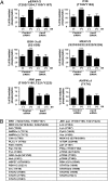
Fig. 1.
Fig. 1.
Human phospho-antibody array analysis reveals that phosphorylation events induced by SII are primarily β-arrestin–dependent. (A) Normalized intensities for six representative phosphoproteins from samples applied to the phospho-antibody array. Lysates were from HEK293 cells transfected with either control or β-arrestin 2 (βarr2) siRNA followed by 30 μM SII stimulation for 5 min [SII(+)] or no stimulation [SII(−)]. The normalized intensity for each antibody was calculated as a percentage of the corresponding SII-stimulated sample treated with control siRNA. The experiment was repeated at least three times, and statistical analysis was performed using a one-way ANOVA. ***P < 0.001. (B) A list of β-arrestin–regulated phosphoproteins revealed by human phospho-antibody array analysis. The residues indicated in parentheses are the phosphorylation sites.
Fig. 2.
Fig. 2.
Protein kinases involved in β-arrestin–mediated cellular signaling downstream of AT1aR. A comparison of experimentally identified, bioinformatically predicted, and previously reported protein kinases involved in β-arrestin–mediated signaling. Bioinformatic predictions were performed using Motif-X analysis and KEA as described in the text.
Fig. 3.
Fig. 3.
A β-arrestin–dependent cytoskeletal reorganization network downstream of AT1aR. β-Arrestin–mediated phosphoproteome and β-arrestin interactome revealed that many proteins in cytoskeletal reorganization pathways were regulated downstream of AT1aR/β-arrestin, suggesting a critical role of β-arrestins in cytoskeletal reorganization. The β-arrestin–interacting proteins were identified in the previous β-arrestin interactome study (8) or in biochemical experiments in the current study.
Fig. 4.
Fig. 4.
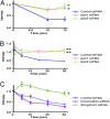
SII stimulation leads to β-arrestin–mediated dephosphorylation of slingshot and cofilin downstream of AT1aR. HEK293 cells stably overexpressing AT1aR were stimulated with 30 μM SII for the indicated time periods. (A) β-Arrestin–dependent dephosphorylation of slingshot. The cell lysates were probed with the slingshot pSer-978 antibody in the presence of control, β-arrestin1 (βarr1), or β-arrestin 2 (βarr2) siRNAs. The ratio of phospho-slingshot/total slingshot was calculated for each data point and plotted as fold decrease from the nonstimulated sample. (B) β-Arrestin–dependent dephosphorylation of cofilin. The cell lysates were probed using cofilin pSer-3 antibody. The ratio of phospho-cofilin/total cofilin was calculated for each data point and plotted as fold decrease from the nonstimulated sample. (C) Cofilin pSer-3 dephosphorylation downstream of AT1aR is mediated via slingshot and not via chronophin. HEK293 cells stably overexpressing AT1aRs were transfected with control, chronophin, or slingshot1/2 siRNA. Cofilin dephosphorylation was probed with phospho-cofilin Ser-3. The phospho-cofilin/total cofilin ratio was calculated for each data point and plotted as fold decrease from the nonstimulated sample. The data presented here are from at least three independent experiments. Statistical analysis was done using two-way ANOVA. *P < 0.05; **P < 0.01.
Fig. 5.
Fig. 5.
β-arrestin–dependent kinase network downstream of AT1aR revealed by the β-arrestin–mediated phosphoproteome and β-arrestin interactome. The network was generated by an inference algorithm (24) and literature-based kinome network (17) as described in the text and SI Text. This network covers a broad range of cellular functions including MAPK and PI3K/AKT signaling, cytoskeletal reorganization, cellular adhesion, cell-to-cell communication, and cell cycle and development. Green rectangle, identified in β-arrestin–mediated phosphoproteome; orange ellipse, β-arrestin–interacting proteins; pink hexagon, identified in β-arrestin–mediated phosphoproteome and β-arrestin interactome; gray rectangle, ”hub proteins” important for the signaling network but not identified in either β-arrestin–mediated phosphoproteome or β-arrestin interactome; red arrow, regulation of phosphorylation/dephosphorylation; black line, interaction with β-arrestin; dashed line, regulation of hub proteins. Several known β-arrestin–interacting proteins (ARAF, MEK1, p38, and JNK) that were not in our β-arrestin interactome are indicated also.

References

    1. Pierce KL, Premont RT, Lefkowitz RJ. Seven-transmembrane receptors. Nat Rev Mol Cell Biol. 2002;3:639–650. - PubMed
    1. Lefkowitz RJ, Shenoy SK. Transduction of receptor signals by beta-arrestins. Science. 2005;308:512–517. - PubMed
    1. Lefkowitz RJ, Whalen EJ. Beta-arrestins: Traffic cops of cell signaling. Curr Opin Cell Biol. 2004;16:162–168. - PubMed
    1. Olsen JV, et al. Global, in vivo, and site-specific phosphorylation dynamics in signaling networks. Cell. 2006;127:635–648. - PubMed
    1. Rinschen MM, et al. Quantitative phosphoproteomic analysis reveals vasopressin V2-receptor-dependent signaling pathways in renal collecting duct cells. Proc Natl Acad Sci U S A. 107(8):3882–3887. - PMC - PubMed

Publication types

MeSH terms