. 2010 Sep 1;185(5):2903-17.
doi: 10.4049/jimmunol.0903635. Epub 2010 Aug 4.

Heat shock protein 90 mediates efficient antigen cross presentation through the scavenger receptor expressed by endothelial cells-I

Affiliations

Heat shock protein 90 mediates efficient antigen cross presentation through the scavenger receptor expressed by endothelial cells-I

Ayesha Murshid et al. J Immunol. .

Abstract

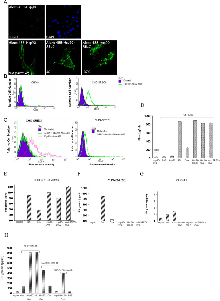
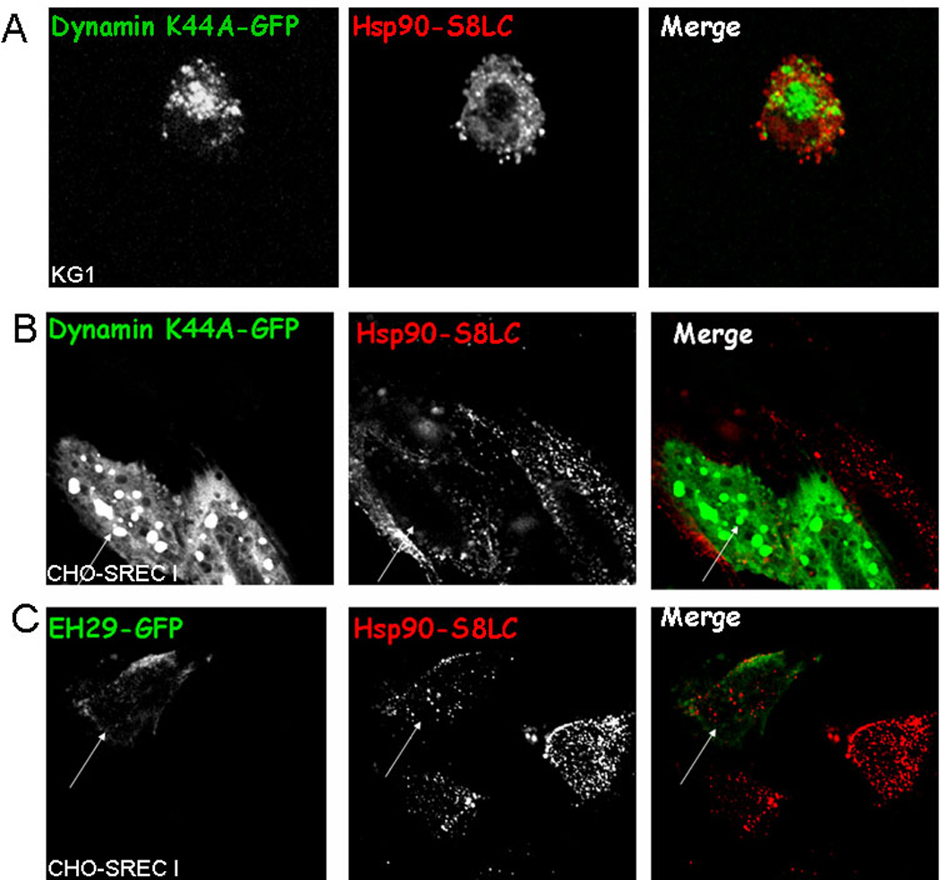
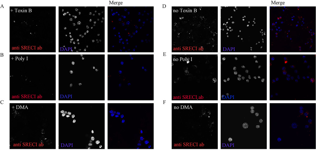
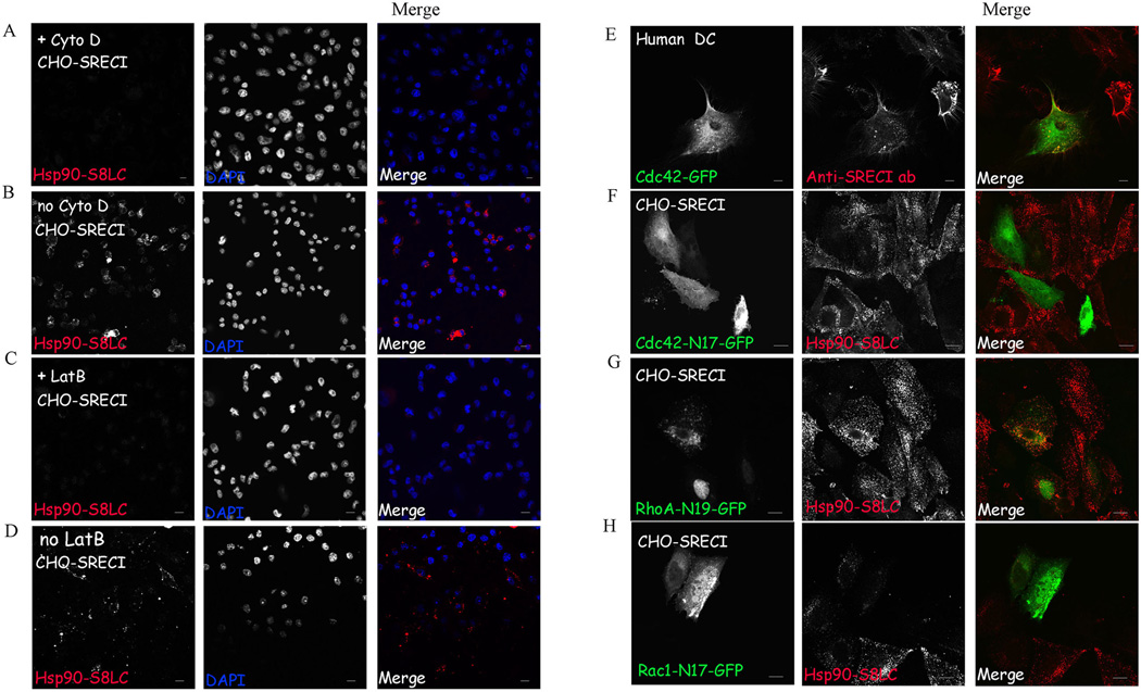
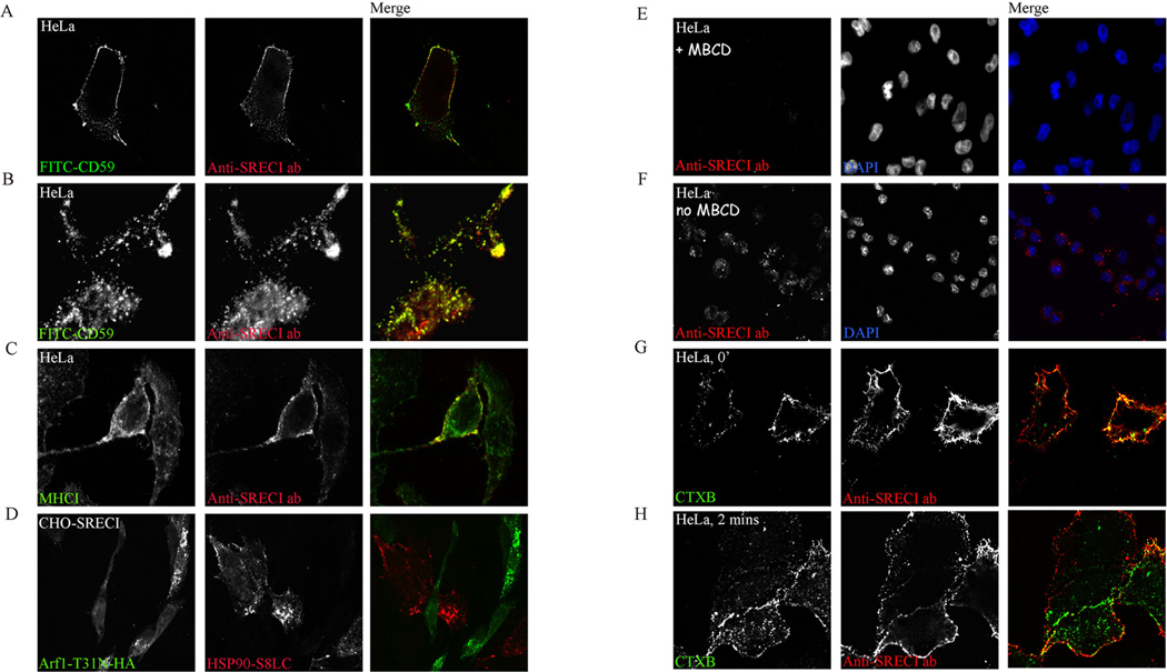
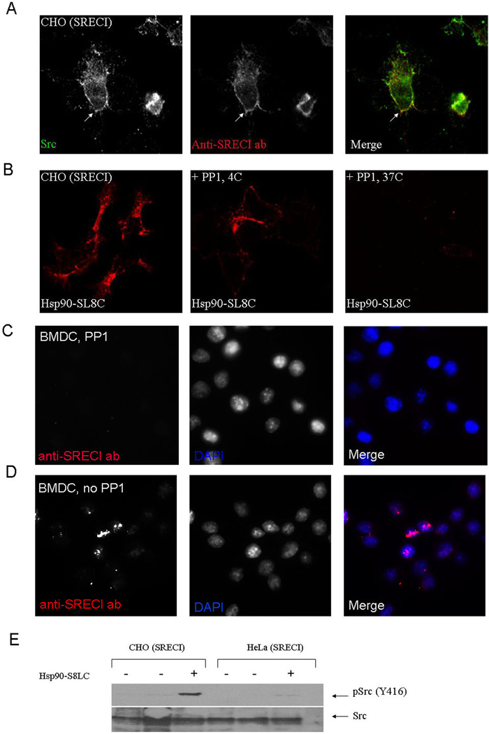
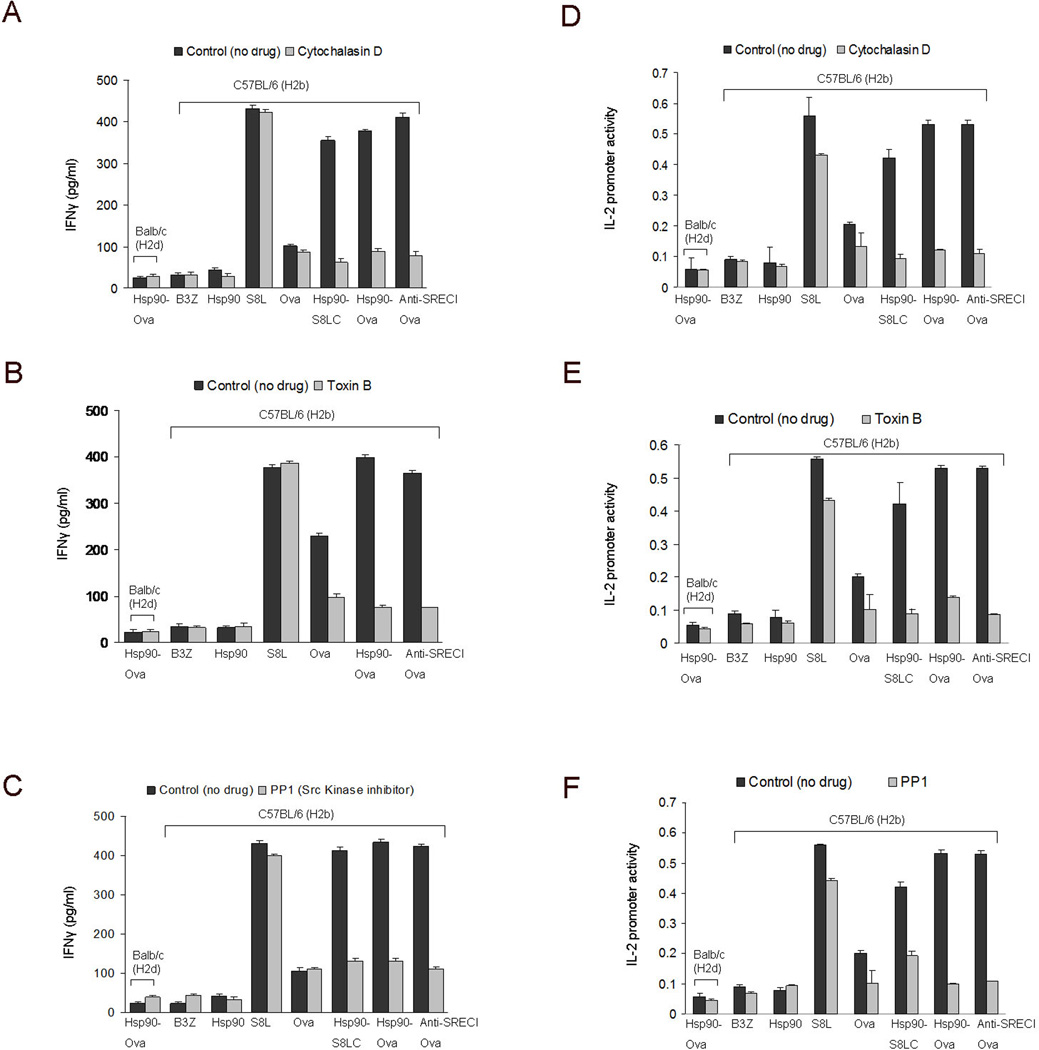
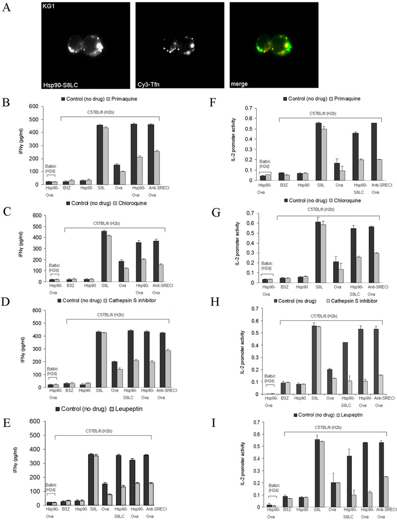
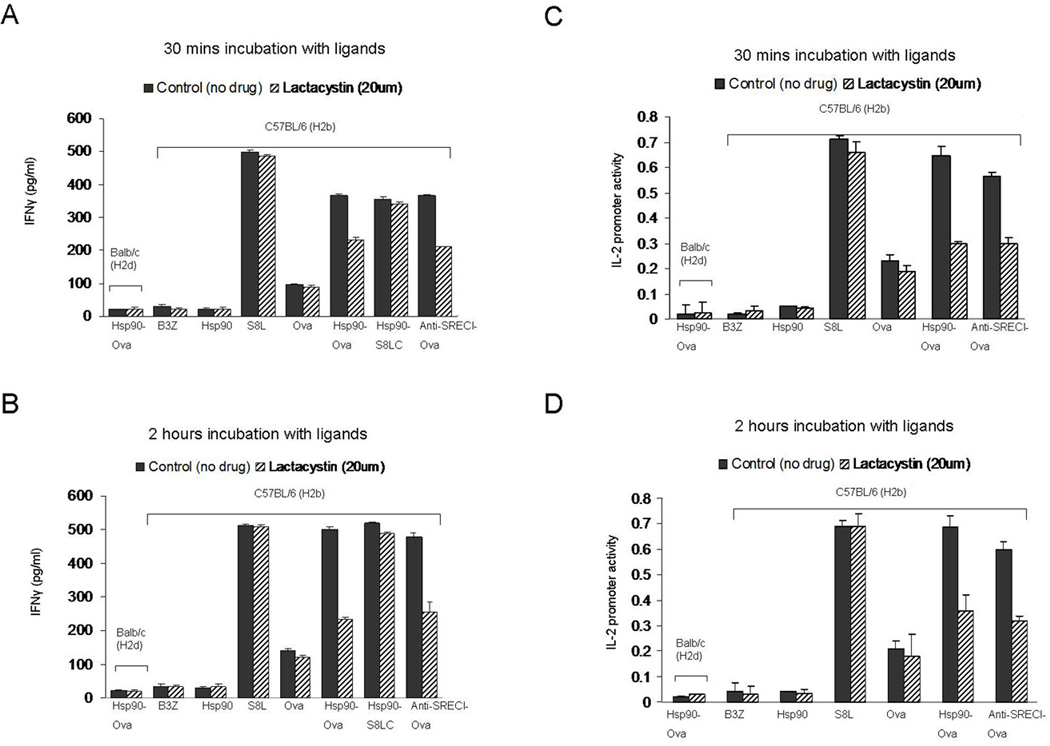
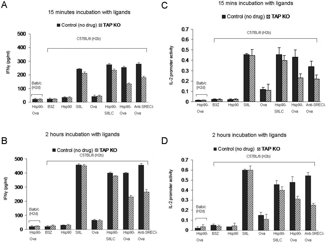
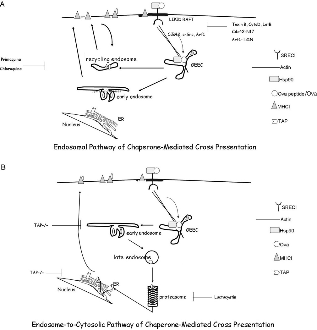
Ag cross presentation is an important mechanism for CD8(+) T cell activation by APCs. We have investigated mechanisms involved in heat shock protein 90 (Hsp90) chaperone-mediated cross presentation of OVA-derived Ags. Hsp90-OVA peptide complexes bound to scavenger receptor expressed by endothelial cells (SREC-I) on the surface of APCs. SREC-I then mediated internalization of Hsp90-OVA polypeptide complexes through a Cdc42-regulated, dynamin-independent endocytic pathway known as the GPI-anchored protein-enriched early endosomal compartment to recycling endosomes. Peptides that did not require processing could then be loaded directly onto MHC class I in endosomes, whereas longer peptides underwent endosomal and cytosomal processing by aminopeptidases and proteases. Cross presentation of Hsp90-chaperoned peptides through this pathway to CD8(+) T cells was highly efficient compared with processing of free polypeptides. In addition, Hsp90 also activated c-Src kinase associated with SREC-I, an activity that we determined to be required for effective cross presentation. Extracellular Hsp90 can thus convey antigenic peptides through an efficient endocytosis pathway in APCs and facilitate cross presentation in a highly regulated manner.


Figures

Figure 1
Figure 1. Hsp90.PC binds to SREC-I and mediates Ova antigen cross presented toB3Z cells after uptake
(A) CHO-K1 and CHO-SREC-I cells were incubated with Alexa 488-Hsp90/Hsp90-S8LC (green) at 4°C for 30 min and counterstained with DAPI to examine cell surface binding. Next, we assayed for internalization of ligands after chase with fresh medium at 37°C for 5–10 min followed by fixation and fluorescence microscopy. (B) CHO-K1 and CHO-SREC-I cells were incubated with Alexa 488 labeled Hsp90 as above and binding at the cell surface quantitated by flow cytometry. (C) CHO-SREC-I cells were incubated with Alexa 488-labeled Hsp90 with or without scavenger receptor agonist mBSA or anti-SREC-I ab. Binding of Hsp90 to the CHO-SREC-I cells was then assayed by flow cytometry. (D) Immature BMDC from either Balbc or C57BL/6J mice were incubated with Hsp90, free Ova, Ova peptide (S8L), Hsp90.Ova, Hsp90.S8LC, Ova peptide complexes and anti SREC-I Ab-Ova complexes for 2 hours at 37°C. Cells were then fixed, washed with 1X PBS and cultured with B3Z overnight before assaying secreted IFNγ by ELISA. As a control, cross presentation assays were carried out using Balbc derived BMDC which lack Ova-specific H2kb (lane 1). (E) CHO-K1 cells were transfected with SREC-I and H2Kb (murine MHC-I) for 22 hours and then chased with Hsp90, free Ova, Ova peptide and Hsp90.PC for 2 hours. Cells were then fixed, washed and cultured with B3Z overnight. The IFNγ secreted by B3Z was assayed by ELISA. (F) In a control experiment for (E) CHO-K1 cells were transfected with H2Kb alone for 22 hrs, and chased with free or Hsp90 conjugated Ova antigens as in (E) for 2 hrs and assayed for antigen cross presentation. (G) Untransfected CHO-K1 cells were chased with ligands as E and F and IFNγ secreted by B3Z assayed for antigen cross presentation. Data shown in D-F are the mean +SD of triplicate wells. The results shown (A–F) are representative of at least three independent experiments. (H) Immature BMDC from C57BL/6J mice were incubated with Hsp90, S8L, free Ova and Hsp90.Ova for 2 hours at 37°C with or without incubation with anti-LOX-1 blocking ab or anti-SREC-I blocking ab. Cells were then fixed, washed with 1X PBS and cultured with B3Z overnight before assaying secreted IFNγ by ELISA.
Figure 2
Figure 2. Hsp90.PC is internalized through a dynamin and clathrin independent endocytosis pathway
(A) KG1 cells were transfected with dynamin (K44A)-GFP (green) for 22 hours and cells then incubated with Alexa 555-Hsp90.PC (red). (B) CHO-SREC-I cells were transfected with dynamin (K44A)-GFP (green) and chased with Alexa 555-Hsp90.PC (red). (C) CHO-SREC-I cells were transfected with EH29-YFP (green) and labeled and chased with Alexa 555-Hsp90.PC (red). Experiments were carried out three times with reproducible results. More than 50 cells were examined in each experiment and representative samples presented.
Figure 3
Figure 3. Anti-SREC-I antibody is internalized in a Rho-GTPase regulated pathway
(A, D) Immature BMDC were incubated without (D) or with (A) Toxin B (2ng/ml). Cells were then pulsed with Alexa 555 labeled anti-SREC-I ab (red) at 4°C then incubated at 37°C for 10 min. Cells were fixed and analyzed using fluorescence microscopy. (B, E) Immature BMDC were treated with (B) or without (E) Poly I (50µg/ml) along with Alexa 555 labeled anti-SREC-I ab and antibody internalization examined under fluorescence microscope. (C, F) Immature BMDC were treated as in A, D, B and E and with (C) or without (F) DMA (500µM) along with Alexa labeled anti-SREC-I ab. Cells were later fixed and analyzed as described in A. Experiments were carried out after pre-blocking with FcR blocking antibodies as described in Materials and Methods. In these experiments BMDC were pre-treated with LPS (10µg/ml) for 2 h to boost expression of SREC-I. Experiments were carried out twice, reproducibly.
Figure 4
Figure 4. Hsp90.PC internalization is actin and Rho GTPase dependent
(A) CHO-SREC-I cells were treated with Cytochalasin D (10µM) before incubating with Alexa 555-Hsp90.PC. at 4°C for 20 minutes, then chasing with medium for 10 minutes at 37°C. (B) CHO-SREC-1 cells were incubated without Cytochalasin D and with Alexa 555-Hsp90.PC at 4°C for 20 minutes, then at 37°C in growth media for 10 minutes. (C) CHO-SREC-I cells were treated with latrunculin B (1µM) and then labeled with Alexa 555-Hsp90.PC (red) at 4°C for 20 minutes, then incubated at 37°C with media for 10 minutes. (D) Control CHO-SREC-I cells were incubated without Lat B and with Alexa 555-Hsp90.PC at 4°C for 20 minutes, then at 37°C as described in (A) (E) Human myeloid DC were transfected with Cdc42-GFP (green) for 22 hours and then incubated with Alexa 555 anti SREC-I ab (red) in the presence of Hsp90.PC. (F–H) CHO-SREC-I cells were transfected with (F) Cdc42 (N17)-GFP (green) (G) RhoA (N19)-GFP (green) and (H) Rac1 (N17)-GFP (green) for 22 hours and then labeled with Alexa 555-Hsp90.PC (red) for 20 minutes at 4°C. Later the cells were chased with normal growth media for 10 minutes at 37°C. Experiments were carried out twice with reproducible results. More than 50 cells were examined in each experiment and representative samples shown.
Figure 5
Figure 5. Anti SREC-I Ab uptake into the CD59-marked GEEC compartment requires cholesterol and is regulated by ARF 1 GTPase
(A) HeLa -SREC-I cells were labeled with Alexa 555 labeled anti SREC-I Ab (red) and FITC-CD59 (green) at 4°C for 20 minutes. (B) HeLa-SREC-I cells were labeled with anti MHC-I Ab (red) and FITC-CD59 (green) at 4°C for 20 minutes and chased at 37°C for 2 minutes. (C) HeLa -SREC-I cells were labeled with Alexa 555 labeled anti SREC-I Ab (red) at 4°C for 20 minutes and then chased at 37°C as in B. Cells were then stained for MHC-I with anti-MHC-I ab (green). (D) CHO-SREC-I cells were transfected with ARF 1 (T31N)-HA (green) for 22 hours, labeled with Hsp90.PC at 4°C for 20 minutes then chased at 37°C for 10 minutes. (E) HeLa-SREC-I cells were treated with MβCD (10 mM) and then incubated with Alexa 555-anti SREC-I Ab (red). (F) Control HeLa-SREC-I cells were incubated without MβCD and with Alexa 555-anti SREC-I Ab (red) as in E. (G–H) HeLa-SREC-I cells were incubated with CTXB (green) and Alexa 555- anti SREC-I Ab (red) (G) at 4°C for 20 minutes and (H) later chased for 2 minutes at 37°C with fresh growth media. Experiments were carried out twice with reproducible results. More than 50 cells were examined in each experiment.
Figure 6
Figure 6. Hsp90.PC internalization via SREC-I requires Src kinase activity
(A) CHO-SREC-I cells were stained with anti-c-src antibody (green) and Alexa 555 labeled anti-SREC-I Ab (red) at 4°C for 20 minutes and analyzed by confocal microscopy as in Materials and Methods. (B) CHO-SREC-I cells were not treated or treated with c-src kinase inhibitor PP1 (10 µM), then incubated with Alexa 555 Hsp90.S8LC complexes (red) at 4°C or 37°C as indicated. (C–D) Immature BMDC were not treated (C) or treated with PP1 (10 µM) (D), then pulsed with Alexa 555 labeled anti-SREC-I ab (red) for 20 minutes at 4°C and later chased with fresh medium for 10 minutes at 37°C. Cells were later fixed and stained with nuclear dye, DAPI (blue). (E) CHO-SREC-I and HeLa-SREC-I cells were incubated with Hsp90.S8LC and assayed for c-src phosphorylation. Levels of total c-src and c-src-phospho-Y416 were examined in the lysates from control cells and cells incubated with Hsp90.S8LC by immunoblot. Experiments were done twice with reproducible outcomes.
Figure 7
Figure 7. Cross presentation of Hsp90 chaperoned peptides after binding SREC-I requires actin, Rho GTPase activity and c-src activation
(A–C) Immature BMDC were treated with (A, D) cytochalasin D (10 µM) (B, E) Toxin B (2 ng/ml), and (C, F) PP1 (10µM) and incubated at 37°C. Cells were then pulsed with Ova polypeptides, Hsp90.PC complexes and anti SREC-I Ab-Ova for 2 hours at 37°C, fixed and washed then cultured with B3Z T cell hybridoma cells overnight. As a control, immature BMDC from Balbc mice were pulsed with Hsp90-Ova in each experiment. IFNγ secreted by B3Z was measured by sandwich ELISA using anti IFNγ Ab (A, B, C). IL-2 promoter induction in B3Z cells was assayed using an endogenous IL-2 promoter–LacZ reporter construct, with intracellular β-galactosidase activity employed as readout. Data shown are the means +SD of triplicate assays. The results shown are representative of at least three independent experiments.
Figure 8
Figure 8. Hsp90-mediated Ova peptide cross presentation requires endosomal protease, cathepsin S and is inhibited by leupeptin
(A) KG1 cells were labeled with Alexa-555-Hsp90.S8LC complexes and Cy3 Tfn at 37°C. (B–C) Immature BMDC were treated with either (B, F) primaquine (20 µM) or (C, G) chloroquine (20 µM) at 37°C for 2 hours before incubation with Hsp90-Ova peptide complexes. Cells were then fixed, washed and cultured with B3Z T cells overnight and assayed for IFNγ release. (D–E) Immature BMDC were treated with (D, H) cathepsin S inhibitor (200 µM) or (E, I) (leupeptin 5 µM) at 37°C for 2 hours and then incubated with Ova peptides, free Ova, Hsp90-Ova complexes and anti SREC-I-Ab-Ova. As a control, for non-specific activation, immature BMDC from Balbc mice were pulsed with Hsp90-Ova in each experiment. Cells were then fixed, washed and cultured with B3Z T cell. As control in each experiment, we also examined BMDC activation by Hsp90 or the activity of B3Z cells alone (in 7, 8, 9). B3Z activation was assayed either by IFNγ secretion or IL-2 promoter activity as in Fig. 7. Data shown are the mean +SD of triplicate wells. The results shown are representative of at least three independent experiments.
Figure 9
Figure 9. Role of proteasome activity in cross presentation of hsp90-chaperoned peptides
(A–B) Immature BMDC were treated with lactacystin (20µM) before incubating with Ova peptides, free Ova, Hsp90-Ova complexes and anti SREC-I-ab-Ova for (A, C) 30 minutes and (B, D) 2 hours. Cells were then fixed, washed and cultured with B3Z T cells overnight. For control, immature BMDC from Balbc mice were pulsed with Hsp90-Ova in all cases. B3Z activation was assayed either by IFNγ secretion or IL-2 promoter activity as in Fig. 7. Data are represented as the mean +SD of triplicate assays. The results shown are representative of at least three independent experiments.
Figure 10
Figure 10. Role of TAP in Ova antigen cross presentation via the Hsp90-peptide-SREC-1 pathway
Immature BMDC from TAP−/− mice and C57 BL/6 (H-2b) mice were incubated with Hsp90, free precursor peptide (S8L), free Ova, Hsp90.Ova and anti SREC-I Ab. Ova complexes at 37°C for (A, C) 15mins and (B, D) 2 hours. Cells were then fixed, washed and cultured with B3Z T cell hybridoma overnight. As a control, immature BMDC from Balbc mice were pulsed with Hsp90-Ova in each experiment. B3Z activation was assayed either by IFNγ secretion or IL-2 promoter activity as in Fig. 7. Mean IFNγ levels +SD of triplicate assays are indicated. The results shown are representative of at least three independent experiments.
Figure 11
Figure 11. Schematic diagram of Hsp90-mediated antigen cross presentation
(A) Endosomal pathway of cross presentation. Hsp90.Ova/Ova peptide complexes are internalized by BMDC in a SREC-I receptor-mediated, Cdc42-regulated pinocytic pathway into the GEEC compartment which is regulated by the activity of Arf1 and c-src. Ova peptides then traverse early endosomes complexed to Hsp90 and SREC-I and can be processed and loaded onto MHC-I molecules in recycling endosomes. MHC-I-Ova peptide complexes can then recycle to the plasma membrane and encounter CD8+ T cells. (B) Endosome-to-cytosolic pathway of cross presentation. In an additional pathway, observed with Hsp90 complexes containing full length Ova, the Ova traverses the SREC-I/GEEC pathway defined in this study and is partially processed at an endosomal site as in (A). However, a proportion of the peptides evidently enter the cytosol, are processed by the proteasome and enter compartments containing TAP such as endosomes and loaded onto MHC-I or the ER where they may be further processed by ERAP (Endoplasmic Reticulum amino peptidases) and loaded onto newly synthesized MHC-I molecules. MHC-I-processed Ova can then travel from the ER (or other TAP-containing compartments) to the plasma membrane for activation of CD8+ T cells.

Similar articles

Cited by

References

    1. Lindquist S, Craig EA. The heat shock proteins. Ann. Rev. Genet. 1988;22:631–637. - PubMed
    1. Wegele H, Muller L, Buchner J. Hsp70 and Hsp90--a relay team for protein folding. Rev Physiol Biochem Pharmacol. 2004;151:1–44. - PubMed
    1. Srivastava P. Interaction of heat shock proteins with peptides and antigen presenting cells: chaperoning of the innate and adaptive immune responses. Annu Rev Immunol. 2002;20:395–425. - PubMed
    1. Mambula SS, Calderwood SK. Heat induced release of Hsp70 from prostate carcinoma cells involves both active secretion and passive release from necrotic cells. Int J Hyperthermia. 2006;22:575–585. - PubMed
    1. Mambula SS, Calderwood SK. Heat shock protein 70 is secreted from tumor cells by a nonclassical pathway involving lysosomal endosomes. J Immunol. 2006;177:7849–7857. - PubMed

Publication types

MeSH terms

Substances