

Selective deletion of Connexin 40 in renin-producing cells impairs renal baroreceptor function and is associated with arterial hypertension
- PMID: 20686449
- PMCID: PMC3033195
- DOI: 10.1038/ki.2010.257
Selective deletion of Connexin 40 in renin-producing cells impairs renal baroreceptor function and is associated with arterial hypertension
Abstract
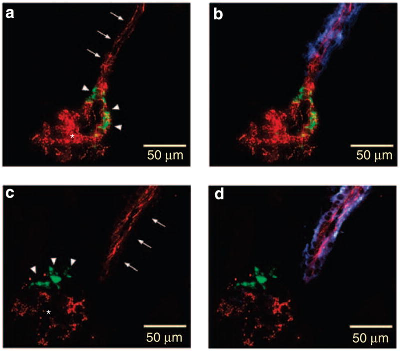
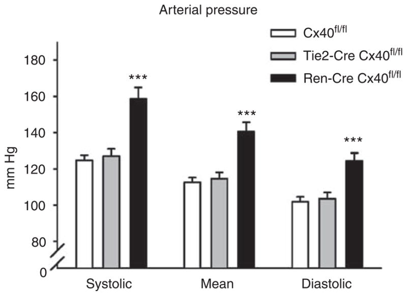
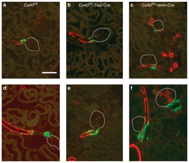
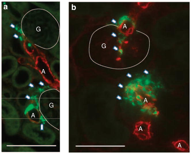
Renin-producing juxtaglomerular cells are connected to each other and to endothelial cells of afferent arterioles by gap junctions containing Connexin 40 (Cx40), abundantly expressed by these two cell types. Here, we generated mice with cell-specific deletion of Cx40 in endothelial and in renin-producing cells, as its global deletion caused local dissociation of renin-producing cells from endothelial cells, renin hypersecretion, and hypertension. In mice lacking endothelial Cx40, the blood pressure, renin-producing cell distribution, and the control of renin secretion were similar to wild-type mice. In contrast, mice deficient for Cx40 in renin-producing cells were hypertensive and these cells were ectopically localized. Although plasma renin activity and kidney renin mRNA levels of these mice were not different from controls, the negative regulation of renin secretion by pressure was inverted to a positive feedback in kidneys lacking Cx40 in renin-producing cells. Thus, our findings show that endothelial Cx40 is not essential for the control of renin expression and/or release. Cx40 in renin-producing cells is required for their correct positioning in the juxtaglomerular area and the control of renin secretion by pressure.
Conflict of interest statement
All the authors declared no competing interests.
Figures


Similar articles
-
Connexin 40 is dispensable for vascular renin cell recruitment but is indispensable for vascular baroreceptor control of renin secretion.Pflugers Arch. 2015 Aug;467(8):1825-34. doi: 10.1007/s00424-014-1615-y. Epub 2014 Sep 23. Pflugers Arch. 2015. PMID: 25241776
-
Replacement of connexin 40 by connexin 45 causes ectopic localization of renin-producing cells in the kidney but maintains in vivo control of renin gene expression.Am J Physiol Renal Physiol. 2009 Aug;297(2):F403-9. doi: 10.1152/ajprenal.00176.2009. Epub 2009 May 27. Am J Physiol Renal Physiol. 2009. PMID: 19474190
-
Connexin 37 is dispensable for the control of the renin system and for positioning of renin-producing cells in the kidney.Pflugers Arch. 2009 Nov;459(1):151-8. doi: 10.1007/s00424-009-0707-6. Epub 2009 Aug 13. Pflugers Arch. 2009. PMID: 19672618
-
Connexins, renin cell displacement and hypertension.Curr Opin Pharmacol. 2015 Apr;21:1-6. doi: 10.1016/j.coph.2014.11.009. Epub 2014 Dec 5. Curr Opin Pharmacol. 2015. PMID: 25483714 Review.
-
Function of connexins in the renal circulation.Kidney Int. 2008 Mar;73(5):547-55. doi: 10.1038/sj.ki.5002720. Epub 2007 Dec 12. Kidney Int. 2008. PMID: 18075497 Review.
Cited by
-
Activation of KCa3.1 by SKA-31 induces arteriolar dilatation and lowers blood pressure in normo- and hypertensive connexin40-deficient mice.Br J Pharmacol. 2013 Sep;170(2):293-303. doi: 10.1111/bph.12267. Br J Pharmacol. 2013. PMID: 23734697 Free PMC article.
-
The renin angiotensin aldosterone system.Pflugers Arch. 2024 May;476(5):705-713. doi: 10.1007/s00424-024-02908-1. Epub 2024 Jan 17. Pflugers Arch. 2024. PMID: 38233636 Free PMC article. Review.
-
Hypertensive Nephropathy: Unveiling the Possible Involvement of Hemichannels and Pannexons.Int J Mol Sci. 2022 Dec 14;23(24):15936. doi: 10.3390/ijms232415936. Int J Mol Sci. 2022. PMID: 36555574 Free PMC article. Review.
-
Renin cells in homeostasis, regeneration and immune defence mechanisms.Nat Rev Nephrol. 2018 Apr;14(4):231-245. doi: 10.1038/nrneph.2017.186. Epub 2018 Jan 30. Nat Rev Nephrol. 2018. PMID: 29380818 Free PMC article. Review.
-
Connexin45 is expressed in vascular smooth muscle but its function remains elusive.PLoS One. 2012;7(7):e42287. doi: 10.1371/journal.pone.0042287. Epub 2012 Jul 27. PLoS One. 2012. PMID: 22848755 Free PMC article.
References
-
- Hwan Seul K, Beyer EC. Heterogeneous localization of connexin 40 in the renal vasculature. Microvasc Res. 2000;59:140–148. - PubMed
-
- Haefliger JA, Demotz S, Braissant O, et al. Connexins 40 and 43 are differentially regulated within the kidneys of rats with renovascular hypertension. Kidney Int. 2001;60:190–201. - PubMed
-
- Zhang J, Hill CE. Differential connexin expression in preglomerular and postglomerular vasculature: accentuation during diabetes. Kidney Int. 2005;68:1171–1185. - PubMed
-
- Wagner C, de Wit C, Kurtz L, et al. Connexin40 is essential for the pressure control of renin synthesis and secretion. Circ Res. 2007;100:556–563. - PubMed
Publication types
MeSH terms
Substances
Grants and funding
LinkOut - more resources
Full Text Sources
Other Literature Sources
Molecular Biology Databases
Miscellaneous
