. 2010 Jul 29;6(7):e1001024.
doi: 10.1371/journal.ppat.1001024.

Discovery of a new human polyomavirus associated with trichodysplasia spinulosa in an immunocompromized patient

Affiliations

Discovery of a new human polyomavirus associated with trichodysplasia spinulosa in an immunocompromized patient

Els van der Meijden et al. PLoS Pathog. .

Abstract

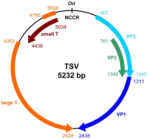
The Polyomaviridae constitute a family of small DNA viruses infecting a variety of hosts. In humans, polyomaviruses can cause infections of the central nervous system, urinary tract, skin, and possibly the respiratory tract. Here we report the identification of a new human polyomavirus in plucked facial spines of a heart transplant patient with trichodysplasia spinulosa, a rare skin disease exclusively seen in immunocompromized patients. The trichodysplasia spinulosa-associated polyomavirus (TSV) genome was amplified through rolling-circle amplification and consists of a 5232-nucleotide circular DNA organized similarly to known polyomaviruses. Two putative "early" (small and large T antigen) and three putative "late" (VP1, VP2, VP3) genes were identified. The TSV large T antigen contains several domains (e.g. J-domain) and motifs (e.g. HPDKGG, pRb family-binding, zinc finger) described for other polyomaviruses and potentially involved in cellular transformation. Phylogenetic analysis revealed a close relationship of TSV with the Bornean orangutan polyomavirus and, more distantly, the Merkel cell polyomavirus that is found integrated in Merkel cell carcinomas of the skin. The presence of TSV in the affected patient's skin was confirmed by newly designed quantitative TSV-specific PCR, indicative of a viral load of 10(5) copies per cell. After topical cidofovir treatment, the lesions largely resolved coinciding with a reduction in TSV load. PCR screening demonstrated a 4% prevalence of TSV in an unrelated group of immunosuppressed transplant recipients without apparent disease. In conclusion, a new human polyomavirus was discovered and identified as the possible cause of trichodysplasia spinulosa in immunocompromized patients. The presence of TSV also in clinically unaffected individuals suggests frequent virus transmission causing subclinical, probably latent infections. Further studies have to reveal the impact of TSV infection in relation to other populations and diseases.


Conflict of interest statement

The authors have declared that no competing interests exist.

Figures

Figure 1
Figure 1. Clinical appearance and histology of the trichodysplasia spinulosa patient.
Facial appearance at presentation is shown in panel A. Note the thickened skin, particularly on the nose and in the eyebrow region accompanied by central alopecia. Apart from the eyebrows and nose, papules are seen on the cheeks, chin, forehead and ears. Especially on the nose, but occasionally also in cheeks and chin, keratotic spicules protruded from the enlarged follicular orifices. Panel B shows a close-up of the nose at presentation with numerous papules and spicules. In panel C is shown a section of a formalin-fixed, paraffin-embedded biopsy of a hyperkeratotic follicular papule from the forehead. The epidermis reveals enlarged, hyperplastic hair bulbs and hypercornification within a distended follicular infundibulum (HE stain, 10×). Panel D shows a detail of the nose 3 months after topical cidofovir treatment. Papules and spicules have largely resolved and hairs have regained growth.
Figure 2
Figure 2. Restriction analysis of the RCA product.
EcoRV and XbaI digestion revealed one band of around 5000 bp. EcoRI digestion produced two bands of around 3600 and 1600 bp. After HindIII digestion 3 bands were visible of around 3000, 1500 and 400 bp. Sequence analysis later showed that HindIII digestion in fact produced four fragments of which the smallest, 378 and 364 bp, coincided on gel.
Figure 3
Figure 3. Genome map of TSV.
Indicated are the five identified ORFs representing the putative “early” genes encoding small and large T antigen, and the putative “late” genes encoding VP1, VP2 and VP3. The NCCR is placed on top and contains the putative ori. Nucleotide position 1 was chosen within the NCCR in the large T binding region. For a detailed view of the NCCR, see Figure 4A.
Figure 4
Figure 4. Detail of the TSV non-coding control region.
Indicated are the putative large T-bindings sites located on both strands (gray-shaded boxes), the putative ori and putative TATA box (A/T-rich elements). Downstream of this area another two TSV putative large T-bindings sites are located, as well as one from KIV . Nucleotide positions 1 are shown in white and underlined, except for MCV where the position 1 is shown in bold and underlined. For MCV two isolates are shown, MCV 339 and MCV350 .
Figure 5
Figure 5. Amino acid sequence of the TSV large T protein.
Indicated are three major domains found in the large T amino acid sequence; the J-domain (blue-shaded box), the ori DNA-binding domain (yellow-shaded box) and the helicase domain (green-shaded box). Within the J-domain the putative locations of conserved region 1 and the HPDKGG motif are depicted. Downstream of the J-domain, a pRb family-binding motif and nuclear localization signal are located. In the helicase domain, a zinc finger motif, NTPase-binding ‘Walker’ motifs A and B and an helicase SF3 (superfamily 3) motif C are located.
Figure 6
Figure 6. Phylogenetic analysis of known polyomaviruses and TSV.
Bayesian posterior probability trees are shown for VP1 (A), VP2 (B), large T antigen (C) and a concatenation of all three (D). Numbers at branch points represent posterior probability support values and the scale bar is given in average number of substitutions per amino acid position. Major clades in the trees are highlighted using colored triangles. The following viruses are shown: Simian virus 40 (SV40), Goose hemorrhagic polyomavirus (GHPyV), Simian virus 12 (SV12), Squirrel monkey polyomavirus (SquiPyV), Finch polyomavirus (FPyV), Crow polyomavirus (CPyV), Bovine polyomavirus (BPyV), Merkel cell polyomavirus (MCPyV), WU Polyomavirus (WUPyV), KI polyomavirus Stockholm 60 (KIPyV), Budgerigar fledgling polyomavirus (BFPyV), African green monkey polyomavirus (AGMPyV), JC polyomavirus (JCPyV), BK polyomavirus (BKPyV), Murine polyomavirus (MPyV), Murine pneumotropic virus (MuPtV), Myotis polyomavirus VM-2008 (MyPyV), Bornean orangutan polyomavirus isolate Bo (OraPyV1), Sumatran orangutan polyomavirus isolate Pi (OraPyV2) and Trichodysplasia spinulosa-associated polyomavirus (TSPyV).

References

    1. Allander T, Andreasson K, Gupta S, Bjerkner A, Bogdanovic G, Persson MA, Dalianis T, Ramqvist T, Andersson B. Identification of a third human polyomavirus. J Virol. 2007;81:4130–4136. - PMC - PubMed
    1. Gaynor AM, Nissen MD, Whiley DM, Mackay IM, Lambert SB, Wu G, Brennan DC, Storch GA, Sloots TP, Wang D. Identification of a novel polyomavirus from patients with acute respiratory tract infections. PLoS Pathog. 2007;3:e64. - PMC - PubMed
    1. Feng H, Shuda M, Chang Y, Moore PS. Clonal integration of a polyomavirus in human Merkel cell carcinoma. Science. 2008;319:1096–1100. - PMC - PubMed
    1. Wieland U, Mauch C, Kreuter A, Krieg T, Pfister H. Merkel cell polyomavirus DNA in persons without merkel cell carcinoma. Emerg Infect Dis. 2009;15:1496–1498. - PMC - PubMed
    1. Abend JR, Jiang M, Imperiale MJ. BK virus and human cancer: Innocent until proven guilty. Seminars in Cancer Biology. 2009;19:252–260. - PMC - PubMed

Publication types