Low-density lipoprotein receptor-related protein 1 (LRP1) mediates neuronal Abeta42 uptake and lysosomal trafficking
- PMID: 20686698
- PMCID: PMC2912373
- DOI: 10.1371/journal.pone.0011884
Low-density lipoprotein receptor-related protein 1 (LRP1) mediates neuronal Abeta42 uptake and lysosomal trafficking
Abstract
Background: Alzheimer's disease (AD) is characterized by the presence of early intraneuronal deposits of amyloid-beta 42 (Abeta42) that precede extracellular amyloid deposition in vulnerable brain regions. It has been hypothesized that endosomal/lysosomal dysfunction might be associated with the pathological accumulation of intracellular Abeta42 in the brain. Our previous findings suggest that the LDL receptor-related protein 1 (LRP1), a major receptor for apolipoprotein E, facilitates intraneuronal Abeta42 accumulation in mouse brain. However, direct evidence of neuronal endocytosis of Abeta42 through LRP1 is lacking.
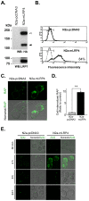
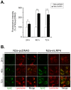
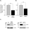
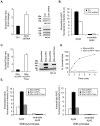
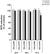
Methodology/principal findings: Here we show that LRP1 endocytic function is required for neuronal Abeta42 uptake. Overexpression of a functional LRP1 minireceptor, mLRP4, increases Abeta42 uptake and accumulation in neuronal lysosomes. Conversely, knockdown of LRP1 expression significantly decreases neuronal Abeta42 uptake. Disruptions of LRP1 endocytic function by either clathrin knockdown or by removal of its cytoplasmic tail decreased both uptake and accumulation of Abeta42 in neurons. Finally, we show that LRP1-mediated neuronal accumulation of Abeta42 is associated with increased cellular toxicity.
Conclusions/significance: These results demonstrate that LRP1 endocytic function plays an important role in the uptake and accumulation of Abeta42 in neuronal lysosomes. These findings emphasize the central function of LRP1 in neuronal Abeta metabolism.
Conflict of interest statement
Figures
Similar articles
-
Heparan sulphate proteoglycan and the low-density lipoprotein receptor-related protein 1 constitute major pathways for neuronal amyloid-beta uptake.J Neurosci. 2011 Feb 2;31(5):1644-51. doi: 10.1523/JNEUROSCI.5491-10.2011. J Neurosci. 2011. PMID: 21289173 Free PMC article.
-
Mitogen-activated protein kinase signaling pathways promote low-density lipoprotein receptor-related protein 1-mediated internalization of beta-amyloid protein in primary cortical neurons.Int J Biochem Cell Biol. 2015 Jul;64:252-64. doi: 10.1016/j.biocel.2015.04.013. Epub 2015 Apr 29. Int J Biochem Cell Biol. 2015. PMID: 25936756
-
Blood-brain barrier-associated pericytes internalize and clear aggregated amyloid-β42 by LRP1-dependent apolipoprotein E isoform-specific mechanism.Mol Neurodegener. 2018 Oct 19;13(1):57. doi: 10.1186/s13024-018-0286-0. Mol Neurodegener. 2018. PMID: 30340601 Free PMC article.
-
LRP in amyloid-beta production and metabolism.Ann N Y Acad Sci. 2006 Nov;1086:35-53. doi: 10.1196/annals.1377.005. Ann N Y Acad Sci. 2006. PMID: 17185504 Review.
-
Functional role of lipoprotein receptors in Alzheimer's disease.Curr Alzheimer Res. 2008 Feb;5(1):15-25. doi: 10.2174/156720508783884675. Curr Alzheimer Res. 2008. PMID: 18288927 Review.
Cited by
-
Altered Met receptor phosphorylation and LRP1-mediated uptake in cells lacking carbohydrate-dependent lysosomal targeting.J Biol Chem. 2017 Sep 8;292(36):15094-15104. doi: 10.1074/jbc.M117.790139. Epub 2017 Jul 19. J Biol Chem. 2017. PMID: 28724630 Free PMC article.
-
Overexpression of Glutamate Decarboxylase in Mesenchymal Stem Cells Enhances Their Immunosuppressive Properties and Increases GABA and Nitric Oxide Levels.PLoS One. 2016 Sep 23;11(9):e0163735. doi: 10.1371/journal.pone.0163735. eCollection 2016. PLoS One. 2016. PMID: 27662193 Free PMC article.
-
Amyloid beta dimers/trimers potently induce cofilin-actin rods that are inhibited by maintaining cofilin-phosphorylation.Mol Neurodegener. 2011 Jan 24;6:10. doi: 10.1186/1750-1326-6-10. Mol Neurodegener. 2011. PMID: 21261978 Free PMC article.
-
Mechanisms of U87 astrocytoma cell uptake and trafficking of monomeric versus protofibril Alzheimer's disease amyloid-β proteins.PLoS One. 2014 Jun 18;9(6):e99939. doi: 10.1371/journal.pone.0099939. eCollection 2014. PLoS One. 2014. PMID: 24941200 Free PMC article.
-
Insulin in the brain: there and back again.Pharmacol Ther. 2012 Oct;136(1):82-93. doi: 10.1016/j.pharmthera.2012.07.006. Epub 2012 Jul 17. Pharmacol Ther. 2012. PMID: 22820012 Free PMC article. Review.
References
-
- Hardy J, Selkoe DJ. The amyloid hypothesis of Alzheimer's disease: progress and problems on the road to therapeutics. Science. 2002;297:353–356. - PubMed
-
- Oddo S, Caccamo A, Shepherd JD, Murphy MP, Golde TE, et al. Triple-transgenic model of Alzheimer's disease with plaques and tangles: intracellular Abeta and synaptic dysfunction. Neuron. 2003;39:409–421. - PubMed
-
- Billings LM, Oddo S, Green KN, McGaugh JL, LaFerla FM. Intraneuronal Abeta causes the onset of early Alzheimer's disease-related cognitive deficits in transgenic mice. Neuron. 2005;45:675–688. - PubMed
Publication types
MeSH terms
Substances
Grants and funding
LinkOut - more resources
Full Text Sources
Other Literature Sources
Research Materials
Miscellaneous

