. 2010 Jul 29;5(7):e11875.
doi: 10.1371/journal.pone.0011875.

Nuclear factor-kappa B family member RelB inhibits human immunodeficiency virus-1 Tat-induced tumor necrosis factor-alpha production

Affiliations

Nuclear factor-kappa B family member RelB inhibits human immunodeficiency virus-1 Tat-induced tumor necrosis factor-alpha production

Michelle Kiebala et al. PLoS One. .

Abstract

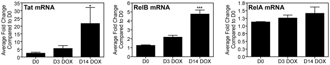
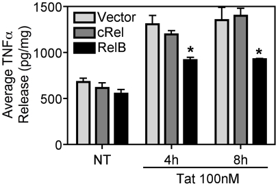
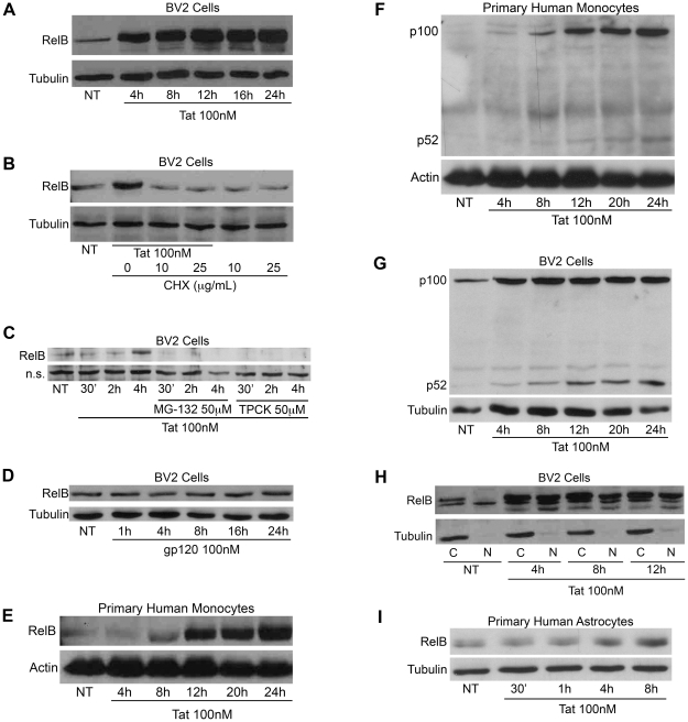
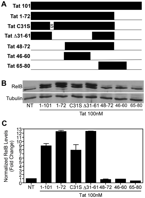
Human Immunodeficiency Virus-1 (HIV-1)-associated neurocognitive disorder (HAND) is likely neuroinflammatory in origin, believed to be triggered by inflammatory and oxidative stress responses to cytokines and HIV protein gene products such as the HIV transactivator of transcription (Tat). Here we demonstrate increased messenger RNA for nuclear factor-kappa B (NF-kappaB) family member, transcription factor RelB, in the brain of doxycycline-induced Tat transgenic mice, and increased RelB synthesis in Tat-exposed microglial cells. Since genetic ablation of RelB in mice leads to multi-organ inflammation, we hypothesized that Tat-induced, newly synthesized RelB inhibits cytokine production by microglial cells, possibly through the formation of transcriptionally inactive RelB/RelA complexes. Indeed, tumor necrosis factor-alpha (TNFalpha) production in monocytes isolated from RelB deficient mice was significantly higher than in monocytes isolated from RelB expressing controls. Moreover, RelB overexpression in microglial cells inhibited Tat-induced TNFalpha synthesis in a manner that involved transcriptional repression of the TNFalpha promoter, and increased phosphorylation of RelA at serine 276, a prerequisite for increased RelB/RelA protein interactions. The Rel-homology-domain within RelB was necessary for this interaction. Overexpression of RelA itself, in turn, significantly increased TNFalpha promoter activity, an effect that was completely blocked by RelB overexpression. We conclude that RelB regulates TNFalpha cytokine synthesis by competitive interference binding with RelA, which leads to downregulation of TNFalpha production. Moreover, because Tat activates both RelB and TNFalpha in microglia, and because Tat induces inflammatory TNFalpha synthesis via NF-kappaB, we posit that RelB serves as a cryoprotective, anti-inflammatory, counter-regulatory mechanism for pathogenic NF-kappaB activation. These findings identify a novel regulatory pathway for controlling HIV-induced microglial activation and cytokine production that may have important therapeutic implications for the management of HAND.


Conflict of interest statement

Competing Interests: The authors have declared that no competing interests exist.

Figures

Figure 1
Figure 1. Transgenic expression of Tat induces RelB synthesis in mouse brain.
6-week old Tat-transgenic mice were induced with doxycycline for the indicated periods of time. Total RNA was extracted from brain tissue, reverse transcribed using oligo-dT primers, and subjected to Real-Time SYBR Green RT-PCR amplification. Fold induction of Tat, RelB and RelA mRNA species was normalized to GAPDH and presented as a function of the expression level in D0 samples. Data represent mean ± SEM of four replicates for D0 and D14 samples, and five replicates for D3 samples. Statistical significance (***, p<0.001 or *, p<0.05) is denoted as compared to D0 samples.
Figure 2
Figure 2. RelB inhibits Tat-induced TNFα and cytokine production in monocytes.
A, Monocytes from RelB+/+ or RelB−/− mice (3×10⁵) treated with Tat (100nM) for 8h and subjected to immunoblot analysis with either RelB-specific (upper panel) or Actin-specific (lower panel) antibodies, confirmed that Tat induced RelB in these cells. Data are representative of results from two separate experiments. B, Levels of TNFα in culture supernatants from these cells were analyzed by ELISA, and C, Levels of IL-1β, IL-6, and MCP-1 were anlayzed by Multi-Plex cytokine array as described in Results. Data is shown as mean ± SEM of values derived from three replicates each from two combined experiments. Statistical significance (p<0.001) is indicated (***), as compared with Tat treated RelB+/+ cells.
Figure 3
Figure 3. RelB inhibits Tat-induced TNFα production in BV-2 microglia.
BV-2 cells (5×10⁶) were transiently transfected with either empty vector (pcDNA3.1-Myc/His-B), cRel, or RelB (10 µg), using Nucleofector (Amaxa/Lonza), and treated with Tat (100nM) for the indicated periods of time. TNFα release was measured by ELISA and normalized to total cellular protein content in the culture wells. Results are shown as mean ± SEM of values derived from two replicates from one representative experiment; two total experiments were performed. Statistical significance (p<0.05) is indicated, as compared to empty vector transfected cells (*).
Figure 4
Figure 4. Tat induces de novo synthesis of RelB.
A, BV-2 cells (1.2×10⁵) were treated with Tat (100nM) for the indicated periods of time and whole cell lysates were subjected to immunoblot analysis using either RelB-specific (upper panel) or α-Tubulin-specific (lower panel) antibodies. B, BV-2 cells (1.2×10⁵) were treated with Tat alone or together with CHX as indicated for 4h. Whole cell lysates were subjected to immunoblot analysis using either RelB-specific (upper panel) or α-Tubulin-specific (lower panel) antibodies. C, BV-2 cells (1.2×10⁵) were treated with Tat (100nM) alone or together with MG-132 or TPCK both at a concentration of 50µM for the indicated periods of time. Whole cell lysates were subjected to immunoblot analysis using a RelB-specific (upper panel) antibody. Levels of a nonspecific (n.s.) band (lower panel) are shown to indicate equal protein loading. D, BV-2 cells (1.2×10⁵) were treated with gp120 (SF162, 100nM) for the indicated periods of time and whole cell lysates were subjected to immunoblot analysis using either RelB-specific (upper panel) or α-Tubulin-specific (lower panel) antibodies. E, Primary human monocytes (2×10⁵) were treated with Tat for the indicated periods of time and whole cell lysates were subjected to immunoblot analysis using either RelB-specific (upper panel) or Actin-specific (lower panel) antibodies. F, Primary human monocytes (2×10⁵) were treated with Tat for the indicated periods of time and whole cell lysates were subjected to immunoblot analysis using either p100/p52-specific (upper panel) or Actin-specific (lower panel) antibodies. G, BV-2 cells (1.2×10⁵) were treated with Tat for the indicated periods of time and whole cell lysates were subjected to immunoblot analysis using either p100/p52-specific (upper panel) or α-Tubulin-specific (lower panel) antibodies. H, BV-2 cells (1.2×10⁵) were treated with Tat as indicated and cytosolic (labeled “C”) and nuclear (labeled “N”) extracts were subjected to immunoblot analysis with either RelB-specific (upper panel) or α-Tubulin-specific (lower panel) antibodies. I, Primary human astrocytes (4×10⁴) were plated 72h prior to treatment. These cells were treated with Tat as indicated and whole cell lysates were subjected to immunoblot analysis using either RelB-specific (upper panel) or α-Tubulin-specific (lower panel) antibodies.
Figure 5
Figure 5. Opposing effects of RelA and RelB on TNFα promoter activity.
A, BV-2 cells (1.2×10⁵) were transiently transfected with 0.25 µg of plasmid DNA containing a luciferase reporter gene under transcriptional control of the mouse TNFα promoter region in the absence or presence of increasing amounts of a RelB-encoding plasmid. 24h later cells were lysed and luciferase activity was determined. Total protein amount, as determined by Bradford assay was used to normalize the samples. Data are presented as fold change compared to cells transfected with the luciferase reporter alone. ***, p<0.001, and **, p<0.01 as compared to cells transfected with the luciferase reporter alone. B, HEK 293 cells (1.8×10⁵) were transiently transfected with the mouse TNFα promoter-luciferase reporter plasmid along with increasing amounts of a plasmid for RelA or RelB. The luciferase (i.e. TNFα promoter) activity in whole cell lysates was determined 24h post-transfection. Samples were subjected to immunoblot analysis using either RelA or RelB-specific antibodies to determine the expressed level of RelA and RelB protein. ***, p<0.001 as compared to cells transfected with the luciferase reporter alone. C, HEK 293 cells (1.8×10⁵) were transiently transfected with the mouse TNFα promoter-luciferase reporter plasmid along with a plasmid for RelA in the absence or presence of increasing amounts of RelB-encoding plasmid. The luciferase activity in whole cell lysates was determined 24h post-transfection. Samples were subjected to immunoblot analysis using either RelA or RelB-specific antibodies to determine the expressed level of RelA and RelB protein. ***, p<0.001 as compared to cells transfected with the luciferase reporter alone. D, HEK 293 cells (1.8×10⁵) were transiently transfected with the mouse TNFα promoter-luciferase reporter along with a plasmid for RelA in the absence or presence of increasing amounts of cRel or β-galactosidase-encoding plasmid. Luciferase activity in whole cell lysates was determined 24h post-transfection.
Figure 6
Figure 6. RelB inhibits NF-κB activation via physical interaction with RelA.
A, BV-2 cells (8×10⁵) were treated with Tat (100nM) for the indicated periods of time. Whole cell lysates were subjected to immunoprecipitation using a RelA-specific antibody and Protein A/G+ agarose beads. Immunocomplexes were separated by 7.5% SDS-PAGE and blotted onto nitrocellulose membrane and subjected to immunoblot analysis with antibodies specific for RelB or RelA. The plot below the bands represents densitometry values for each band, normalized as a ratio of RelA(IP)/RelB(IB) (i.e. top bands): RelA(IP)/RelA(IB) (i.e. bottom bands), to indicate the increase in RelB/RelA interactions at each time point. B, BV-2 cells (1.2×10⁵) were treated with Tat (100nM) for the indicated periods of time and whole cell lysates were subjected to immunoblot analysis using either RelA-specific (upper panel), RelA phospho-serine 276-specific (center panel) or α-Tubulin-specific (lower panel) antibodies. Protein levels were quantified using ImageJ software (bottom graphs). The results of a single representative experiment are shown. C, BV-2 cells (5×10⁶) were transiently transfected using Nucleofector (Amaxa/Lonza) with an NF-κB-dependent luciferase reporter plasmid either alone or together with a RelB-encoding plasmid. 16h post-transfection cells were either left untreated or were treated with Tat (100nM) for 8h. Luciferase activity in whole cell lysates was determined. Results are shown as mean ± SEM of values derived from three replicates from one representative experiment; two total experiments were performed. Statistical significance (p<0.001) is indicated (***). D, BV-2 cells (1.5×10⁵) were transiently transfected using Lipofectamine (Invitrogen) with the NF-κB-luciferase reporter plasmid together with a plasmid for RelA in the absence or presence of increasing amounts of RelB-encoding plasmid. The luciferase activity in whole cell lysates was determined 18h post-transfection. Results are shown as mean ± SEM of values derived from two replicates from one representative experiment; two total experiments were performed.
Figure 7
Figure 7. RelB Rel homology domain required for interaction with RelA and blockade of TNFα promoter activity.
A, B, Diagrams show representation of full length and deletion mutants of RelB used to determine the domains of RelB necessary for interaction with RelA. HEK 293 cells (1.8×10⁵) were transiently transfected with either HA-tagged RelA or Myc-tagged RelB deletion mutants alone or in combination. Interaction was determined by immunoprecipitation with Myc-specific antibody and Protein A/G+ agarose beads followed by immunoblot analysis with HA-specific or Myc-specific antibodies. Interacting (+) and non-interacting (−) mutants are indicated. These RelB deletion mutants were also analyzed using the TNFα promoter-luciferase reporter plasmid co-transfected into HEK 293 cells together with a plasmid for RelA in the absence or presence of increasing amounts of RelB-encoding plasmid (WT or deletion mutants). Inhibition of RelA activation of the TNFα promoter is indicated (+/−). C, HEK 293 cells (1.8×10⁵) were transiently transfected with either HA-tagged RelA or Myc-tagged RelB Δ52–402 alone or in combination. Interaction was determined by immunoprecipitation with Myc-specific antibody and Protein A/G+ agarose beads followed by immunoblot analysis with HA-specific or Myc-specific antibodies. D, RelB Δ52–402 was also analyzed using the TNFα promoter-luciferase reporter plasmid co-transfected into HEK 293 cells along with a plasmid for RelA in the absence or presence of increasing amounts of RelB Δ52–402-encoding plasmid. Statistical significance (p<0.001) as compared to cells transfected with 0.1 µg WT RelB+RelA is indicated (***).
Figure 8
Figure 8. Amino terminal region of Tat is necessary for RelB induction in microglia.
A, Diagram shows a representation of full length, mutated and truncated Tat proteins and peptides used in these experiments. B, BV-2 cells (1.2×10⁵) were treated with Tat (100nM) for 8h and whole cell lysates were subjected to immunoblot analysis using either RelB-specific (upper panel) or α-Tubulin-specific (lower panel) antibodies. These results indicate that standard Tat 1–101, Tat 1–72, Tat C31S, and Tat Δ31–61 all activated RelB, whereas Tat peptides 48–72, 46–60 and 65–80 did not. C, Densitometry quantification of immunoblots shown in B. RelB levels were normalized to Tubulin levels and fold change compared to non-treated (NT) was calculated.

Similar articles

Cited by

References

    1. Resnick L, Berger JR, Shapshak P, Tourtellotte WW. Early penetration of the blood-brain-barrier by HIV. Neurology. 1988;38:9–14. - PubMed
    1. Kaul M, Lipton SA. Mechanisms of neuronal injury and death in HIV-1 associated dementia. Curr HIV Res. 2006;4:307–318. - PubMed
    1. Masliah E, DeTeresa RM, Mallory ME, Hansen LA. Changes in pathological findings at autopsy in AIDS cases for the last 15 years. AIDS. 2000;14:69–74. - PubMed
    1. Nath A, Sacktor N. Influence of highly active antiretroviral therapy on persistence of HIV in the central nervous system. Curr Opin Neurol. 2006;19:358–361. - PubMed
    1. Tozzi V, Balestra P, Bellagamba R, Corpolongo A, Salvatori MF, et al. Persistence of neuropsychologic deficits despite long-term highly active antiretroviral therapy in patients with HIV-related neurocognitive impairment: prevalence and risk factors. J Acquir Immune Defic Syndr. 2007;45:174–182. - PubMed

Publication types

MeSH terms

Substances