. 2010 Jul 29;6(7):e1001040.
doi: 10.1371/journal.pgen.1001040.

Calpain 8/nCL-2 and calpain 9/nCL-4 constitute an active protease complex, G-calpain, involved in gastric mucosal defense

Affiliations

Calpain 8/nCL-2 and calpain 9/nCL-4 constitute an active protease complex, G-calpain, involved in gastric mucosal defense

Shoji Hata et al. PLoS Genet. .

Abstract

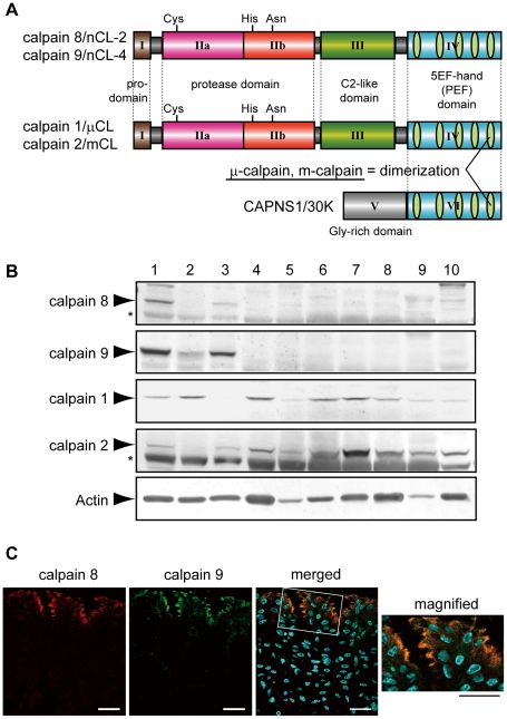
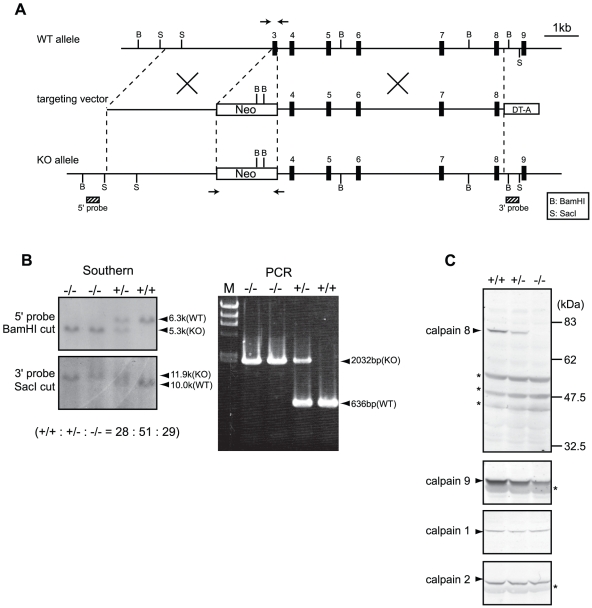
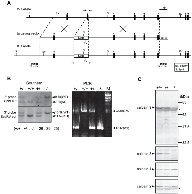
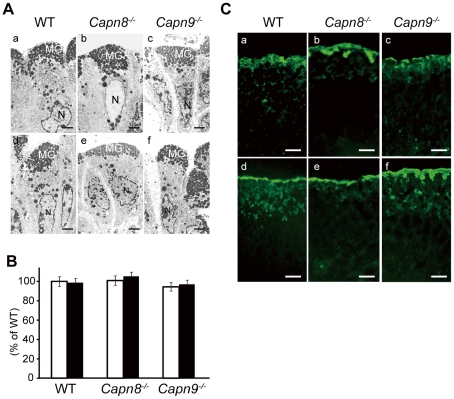
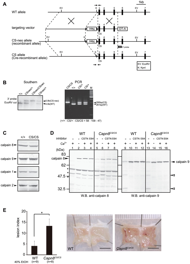
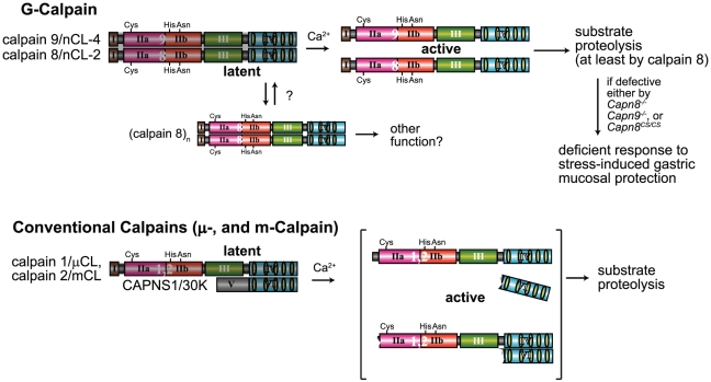
Calpains constitute a superfamily of Ca2+-dependent cysteine proteases, indispensable for various cellular processes. Among the 15 mammalian calpains, calpain 8/nCL-2 and calpain 9/nCL-4 are predominantly expressed in the gastrointestinal tract and are restricted to the gastric surface mucus (pit) cells in the stomach. Possible functions reported for calpain 8 are in vesicle trafficking between ER and Golgi, and calpain 9 are implicated in suppressing tumorigenesis. These highlight that calpains 8 and 9 are regulated differently from each other and from conventional calpains and, thus, have potentially important, specific functions in the gastrointestinal tract. However, there is no direct evidence implicating calpain 8 or 9 in human disease, and their properties and physiological functions are currently unknown. To address their physiological roles, we analyzed mice with mutations in the genes for these calpains, Capn8 and Capn9. Capn8(-/-) and Capn9(-/-) mice were fertile, and their gastric mucosae appeared normal. However, both mice were susceptible to gastric mucosal injury induced by ethanol administration. Moreover, the Capn8(-/-) stomach showed significant decreases in both calpains 9 and 8, and the same was true for Capn9(-/-). Consistent with this finding, in the wild-type stomach, calpains 8 and 9 formed a complex we termed "G-calpain," in which both were essential for activity. This is the first example of a "hybrid" calpain complex. To address the physiological relevance of the calpain 8 proteolytic activity, we generated calpain 8:C105S "knock-in" (Capn8(CS/CS)) mice, which expressed a proteolytically inactive, but structurally intact, calpain 8. Although, unlike the Capn8(-/-) stomach, that of the Capn8(CS/CS) mice expressed a stable and active calpain 9, the mice were susceptible to ethanol-induced gastric injury. These results provide the first evidence that both of the gastrointestinal-tract-specific calpains are essential for gastric mucosal defense, and they point to G-calpain as a potential target for gastropathies caused by external stresses.


Conflict of interest statement

The authors have declared that no competing interests exist.

Figures

Figure 1
Figure 1. Calpains 8 and 9 showed the same tissue distribution and localization in the stomach.
(A) Schematic illustrations of mammalian calpains 1, 2, 8, and 9. Calpains 8 and 9 have a typical domain structure like calpains 1 and 2: a regulatory N-terminal pro-domain (domain I), protease domain (subdomains IIa and IIb, also called subdomains I and II), C2-like Ca2+/phospholipid-binding domain (domain III), and penta-EF-hand (PEF) domain (domain IV). CAPNS1 includes a Glycine-rich domain (domain V) and PEF domain (domain VI). Calpain 8 shows high similarity to calpain 2 over the whole molecule (amino acid identities of the full-length protein and of domains II and IV are 61.5%, 73.4%, and 51.8%, respectively). Calpain 1 (or 2) forms a heterodimer with CAPNS1 to be μ-calpain (or m-calpain). (B) Western blot analysis of mouse tissues using antibodies against calpain 8, calpain 9, calpain 1, calpain 2, and β-actin. Twenty micrograms of tissue homogenate was used for each lane except for β-actin, for which 2 µg of homogenate was used. Lanes: 1, gastric mucosa; 2, jejunum; 3, colon; 4, spleen; 5, liver; 6, kidney; 7, lung; 8, uterus; 9, heart; 10, brain. Asterisks indicate non-specific signals originating from the secondary antibody. (C) Double-immunostaining of the mouse stomach was performed using antibodies against calpain 8 (red) and calpain 9 (green). The right panel is a magnified view of the boxed area. Blue signals represent DAPI-stained nuclei. Bars, 50 µm.
Figure 2
Figure 2. Generation of Capn8−/− mice.
(A) Schematic representation of the targeting vector and WT and knock-out (KO) alleles of mouse Capn8. Exons 3 to 9 are indicated by black boxes with exon numbers. The probes for Southern blotting are shown as boxes with hatched lines. The PCR primer positions for genotyping are shown by arrows. Neo, neomycin-resistance gene; DT-A, diphtheria toxin A fragment. (B) Southern blot (left) and PCR (right) analyses of genomic DNA extracted from the tail of WT (+/+), Capn8+/− (+/−), and Capn8−/− (−/−) mice. The intercrossing of heterozygous mice generated WT, heterozygous, and homozygous mice at a ratio not significantly different from the expected Mendelian ratio. M, DNA marker. (C) Western blot analysis of gastric mucosal proteins prepared from WT (+/+), Capn8 +/− (+/−), and Capn8 −/− (−/−) littermate mice. Twenty micrograms of sample was used for each lane. Asterisks indicate non-specific signals.
Figure 3
Figure 3. Generation of Capn9−/− mice.
(A) Schematic representation of the targeting vector and WT and knock-out (KO) alleles of mouse Capn9. Exons 2 to 9 are indicated by black boxes with exon numbers. The probes for Southern blotting are shown as boxes with hatched lines. The PCR primer positions for genotyping are shown by arrows. Neo, neomycin-resistance gene; DT-A, diphtheria toxin A fragment. (B) Southern blot (left) and PCR (right) analyses of genomic DNA extracted from the tail of WT (+/+), Capn9+/− (+/−), and Capn9−/− (−/−) mice. Intercrossing of heterozygous mice generated WT, heterozygous, and homozygous mice at a ratio not significantly different from the expected Mendelian ratio. M, DNA marker. (C) Western blot analysis of gastric mucosal proteins prepared from WT (+/+), Capn9 +/− (+/−), and Capn9 −/− (−/−) mice. Twenty micrograms of sample was used for each lane. Asterisks indicate non-specific signals.
Figure 4
Figure 4. Capn8 or Capn9 deficiency increased the susceptibility to ethanol-induced gastric lesions.
(A,B) The gastric mucosa of Capn8 −/−, Capn9 −/−, and WT mice was sectioned and analyzed by hematoxylin-eosin (H-E) staining (A), and periodic acid-Schiff (PAS) staining (B). Bars, 100 µm. (C) Susceptibility of WT mice to ethanol-induced gastric lesions. The indicated concentrations of ethanol were orally administered to WT mice, and the lesion index was determined (see Materials and Methods). Values are the means ± standard error (SEM). (n = 5). (D) (left) WT, Capn8 −/−, and Capn9 −/− mice were orally given 0% (white bars) or 40% (black bars) ethanol, and the lesion indexes were determined (see Materials and Methods). Values are means ± SEM. *, P<0.01 vs. WT; **, P<0.05 vs. WT; Not significant vs. Capn8 −/− mice (***), and vs. WT (****). (right) Representative macroscopic views of the gastric mucosa of each mouse 4 hours after 40% ethanol administration are shown. Bars, 5 mm. (E) The gastric mucosa of Capn8 −/−, Capn9 −/−, and WT mice was sectioned and analyzed by immunostaining using antibodies for calpain 8 (red) and calpain 9 (green). Bars, 100 µm.
Figure 5
Figure 5. Effect of calpain 8 or 9 deficiency on mucous granule formation and secretion.
(A) Electron micrographs of the pit cells of WT, Capn8 −/−, and Capn9 −/− mice before (a–c) and after (d–f) ethanol administration. The gastric lumen is at the top of each photograph. MG, mucous granule; N, nuclei. Bars, 2 µm. (B) Comparison of the number of mucous granules per pit cell for WT, Capn8 −/−, and Capn9 −/− mice before (white bars) and after (black bars) ethanol administration. Top-pit (fully differentiated) cells randomly selected from stomach samples were analyzed for each experimental group (n = 45, 41, and 44 for the WT, Capn8 −/−, and Capn9 −/− mouse stomach groups before ethanol administration, and n = 36, 41, and 42 for those groups after the administration, respectively). The sum of the mucous granules divided by the number of cells analyzed was defined as the average number of granules per cell. Each mean value was standardized to that for the WT group before ethanol administration, which was defined as 100%. Values are means ± SEM. (C) Immunostaining of WT, Capn8 −/−, and Capn9 −/− mouse stomachs before (a–c) and after (d–f) ethanol administration using an antibody for Muc5AC (green). Bars, 50 µm.
Figure 6
Figure 6. Calpains 8 and 9 form a complex.
(A) RT-PCR analysis of calpains 8 and 9 mRNA in the WT, Capn8 +/−, Capn8 −/−, Capn9 +/−, and Capn9 −/− gastric mucosa using primer pairs: calpain 8-5′/-3′ for calpain 8, calpain 9-5′/-3′ for calpain 9, and β-actin-5′/-3′ (see Table 2). (B) Gastric mucosal homogenate was immunoprecipitated without (lane 3) or with anti-calpain 8 (lane 2), anti-calpain 9 (lane 5), or absorbed-anti-calpain 9 (lane 6) antibodies, and subjected to western blotting using anti-calpain 8, anti-calpain 9, or anti-m-calpain (for calpain 2 and CAPNS1) antibodies. Lanes 1 and 4, 2% of the input used for immunoprecipitation. (C) Elution profile of the gastric mucosal proteins of WT, Capn8 −/−, and Capn9 −/− mice from a Superdex 200 gel filtration column (solid line, A280). Fractions were subjected to western blotting using anti-calpain 8, anti-calpain 9, or anti-calpain 2 antibodies (lower panels). Asterisks indicate non-specific signals. (D) Gastric mucosal homogenates from WT (lanes 1–8) and Capn8 −/− (lanes 9–12) mice were incubated with or without Ca2+ and inhibitors (CSTN, recombinant human calpastatin domain 1 fragment; E64, E64c). The samples were subjected to western blotting using anti-calpain 8 (lanes 1–4) or anti-calpain 9 (lanes 5–12) antibodies. Open arrowheads and asterisks indicate proteolytic fragments of calpain 9 and non-specific signals, respectively. (E) Mouse calpain 8 (lanes 1–4) was co-expressed with empty vector (lanes 1 and 3) or mouse calpain 9 (lanes 2 and 4) in COS7 cells, and mouse calpain 9 (lanes 5–8) was co-expressed with empty vector (lanes 5 and 7) or mouse calpain 8 (lanes 6 and 8). Soluble lysates (sup) were recovered by ultracentrifugation of the total lysates (total) of the treated COS7 cells. The same amounts of lysate were subjected to western blot analysis using anti-calpain 8 (lanes 1–4) or anti-calpain 9 (lanes 5–8) antibodies.
Figure 7
Figure 7. Capn8CS/CS mice are susceptible to ethanol-induced mucosal lesions.
(A) Schematic representation of the targeting vector and WT, CS-neo, and CS alleles of mouse Capn8. Exons 3 to 7 are indicated by black boxes with exon numbers. The 3′-probe for Southern blotting is shown as a box with hatched lines. The PCR primer positions for the genotyping of Cre-recombinant mice are shown by arrows. Neo, neomycin-resistance gene; DT-A, diphtheria toxin A fragment. (B) (left) Southern blot analysis of genomic DNA extracted from the tail of WT, Capn8CSneo/+ (CSneo/+), and Capn8CSneo/CSneo (CSneo/CSneo) mice. (right) PCR analysis of genomic DNA extracted from the tail of WT (+/+), Capn8CS/+ (CS/+), and Capn8CS/CS (CS/CS) mice. Intercrossing of heterozygous mice generated wild-type, heterozygous, and homozygous mice at a ratio not significantly different from the expected Mendelian ratio. M, DNA marker. (C) Western blot analysis of the gastric mucosal homogenates (20 µg) prepared from WT and Capn8CS/CS (CS/CS) mice. (D) The gastric mucosal homogenates from WT (lanes 1–4 and 9–12) and Capn8CS/CS (lanes 5–8 and 13–16) mice were incubated with or without Ca2+ and inhibitors, as indicated (CSTN, recombinant human calpastatin domain 1 fragment; E64, E64c). The samples were subjected to western blot analysis using an anti-calpain 8 (lanes 1–8) or anti-calpain 9 (lanes 9–16) antibody. Open arrowheads and asterisks indicate proteolytic fragments of calpain 9 and non-specific signals, respectively. (E) (left) WT and Capn8CS/CS mice were orally given 40% ethanol, and the lesion index was determined. Values are the means ± SEM. *, P<0.05 vs. WT. (right) Representative macroscopic views of the gastric mucosa of WT and Capn8CS/CS mice 4 hours after ethanol administration. Bars, 5 mm.
Figure 8
Figure 8. Hypothetical scheme for calpain activation in the stomach pit cells.
(Upper) In the stomach pit cells, in addition to the conventional μ- and m-calpains, G-calpain is predominantly expressed. To be activated by Ca2+, G-calpain requires both catalytic subunits, which intramolecularly and intermolecularly autolyze, probably dissociating from each other. The proteolytic activity of at least calpain 8 is essential for the physiological function of G-calpain, that is, stress-induced gastric mucosal protection. We previously found that calpain 8, when transiently expressed without calpain 9 in cultured cells or in E. coli, forms homo-oligomers . Although the physiological significance of this homo-oligomerization is unclear, it may play a role in modulating the activation of G-calpain under certain conditions. (Lower) In contrast, conventional μ- and m-calpains require a regulatory subunit, CAPNS1, but not other catalytic subunits, to be Ca2+-dependently activated to proteolyze their substrates, although it remains controversial whether or not their activation involves the dissociation of subunits, as illustrated here –. Since the stomach is under frequent stress, an extra calpain system in addition to the conventional one may have been required to respond swiftly to stresses.

Similar articles

Cited by

References

    1. Goll DE, Thompson VF, Li H, Wei W, Cong J. The calpain system. Physiol Rev. 2003;83:731–801. - PubMed
    1. Suzuki K, Hata S, Kawabata Y, Sorimachi H. Structure, activation, and biology of calpain. Diabetes. 2004;53:S12–18. - PubMed
    1. Horikawa Y, Oda N, Cox NJ, Li X, Orho-Melander M, et al. Genetic variation in the gene encoding calpain-10 is associated with type 2 diabetes mellitus. Nat Genet. 2000;26:163–175. - PubMed
    1. Richard I, Broux O, Allamand V, Fougerousse F, Chiannilkulchai N, et al. Mutations in the proteolytic enzyme calpain 3 cause limb-girdle muscular dystrophy type 2A. Cell. 1995;81:27–40. - PubMed
    1. Russo I, Oksman A, Vaupel B, Goldberg DE. A calpain unique to alveolates is essential in Plasmodium falciparum and its knockdown reveals an involvement in pre-S-phase development. Proc Natl Acad Sci USA. 2009;106:1554–1559. - PMC - PubMed

Publication types