

doi: 10.1007/s00247-010-1767-7.
Epub 2010 Aug 5.
Imaging of round pneumonia and mimics in children
Affiliations
- PMID: 20686763
- PMCID: PMC7080009
- DOI: 10.1007/s00247-010-1767-7
Item in Clipboard
Imaging of round pneumonia and mimics in children
Pediatr Radiol.
2010 Dec.
Abstract
Various diseases in the pediatric age group can present as an intrathoracic rounded opacity on a chest radiograph. The purpose of this pictorial essay is to emphasize the imaging appearance of round pneumonia, an entity that occurs especially in the pediatric population. Additional pathologies with similar chest radiographic appearances are also presented. The diagnosis of round pneumonia should be made in children who have the typical clinical presentation along with chest radiographs demonstrating the characteristic findings.
Figures
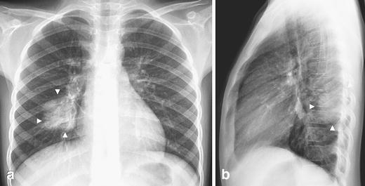
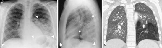
Typical round pneumonia in a 7-year-old boy with cough and fever. a Frontal and (b) lateral chest radiographs show the appearance of a typical round pneumonia (arrowheads)—a well-defined round opacity in the right lower lobe touching the pleura posteriorly
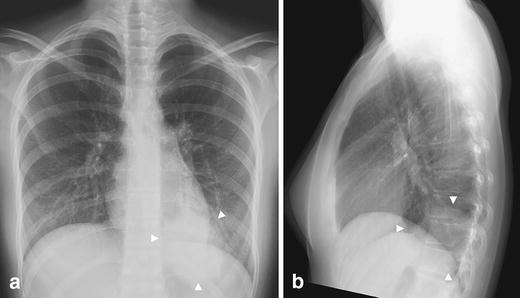
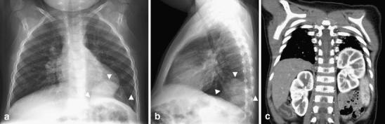
Round pneumonia in a 16-year-old girl with fever and cough. a Frontal and (b) lateral chest radiographs demonstrate a round opacity in the basal segment of the left lower lobe with poorly defined margins (arrowheads). In older children, the margins of the lesions are less sharp because of maturation of the mechanisms of collateral air drift. Follow-up study obtained at 6 weeks revealed complete resolution (not shown)
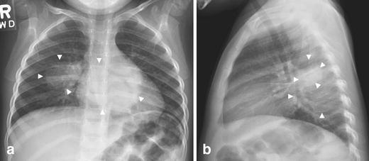
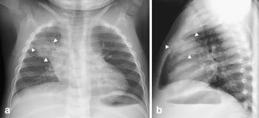
Multiple round pneumonias in a 3-year-old boy with fever and vomiting. Chest radiographs (a=frontal; b=lateral) show round opacities (arrowheads) in the superior segment of right lower lobe and in the left lower lobe posteriorly. Follow-up radiographs demonstrated complete resolution (images not shown)
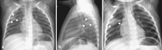
Evolution of an atypical round pneumonia in a 2-year-old with fever and cough. a-b Chest radiograph shows a round opacity in the posterior segment of the right upper lobe. c Follow-up radiograph obtained a few days later demonstrates rapid change. A radiograph obtained several weeks later after antibiotic treatment showed complete resolution (not shown). Note that on the first exam the lesion is in the right upper lobe and surrounded by lung. The clinical presentation and the age of the child suggested round pneumonia as the first possibility. The atypical features prompted the follow-up evaluation
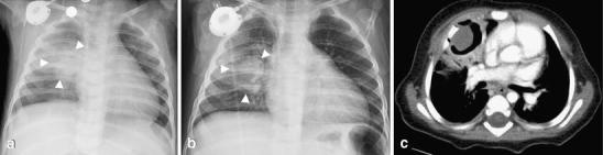
Aspergilloma in a 10-month-old child with leukemia who presented with a fever. a Initial chest radiograph demonstrates a round opacity (arrowheads) in the right parahilar region. Note the presence of a right chest Port-A-Cath. b Frontal chest radiograph obtained several days later shows a well-defined cavitary lesion (arrowheads) in the right upper lobe surrounded by air. c Contrast-enhanced CT on the same day shows an intracavitary lesion consistent with a fungal ball. No other lesions were present. The final diagnosis was an aspergilloma by cultures and histological analysis after surgical removal
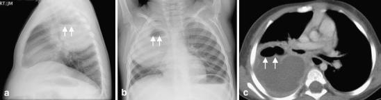
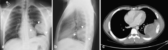
Lung abscess in a 2-year-old boy with fever and cough. a Lateral and (b) frontal chest radiographs reveal a large round opacity (arrowheads) with an air-fluid level (arrowheads) located posteriorly in the right upper lobe. c Contrast-enhanced CT scan obtained later shows a large rim-enhancing lesion with an air-fluid (arrowheads) level consistent with a lung abscess
Tuberculosis mimicking a round pneumonia in a 13-month-old asymptomatic child with a positive PPD test. Chest radiographs (a, b) demonstrate a round opacity (arrowheads) in the anterior segment of the right upper lobe. Chest radiographs obtained a year later following anti-tuberculous treatment revealed only a residual fibrotic scar (not shown). Note the upper lobe location and indistinct margins
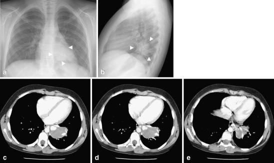
Intralobar sequestration in a 12-year-old girl with chronic cough. a Frontal and (b) lateral chest radiographs show a round opacity (arrowheads) with well-defined margins in the left lower lobe. c–e Contrast-enhanced CT demonstrates a round soft-tissue mass (arrows) in the posteromedial left lower lobe without tracheobronchial airway communication, but with arterial supply by a branch from the descending thoracic aorta and a draining pulmonary vein into the left atrium. The age of the girl and the symptoms should alert the radiologist about a possible underlying lesion. Compare these images with those in Fig. 2 (round pneumonia)
Congenital pulmonary airway malformation (CPAM) in a 13-year-old boy with recurrent respiratory tract infections. a Frontal and (b) lateral chest radiographs show a large opacity (arrowheads) in the left lower lobe. On the frontal projection, the margins are indistinct. However, on the lateral radiograph, the lesion has well-defined margins anteriorly and superiorly. c Because of the boy’s age, the clinical history and the radiographic appearance, a CT angiogram was obtained; showing a large lesion with multiple air-filled cystic spaces and no systemic arterial supply. The diagnosis of CPAM was confirmed after surgery
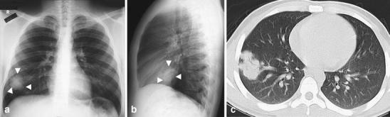
Lymphoma presenting as a round opacity in a 17-year-old boy with malaise, fatigue and cervical adenopathy. a Frontal and (b) lateral chest radiographs show a round lobulated opacity (arrowheads) in the right lower lobe. c Contrast-enhanced CT shows the lesion as a well-defined lobulated opacity with air bronchograms within and barely touching the pleura. Multifocal adenopathy was also present, though not in the mediastinum (not shown). Pathological diagnosis of lymphoma was made after cervical surgical lymph node resection
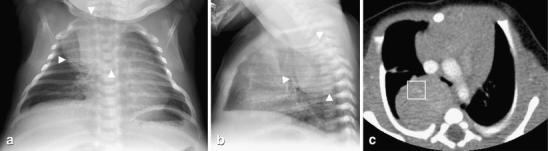
Neuroblastoma in a 1-month-old with shortness of breath and stridor. a Frontal and (b) lateral chest radiographs show a large, round well-defined right posterior mediastinal mass (arrowheads) with mass effect upon the airway. c Contrast-enhanced CT obtained the same day shows a well-defined heterogeneously enhancing posterior mediastinal mass with faint calcifications (within square box) and mass effect on the carina. The pathological diagnosis of neuroblastoma was made by surgical resection
PNET of the chest wall in a 15-year-old boy with left chest wall pain for several weeks. a Frontal and (b) lateral chest radiographs show a large round opacity (arrowheads) with well-defined smooth medial and anterior borders but with obtuse borders at the pleural margins. Subtle widening of the left 7th rib with associated permeative destruction and periosteal reaction are seen. These findings are suggestive of a chest wall mass. c Corresponding CT study shows a large heterogeneously enhancing mass arising from the chest wall. The left 7th rib is expanded with periosteal reaction. The final pathological diagnosis was a peripheral PNET
Diaphragmatic eventration in a 22-month-old with fever and cough. a Frontal and (b) lateral chest radiographs show an opacity (arrowheads) in the left lower lobe with well-defined superior margins. The opacity remained unchanged on radiographs obtained 6 weeks later when the child had clinically shown improvement (not shown). c Contrast-enhanced CT study reveals eventration of the left hemidiaphragm posteromedially with the left kidney at a high ectopic location
Similar articles
-
Computer-aided diagnosis for World Health Organization-defined chest radiograph primary-endpoint pneumonia in children.Pediatr Radiol. 2020 Apr;50(4):482-491. doi: 10.1007/s00247-019-04593-0. Epub 2020 Jan 13. Pediatr Radiol. 2020. PMID: 31930429
-
Pseudolesion of the chest. A conglomerate shadow on the lateral radiograph.Chest. 1985 Apr;87(4):541-3. doi: 10.1378/chest.87.4.541. Chest. 1985. PMID: 3979146
-
Evaluation of pneumonia in children: comparison of MRI with fast imaging sequences at 1.5T with chest radiographs.Acta Radiol. 2011 Oct 1;52(8):914-9. doi: 10.1258/ar.2011.100429. Epub 2011 Aug 4. Acta Radiol. 2011. PMID: 21816896
-
[Noninfectious differential diagnoses of pneumonia].Radiologe. 2017 Jan;57(1):35-42. doi: 10.1007/s00117-016-0196-5. Radiologe. 2017. PMID: 27995287 Free PMC article. Review. German.
-
A radiologic update on medical diseases of the newborn chest.Pediatr Radiol. 1995;25(8):631-7. doi: 10.1007/BF02011835. Pediatr Radiol. 1995. PMID: 8570317 Review.
Cited by
-
Pediatric round pneumonia.Pediatr Neonatol. 2014 Dec;55(6):491-4. doi: 10.1016/j.pedneo.2013.01.014. Epub 2013 Mar 13. Pediatr Neonatol. 2014. PMID: 23597522 Free PMC article.
-
[Specific characteristics of chest X‑ray in childhood : Basics for radiologists].Radiologe. 2018 Apr;58(4):359-376. doi: 10.1007/s00117-018-0374-8. Radiologe. 2018. PMID: 29556698 German.
-
Round pneumonia due to Chlamydia pneumoniae in a child.Radiol Case Rep. 2019 Jan 18;14(4):436-438. doi: 10.1016/j.radcr.2019.01.010. eCollection 2019 Apr. Radiol Case Rep. 2019. PMID: 30701012 Free PMC article.
-
The Echo of Pulmonary Tuberculosis: Mechanisms of Clinical Symptoms and Other Disease-Induced Systemic Complications.Clin Microbiol Rev. 2020 Jul 1;33(4):e00036-20. doi: 10.1128/CMR.00036-20. Print 2020 Sep 16. Clin Microbiol Rev. 2020. PMID: 32611585 Free PMC article. Review.
-
ESR Essentials: imaging of common paediatric pulmonary diseases-practice recommendations by the European Society of Paediatric Radiology.Eur Radiol. 2025 Aug;35(8):5037-5052. doi: 10.1007/s00330-024-11268-4. Epub 2025 Jan 29. Eur Radiol. 2025. PMID: 39881039 Free PMC article. Review.
References
MeSH terms
LinkOut - more resources
Full Text Sources
Medical
