

Age-related changes in the corpus callosum in early-onset bipolar disorder assessed using volumetric and cross-sectional measurements
- PMID: 20686873
- PMCID: PMC3711475
- DOI: 10.1007/s11682-010-9101-4
Age-related changes in the corpus callosum in early-onset bipolar disorder assessed using volumetric and cross-sectional measurements
Abstract
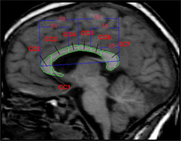
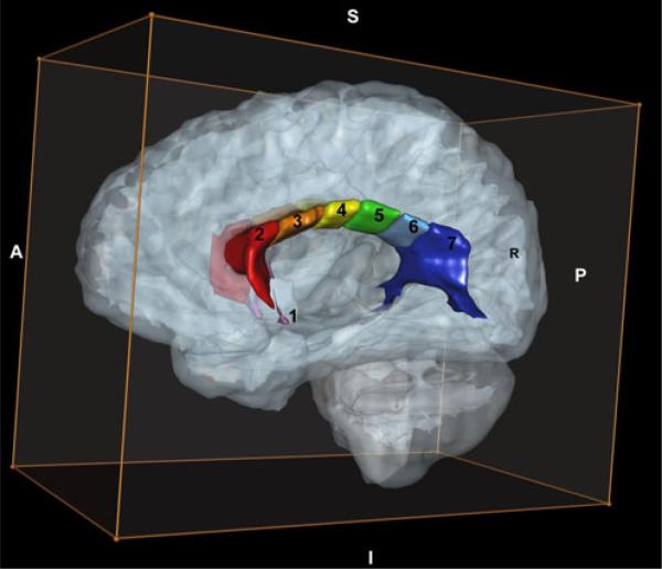
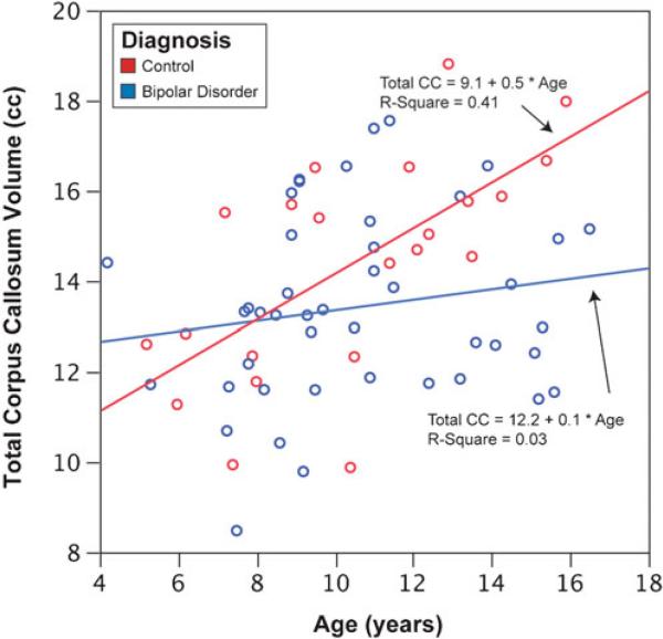
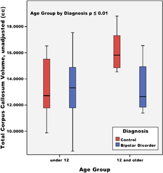
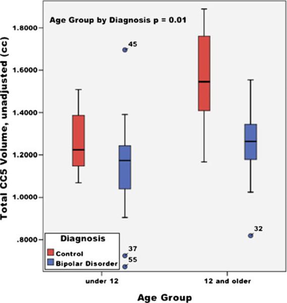
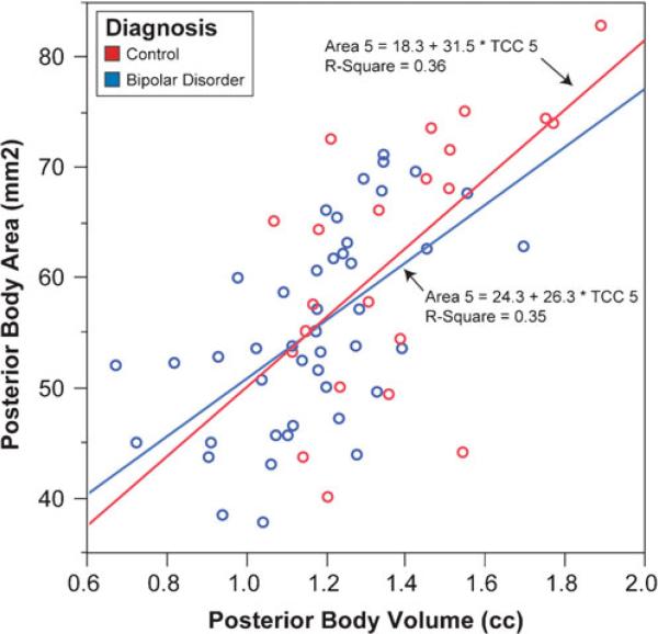
Corpus callosum (CC) area abnormalities have been reported in magnetic resonance imaging (MRI) studies of adults and youths with bipolar disorder (BPD), suggesting interhemispheric communication may be abnormal in BPD and may be present early in the course of illness and affect normal neuromaturation of this structure throughout the lifecycle. Neuroimaging scans from 44 youths with DSM-IV BPD and 22 healthy controls (HC) were analyzed using cross-sectional area measurements and a novel method of volumetric parcellation. Univariate analyses of variance were conducted on CC subregions using both volume and traditional area measurements. Youths with BPD had smaller middle and posterior callosal regions, and reduced typical age-related increases in CC size. The cross-sectional area and novel volumetric methodologies resulted in similar findings. Future longitudinal assessments of CC development would track the evolution of callosal abnormalities in youths with BPD and allow exploration of the functional significance of these findings.
Figures
Similar articles
-
Corpus callosum area in patients with bipolar disorder with and without psychotic features: an international multicentre study.J Psychiatry Neurosci. 2015 Sep;40(5):352-9. doi: 10.1503/jpn.140262. J Psychiatry Neurosci. 2015. PMID: 26151452 Free PMC article.
-
Corpus callosum signal intensity in patients with bipolar and unipolar disorder.J Neurol Neurosurg Psychiatry. 2004 Feb;75(2):221-5. J Neurol Neurosurg Psychiatry. 2004. PMID: 14742592 Free PMC article.
-
Corpus callosal morphology in youth with bipolar depression.Bipolar Disord. 2014 Dec;16(8):889-93. doi: 10.1111/bdi.12247. Epub 2014 Aug 28. Bipolar Disord. 2014. PMID: 25164210
-
Corpus callosum changes in euthymic bipolar affective disorder.Br J Psychiatry. 2014 Feb;204(2):129-36. doi: 10.1192/bjp.bp.112.123687. Epub 2013 Dec 19. Br J Psychiatry. 2014. PMID: 24357572
-
Meta-analysis of magnetic resonance imaging studies of the corpus callosum in bipolar disorder.Acta Psychiatr Scand. 2008 Nov;118(5):357-62. doi: 10.1111/j.1600-0447.2008.01229.x. Epub 2008 Jul 14. Acta Psychiatr Scand. 2008. PMID: 18644004
Cited by
-
Corpus callosum size is highly heritable in humans, and may reflect distinct genetic influences on ventral and rostral regions.PLoS One. 2014 Jun 26;9(6):e99980. doi: 10.1371/journal.pone.0099980. eCollection 2014. PLoS One. 2014. PMID: 24968245 Free PMC article.
-
Differential impairment of interhemispheric transmission in bipolar disease.Exp Brain Res. 2013 Oct;230(2):175-85. doi: 10.1007/s00221-013-3642-x. Epub 2013 Jul 16. Exp Brain Res. 2013. PMID: 23857169
-
Cortico-cerebellar abnormalities in adolescents with heavy marijuana use.Psychiatry Res. 2012 Jun 30;202(3):224-32. doi: 10.1016/j.pscychresns.2011.11.005. Epub 2012 Jul 24. Psychiatry Res. 2012. PMID: 22835865 Free PMC article.
-
Corpus callosum area in patients with bipolar disorder with and without psychotic features: an international multicentre study.J Psychiatry Neurosci. 2015 Sep;40(5):352-9. doi: 10.1503/jpn.140262. J Psychiatry Neurosci. 2015. PMID: 26151452 Free PMC article.
-
White-matter microstructure and gray-matter volumes in adolescents with subthreshold bipolar symptoms.Mol Psychiatry. 2014 Apr;19(4):462-70. doi: 10.1038/mp.2013.44. Epub 2013 Apr 30. Mol Psychiatry. 2014. PMID: 23628983 Free PMC article.
References
-
- Adler CM, Delbello MP, Mills NP, Schmithorst V, Holland S, Strakowski SM. Comorbid ADHD is associated with altered patterns of neuronal activation in adolescents with bipolar disorder performing a simple attention task. Bipolar Disorders. 2005;7(6):577–588. - PubMed
-
- American Psychiatric Association . Diagnostic and statistical manual of mental disorders. 4th ed. American Psychiatric Association; Washington: 1994.
-
- Atmaca M, Ozdemir H, Cetinkaya S, Parmaksiz S, Belli H, Poyraz AK, et al. Cingulate gyrus volumetry in drug free bipolar patients and patients treated with valproate or valproate and quetiapine. Journal of Psychiatric Research. 2007a;41(10):821–827. - PubMed
-
- Atmaca M, Ozdemir H, Yildirim H. Corpus callosum areas in first-episode patients with bipolar disorder. Psychological Medicine. 2007b;37(5):699–704. - PubMed
-
- Barkovich AJ. Normal development of the neonatal and infant brain. In: Barkovich AJ, editor. Pediatric neuroimaging. Raven Press; New York: 1990. pp. 5–34.
Publication types
MeSH terms
Grants and funding
LinkOut - more resources
Full Text Sources
Medical
