. 2010 Oct;19(10):1917-31.
doi: 10.1002/pro.479.

Thermal unfolding studies show the disease causing F508del mutation in CFTR thermodynamically destabilizes nucleotide-binding domain 1

Affiliations

Thermal unfolding studies show the disease causing F508del mutation in CFTR thermodynamically destabilizes nucleotide-binding domain 1

Irina Protasevich et al. Protein Sci. 2010 Oct.

Abstract

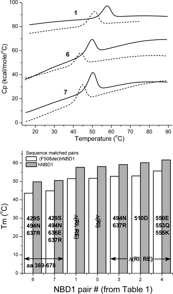
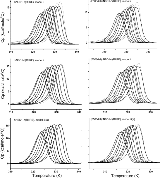
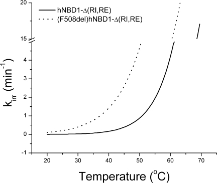
Misfolding and degradation of CFTR is the cause of disease in patients with the most prevalent CFTR mutation, an in-frame deletion of phenylalanine (F508del), located in the first nucleotide-binding domain of human CFTR (hNBD1). Studies of (F508del)CFTR cellular folding suggest that both intra- and inter-domain folding is impaired. (F508del)CFTR is a temperature-sensitive mutant, that is, lowering growth temperature, improves both export, and plasma membrane residence times. Yet, paradoxically, F508del does not alter the fold of isolated hNBD1 nor did it seem to perturb its unfolding transition in previous isothermal chemical denaturation studies. We therefore studied the in vitro thermal unfolding of matched hNBD1 constructs ±F508del to shed light on the defective folding mechanism and the basis for the thermal instability of (F508del)CFTR. Using primarily differential scanning calorimetry (DSC) and circular dichroism, we show for all hNBD1 pairs studied, that F508del lowers the unfolding transition temperature (T(m)) by 6-7°C and that unfolding occurs via a kinetically-controlled, irreversible transition in isolated monomers. A thermal unfolding mechanism is derived from nonlinear least squares fitting of comprehensive DSC data sets. All data are consistent with a simple three-state thermal unfolding mechanism for hNBD1 ± F508del: N(±MgATP) <==> I(T)(±MgATP) → A(T) → (A(T))(n). The equilibrium unfolding to intermediate, I(T), is followed by the rate-determining, irreversible formation of a partially folded, aggregation-prone, monomeric state, A(T), for which aggregation to (A(T))(n) and further unfolding occur with no detectable heat change. Fitted parameters indicate that F508del thermodynamically destabilizes the native state, N, and accelerates the formation of A(T).


Figures

Figure 1
Figure 1
Upper Panel: DSC heat capacity profiles for hNBD1 constructs (solid lines) and the corresponding sequence-matched F508del mutant (dotted lines) in buffer containing 5 mM ATP and 10 mM MgCl2 at 2 K/min scan rate. Buffer scans are subtracted from protein scans and data are normalized to molar concentration. Protein concentrations in the calorimetric cell were 1 mg/mL. Lower Panel: The Tm values are shown for these sequence-matched pairs together with results for the other four pairs presented in Table I. Standard errors in the Tm measurements are given in Table I.
Figure 2
Figure 2
CD spectra of hNBD1-Δ(RI,RE) and (F508del)hNBD1-Δ(RI,RE) in the far-UV region (left panel) and in the near-UV region (right panel). Solid circles are the experimental data for hNBD1-Δ(RI,RE) and open circles are the experimental data for (F508del)hNBD1-Δ(RI,RE). The ATP concentration for far-UV measurements was 0.8 μM for (hNBD1-Δ(RI,RE) and 4 μm ATP for (F508del)hNBD1-Δ(RI,RE); for near-UV measurements, it was 0.125 mM ATP for both proteins. Protein concentration was 0.69 mg/mL in 0.02 cm cuvettes for far-UV CD and 1 mg/mL in 1 cm cuvettes for near-UV CD measurements.
Figure 3
Figure 3
Thermal unfolding of hNBD1-Δ(RI,RE), left panels, and (F508del)hNBD1-Δ(RI,RE), right panels, monitored by CD and DSC at scan rate 2°C/min at 0.125 mM ATP and 0.5 mM MgCl2. CD is monitored at 297 nm (near-UV wavelength region, upper panels) and at 230 nm (far-UV wavelength region, lower panels). Protein concentration in the 1 cm cuvette was 0.125 mg/mL for far-UV and 0.5 mg/mL for near-UV measurements. The unfolding data were normalized to the fractional change observed. No baseline subtraction was performed on the spectral data shown by closed and open circles. DSC unfolding curves (represented by solid lines in the upper panels) were integrated and normalized to the unfolding calorimetric enthalpy value, ΔHTHcal; CD unfolding curves were normalized to maximal change in the CD signal, CDT/(CDinitial-CDfinal), where T = temperature of observed signal. The signal change at 230 nm for both proteins was approximately the same: starting at 20°C, θ = −6 × 10³ deg cm−1 × dmole−1 and ending at 80°C, θ = −2 × 10³ deg cm−1 × dmole−1. The signal change at 297 nm for hNBD1-Δ(RI,RE) was determined from the initial signal at 20°C, θ = 69 × 103 deg cm−1 × dmole−1 and ending at 60°C, θ = −58 × 103 deg cm−1 × dmole−1 ; for (F508del)hNBD1-Δ(RI,RE), initially, θ = 55 × 103 deg cm−1 × dmole−1 and ending with θ = −31 × 103 deg cm−1 × dmole−1. The insets in the upper panels are temperature unfolding of hNBD1-Δ(RI,RE), left panel, and (F508del)hNBD1-Δ(RI,RE), right panel, monitored by intrinsic Trp fluorescence with excitation wavelength at 290 nm and emission wavelength at 340 nm and DSC at scan rate 1°C/min at 0.1 mM ATP and 0.2 mM MgCl2. Trp fluorescence unfolding curves were normalized to maximal change in the Trp fluorescence signal, FLTrpT/(FLTrpinitial-FLTrpfinal), where T = temperature of observed signal. Preunfolding and postunfolding baselines of unfolding profile were subtracted using equations obtained by linear regression to obtain the data shown.
Figure 4
Figure 4
DSC of hNBD1-Δ(RI,RE) and (F508del)hNBD1-Δ(RI,RE) at different concentrations of ATP at scan-rate 2 K/min. The left panel shows the temperature dependence of the molar heat capacity of (hNBD1-Δ(RI,RE) (solid lines) and (F508del)hNBD1-Δ(RI,RE) (dotted lines) at 0 mM, 0.1 mM and 5 mM ATP. The right panel represents results of DSC data analysis for hNBD1-Δ(RI,RE) (solid circles) and (F508del)hNBD1-Δ(RI,RE) (open circles) as ATP concentration dependence of Tm (top panel) and unfolding enthalpy (bottom panel) obtained from 0 mM to 20 mM ATP. Protein concentration was 1 mg/mL. The minimal detectable ATP concentration in the protein solution is 0.5 μM; see Supporting Information Methods for details.
Figure 5
Figure 5
Comparison of global fits of DSC data to Models i, ii, and iii(a). Left panels: Fits for hNBD1-Δ(RI,RE). Right panels: Fit for (508del)hNBD1-Δ(RI,RE). DSC was performed at scan rate 2°C/min in buffer containing 0.088, 0.108, 0.196, 0.3, 0.46, 0.915, 0.97, 1.82, 4.7 mM ATP with 10 mM MgCl2, and 10 mM ATP with 20 mM MgCl2 for hNBD1-Δ(RI,RE) and 0.088, 0.11, 0.21, 0.27, 0.48, 0.85, 1.05, 1.7 and 4.9 mM ATP with 10 mM MgCl2 for (F508del)hNBD1-Δ(RI,RE). Solid lines are experimental data. Dashed lines are best fit curves.
Figure 6
Figure 6
Temperature-dependence of the rate constant, kirr, for the ITAT unfolding step for hNBD1-Δ(RI,RE) and (F508del)hNBD1-Δ(RI,RE). Fitted parameters in Table IV were used to construct the plot.
Figure 7
Figure 7
Plots of ΔG, ΔH, and TΔS versus temperature for the N ⇄ IT transition. The fitted ΔCp values of 1.86 kcal/mol/K, and 2.13 kcal/mol/K, for hNBD1-Δ(RI,RE) and (F508del) hNBD1-Δ(RI,RE), respectively, with other fitted parameters found in Table IV were used to construct the plots.

References

    1. Zielenski J, Tsui LC. Cystic fibrosis: genotypic and phenotypic variations. Annu Rev Genet. 1995;29:777–807. - PubMed
    1. Riordan JR. CFTR function and prospects for therapy. Annu Rev Biochem. 2008;77:701–726. - PubMed
    1. Riordan JR. Assembly of functional CFTR chloride channels. Annu Rev Physiol. 2005;67:701–718. - PubMed
    1. Mornon JP, Lehn P, Callebaut I. Atomic model of human cystic fibrosis transmembrane conductance regulator: membrane-spanning domains and coupling interfaces. Cell Mol Life Sci. 2008;65:2594–2612. - PMC - PubMed
    1. Serohijos AW, Hegedus T, Aleksandrov AA, He L, Cui L, Dokholyan NV, Riordan JR. Phenylalanine-508 mediates a cytoplasmic-membrane domain contact in the CFTR 3D structure crucial to assembly and channel function. Proc Natl Acad Sci USA. 2008;105:3256–3261. - PMC - PubMed

Publication types

MeSH terms

Substances

LinkOut - more resources