Review
. 2010:676:27-55.
doi: 10.1007/978-1-4419-6199-0_3.

Understanding cytokinesis failure

Affiliations
Review

Understanding cytokinesis failure

Guillaume Normand et al. Adv Exp Med Biol. 2010.

Abstract

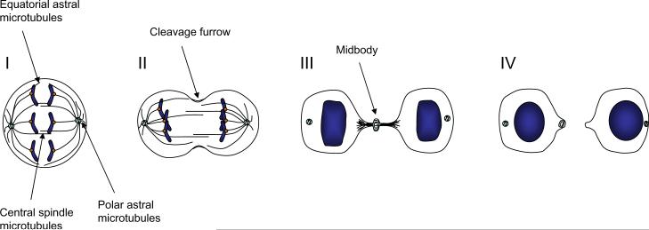
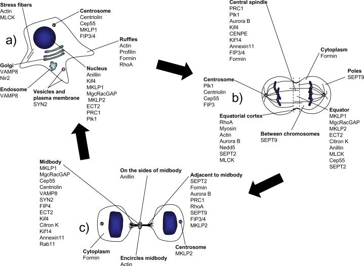
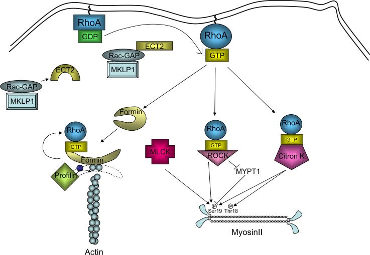
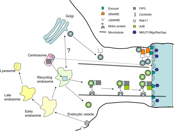
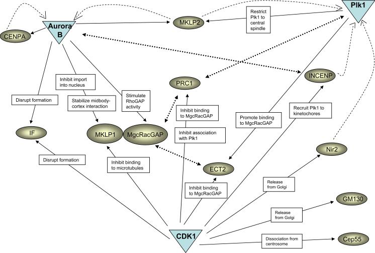
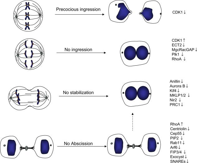
Cytokinesis is the final step in cell division. The process begins during chromosome segregation, when the ingressing cleavage furrow begins to partition the cytoplasm between the nascent daughter cells. The process is not completed until much later, however, when the final cytoplasmic bridge connecting the two daughter cells is severed. Cytokinesis is a highly ordered process, requiring an intricate interplay between cytoskeletal, chromosomal and cell cycle regulatory pathways. A surprisingly broad range of additional cellular processes are also important for cytokinesis, including protein and membrane trafficking, lipid metabolism, protein synthesis and signaling pathways. As a highly regulated, complex process, it is not surprising that cytokinesis can sometimes fail. Cytokinesis failure leads to both centrosome amplification and production of tetraploid cells, which may set the stage for the development of tumor cells. However, tetraploid cells are abundant components of some normal tissues including liver and heart, indicating that cytokinesis is physiologically regulated. In this chapter, we summarize our current understanding of the mechanisms of cytokinesis, emphasizing steps in the pathway that may be regulated or prone to failure. Our discussion emphasizes findings in vertebrate cells although we have attempted to highlight important contributions from other model systems.


Figures

Figure 1
Figure 1. Multiple stages of cytokinesis
Three populations of microtubules first specify the site of cleavage by activating RhoA in a narrow zone between segregating chromosomes (I). Formation and activation of the actomyosin ring next leads to furrow ingression (II). The constricting furrow compacts the central spindle microtubules leading to midbody formation (III). Abscission of the furrow occurs by physically separating the cytoplasm of the daughter cells (IV).
Figure 2
Figure 2. Localization of cytokinesis components
Interphase (a), anaphase (b), and late cytokinesis (c).
Figure 3
Figure 3. Role of the RhoA pathway in furrow initiation
Autoinhibition of ECT2 is suppressed by association of ECT2 with MgcRacGAP. ECT2 then activates RhoA by stimulating exchange of GDP for GTP. Active RhoA then activates formins to stimulate actin nucleation, and binds to ROCK and Citron kinases, stimulating phosphorylation and activation of myosin.
Figure 4
Figure 4. Membrane trafficking in cytokinesis
Secretory vesicles accumulate at the intercellular bridge in a centriolin-dependent manner by SNARE interaction and vesicle tethering by the exocyst complex. Vesicles originating from the recycling endosome and containing the complex Rab11/FIP3 move along microtubules. Interaction of FIP3 with both ARF6 and the exocyst permits vesicle targeting to the midbody. Vesicles then fuse with each other and the plasma membrane, physically separating the daughter cells.
Figure 5
Figure 5. Regulation of cytokinesis by mitotic kinases
A major function of CDK1 is to prevent precocious cytokinesis before proper chromosome segregation. CDK1 thus negatively regulates some of the main players of cytokinesis. At the same time, CDK1 plays a positive role in cytokinesis by releasing cytokinesis proteins from the Golgi apparatus, and by facilitating binding of Plk1 to its substrates. Plk1 and Aurora B phosphorylate substrates that are important for both early and late steps of cytokinesis. Solid arrows indicate phosphorylation; dashed arrows indicate changed protein localization; dotted arrows indicate protein interactions. IF, intermediate filaments.
Figure 6
Figure 6. Summary of different phenotypes resulting from cytokinesis failure
Inhibition (downward arrow) or excessive activation (upward arrow) of different cytokinesis components can give rise to distinct phenotypes, including precocious ingression before the chromosomes have been separated, regression of the furrow giving rise to binucleated cells, or stabilization of the cytoplasmic bridge where daughter cells remain connected. The list is not comprehensive; see text for additional examples.

Similar articles

Cited by

References

    1. Rappaport R. Cytokinesis in animal cells. Int Rev Cytol. 1971;31:169–213. - PubMed
    1. Rappaport R. Establishment of the mechanism of cytokinesis in animal cells. Int Rev Cytol. 1986;105:245–281. - PubMed
    1. Canman JC, Cameron LA, Maddox PS, et al. Determining the position of the cell division plane. Nature. 2003;424:1074–1078. - PubMed
    1. Hiramoto Y. Analysis of cleavage stimulus by means of micromanipulation of sea urchin eggs. Exp Cell Res. 1971;68:291–298. - PubMed
    1. Burgess DR, Chang F. Site selection for the cleavage furrow at cytokinesis. Trends Cell Biol. 2005;15:156–162. - PubMed

Publication types

Substances