Review
. 2010;16(28):3071-88.
doi: 10.2174/138161210793292474.

Ligand-based peptide design and combinatorial peptide libraries to target G protein-coupled receptors

Affiliations
Review

Ligand-based peptide design and combinatorial peptide libraries to target G protein-coupled receptors

Christian W Gruber et al. Curr Pharm Des. 2010.

Abstract

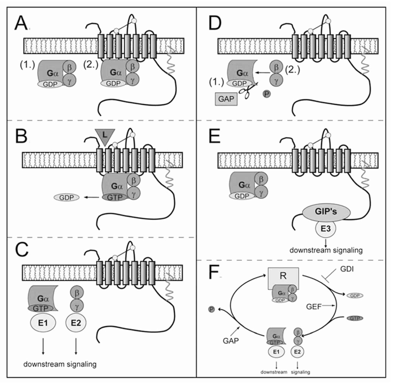
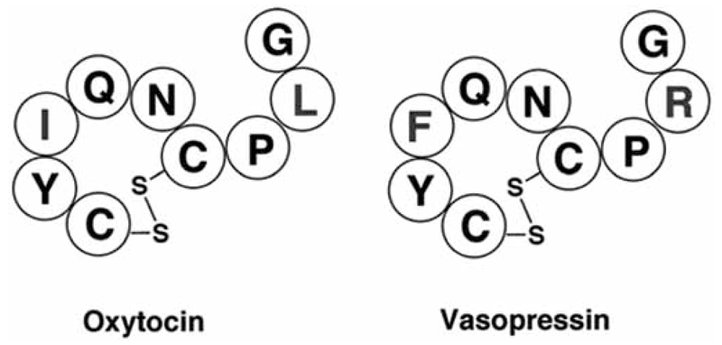
G protein-coupled receptors (GPCRs) are considered to represent the most promising drug targets; it has been repeatedly said that a large fraction of the currently marketed drugs elicit their actions by binding to GPCRs (with cited numbers varying from 30-50%). Closer scrutiny, however, shows that only a modest fraction of (≈60) GPCRs are, in fact, exploited as drug targets, only ≈20 of which are peptide-binding receptors. The vast majority of receptors in the humane genome have not yet been explored as sites of action for drugs. Given the drugability of this receptor class, it appears that opportunities for drug discovery abound. In addition, GPCRs provide for binding sites other than the ligand binding sites (referred to as the "orthosteric site"). These additional sites include (i) binding sites for ligands (referred to as "allosteric ligands") that modulate the affinity and efficacy of orthosteric ligands, (ii) the interaction surface that recruits G proteins and arrestins, (iii) the interaction sites of additional proteins (GIPs, GPCR interacting proteins that regulate G protein signaling or give rise to G protein-independent signals). These sites can also be targeted by peptides. Combinatorial and natural peptide libraries are therefore likely to play a major role in identifying new GPCR ligands at each of these sites. In particular the diverse natural peptide libraries such as the venom peptides from marine cone-snails and plant cyclotides have been established as a rich source of drug leads. High-throughput screening and combinatorial chemistry approaches allow for progressing from these starting points to potential drug candidates. This will be illustrated by focusing on the ligand-based drug design of oxytocin (OT) and vasopressin (AVP) receptor ligands using natural peptide leads as starting points.


Figures

Fig. (1)
Fig. (1)
GPCR signaling, G protein cycle and accessory proteins of signaling. (A) In its basal (non-activated) state the G protein (shown in green) is a heterotrimer composed of three subunits (α, β and γ) and binds the guanine nucleotide diphosphate (shown in pale orange). The G protein(s) interact with their receptors (shown as 7-transmembrane spanning model in grey, with commonly observed motifs pointed out, i.e., disulfide bond in yellow and palmytoylation anchor in red) by (restricted/unrestricted) collision coupling (1→2). (B) Upon activation (i.e., agonist stimulation), the GDP is released from the α–subunit and GTP (red) is bound (see text for explanation). (C) Binding of GTP to the Gα subunit destabilizes the complex, which leads to dissociation of Gα and Gβ/γ subunits from the receptor and their interaction with downstream effector proteins (E1 and E2, shown in yellow). (D) The signal is terminated by the intrinsic GTPase (pale blue) activity of the Gα subunit, which hydrolyses the GTP to GDP. This GTP-GDP “exchange” leads to re-formation (1→2) of the heterotrimer Gαβγ and results in the inactive or basal state of the G protein. (E) In addition, GPCRs can interact with numerous proteins other than their cognate G protein(s). These so-called GPCR interacting proteins (GIPs) can elicit specific cellular responses, independently of the G protein and/or the arrestin-mediated cellular signaling. (F) Several proteins impinge on the G protein cycle by interacting directly with individual G proteins: (i) GTPase activating proteins (GAPs), (ii) non-receptor guanine nucleotide exchange factors (GEFs) and (iii) guanine nucleotide dissociation inhibitors (GDIs).
Fig. (2)
Fig. (2)
Common structural recognition motifs of peptides targeting GPCRs. (A) α-Helix of the human parathyroid hormone [199]. (B) Type II β-turn of deamino-oxytocin [180]. (C) Stromal cell-derived factor-1α (SDF-1α), a member of the chemokine superfamily, exhibiting β-sheets and helices as structural motifs [200].
Fig. (3)
Fig. (3)
Examples of versatile venom peptide scaffolds. (A) The “three-finger” scaffold of snake neurotoxins highlight the target diversity of venom peptides with (i) α-bungaratoxin acting on the nAChR, (ii) γ-bungaratoxin targeting both, nicotinic and muscarinic acetylcholine receptors, and (iii) muscarinic toxin 2 modulating the muscarinic acetylcholine and α-adrenergic receptors. (B) RgIA and Vc1.1, two α-conotxin that are α9α10 nicotinic acetylcholine receptor antagonists, but act also via the GABAB receptor. (C) Mastoparan, a G-protein activating wasp venom peptide that uses its α-helical secondary structure to penetrate cell membranes and to mimic the G protein-activating portion of GPCRs. (D) TIA, an example of the ρ-conotoxin superfamily that targets α1–adrenergic receptors.
Fig. (4)
Fig. (4)
Structure, diversity and oxytocin-like sequence motif of plant cyclotides. (A) Cyclotides comprise the typical structural cyclic-cystine-knot motif, characterized by three disulfide bonds (shown in yellow) in a knotted arrangement (two disulfide bonds and the adjacent backbone segments from a ring that is threaded by the third disulfide bond). Cys-residues are numbered with Roman numerals, loops are indicated in red. The characteristic anti-parallel beta sheets are colored in cyan. (B) The diversity of cyclotides is pointed out by the number of to sequence permutations discovered to date. The wheel diagram shows the sequence of the typical cyclotide kalata B1; residues, disulfide bonds, and loops are colored/labeled according to (A). (C) Sequence alignment of human oxytocin with selected cyclotides (www.cybase.com.au). Some cyclotides and oxytocin share one or both of the activity-bearing sequence elements YxxN and CxxG.
Fig. (5)
Fig. (5)
Amino acid sequences of oxytocin and vasopressin. The amino acid of mature oxytocin and vasopressin peptide hormones is presented. The two differences in each of the peptides are highlighted in blue. Disulfide bonds are indicated.
Fig. (6)
Fig. (6)
Workflow for the search for GPCR agonists and antagonists. The identification of agonists and antagonists works generally via the same methodological workflow (1) Identification of the structural motifs required for binding, (2) systematic modifications of single residues not important for receptor recognition, (3) biological characterization, (4) combined modifications guided by the results obtained, (5) biological characterization and (6) peptidomimetic design and improvement of bioavailability.

References

    1. Kroeze WK, Sheffler DJ, Roth BL. G-protein-coupled receptors at a glance. J Cell Sci. 2003;116:4867–9. - PubMed
    1. Oldham WM, Hamm HE. Heterotrimeric G protein activation by G-protein-coupled receptors. Nat Rev Mol Cell Biol. 2008;9:60–71. - PubMed
    1. Perez DM. From plants to man: the GPCR “tree of life”. Mol Pharmacol. 2005;67:1383–4. - PubMed
    1. Marinissen MJ, Gutkind JS. G-protein-coupled receptors and signaling networks: emerging paradigms. Trends Pharmacol Sci. 2001;22:368–76. - PubMed
    1. Bjarnadottir TK, Gloriam DE, Hellstrand SH, et al. Comprehensive repertoire and phylogenetic analysis of the G protein-coupled receptors in human and mouse. Genomics. 2006;88:263–73. - PubMed

Publication types

LinkOut - more resources