. 2010;12(4):R60.
doi: 10.1186/bcr2622. Epub 2010 Aug 5.

Breast tumor kinase and extracellular signal-regulated kinase 5 mediate Met receptor signaling to cell migration in breast cancer cells

Affiliations

Breast tumor kinase and extracellular signal-regulated kinase 5 mediate Met receptor signaling to cell migration in breast cancer cells

Nancy E Castro et al. Breast Cancer Res. 2010.

Abstract

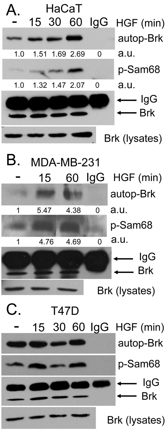
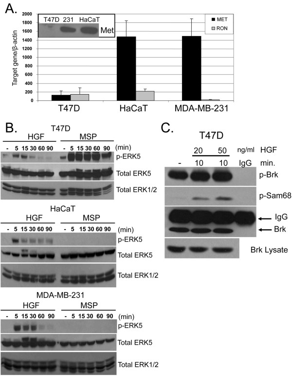
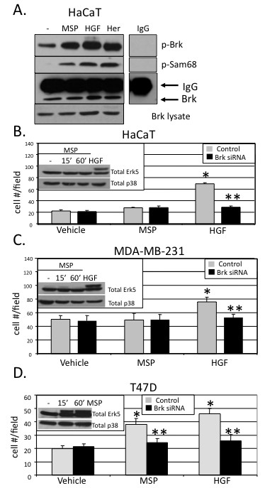
Introduction: Breast tumor kinase (Brk/protein tyrosine kinase 6 (PTK6)) is a nonreceptor, soluble tyrosine kinase overexpressed in the majority of breast tumors. Previous work has placed Brk downstream of epidermal growth factor receptor (ErbB) activation and upstream of extracellular signal-regulated kinase 5 (ERK5) and p38 mitogen-activated protein (MAP) kinases. Herein we investigate the regulation of Brk kinase activity and cell migration in response to treatment of keratinocytes (HaCaT cells) and breast cancer cell lines (MDA-MB-231 and T47D cells) with hepatocyte growth factor (HGF) and macrophage stimulating protein (MSP), peptide ligands for Met and Ron receptors, respectively.

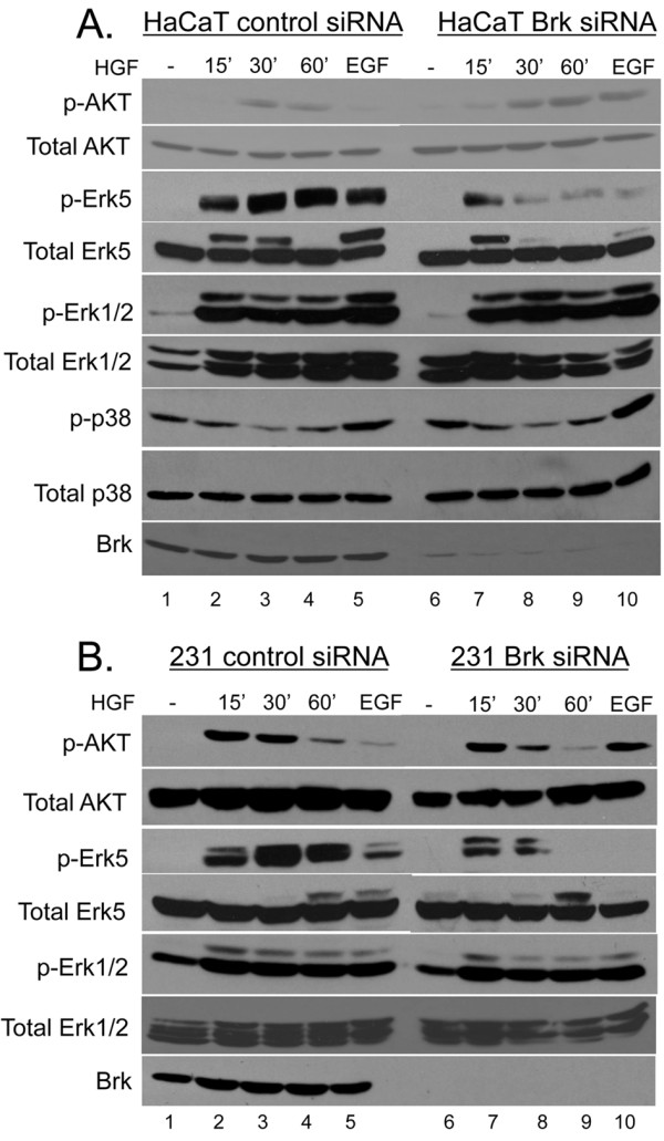
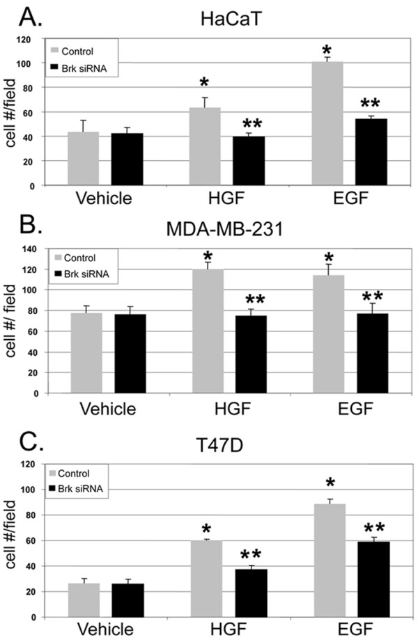
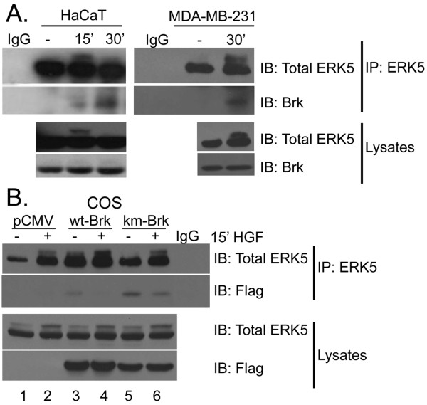
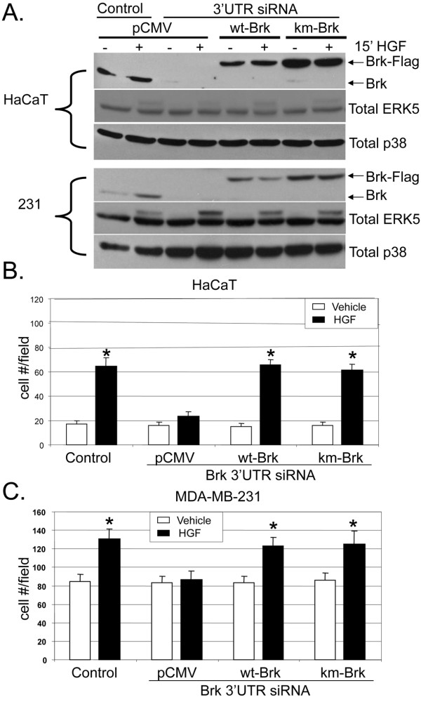
Methods: In vitro kinase assays were performed to directly measure Brk kinase activity in response to MET and RON ligands. Transfection of Brk-targeted RNAi was used to knock down endogenous Brk or ERK5 in multiple cell lines. Kinase activities (downstream of MET signaling) were assayed by Western blotting using total and phospho-specific antibodies. Boyden chamber assays were used to measure cell migration in response to manipulation of Brk and downstream MET effectors. Rescue experiments were performed by knock down of endogenous Brk using RNAi (targeting the untranslated region (3'-UTR)) and transient transfection (re-expression) of either wild-type or kinase-inactive Brk.

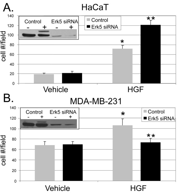
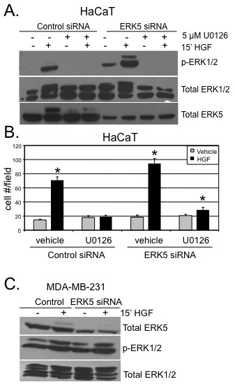
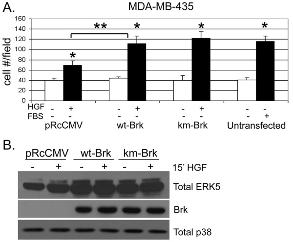
Results: Brk gene silencing revealed that HGF, but not MSP, induced robust Brk-dependent cell migration. Brk and ERK5 copurified in HGF-induced protein complexes, and Brk/ERK5 complexes formed independently of Brk kinase activity. ERK5 was required for breast cancer cell but not keratinocyte cell migration, which became ERK1/2-dependent upon ERK5 knockdown. Notably, rescue experiments indicated that the kinase activity of Brk was not required for HGF-induced cell migration. Further, expression of either wild-type or kinase-inactive Brk in Brk-null MDA-MB-435 cells activated ERK5 and conferred increased HGF-induced cell migration.

Conclusions: These results have identified Brk and ERK5 as important downstream effectors of Met signaling to cell migration. Targeting ERK5 kinase activity or inhibiting the formation of Brk/ERK5 complexes may provide an additional means of blocking cell migration associated with breast cancer progression to metastasis.


Figures

Figure 1
Figure 1
HGF activates Brk. (a). In vitro kinase assays were performed using HaCaT cells. Serum-starved HaCaT cells were treated with either vehicle (water) or 50 ng/ml HGF for 15 to 60 min. Whole cell lysates were subjected to Brk immunoprecipitation and in vitro kinase assays using purified recombinant Sam68 as a substrate. Densitometry of bands represent phosphorylated Brk and Sam68 proteins indicated by arbitrary units (a.u.). Brk kinase assays were performed under similar conditions and treatments as in (a) using MDA-MB-231 (b) and T47D (c) breast cancer cells.
Figure 2
Figure 2
Brk mediates Met receptor signaling to ERK5. HaCaT (a) and MDA-MB-231 (b) cells were transiently transfected with a nontargeting siRNA (control) or Brk siRNA and serum-starved for 24 hr. Cells were then treated with vehicle (water) or 50 ng/ml HGF for 15 to 60 min. Whole cell lysates were harvested and subjected to Western blotting with phospho- and total-specific antibodies to AKT, ERK5, ERK1/2, p-38, and total Brk. Cells were stimulated with 20 ng/ml EGF for 15 min as a positive control for kinase activation.
Figure 3
Figure 3
Signaling specificity of Brk-dependent cell migration. HaCaT (a), MDA-MB-231 (b) and T47D (c) cells were transiently transfected with non-targeting (control) siRNA or Brk siRNA and either vehicle (water) or 50 ng/ml HGF was used as the chemoattractant for each Boyden chamber migration assay. As described in the Materials and Methods, migration assays were performed for 6 hr. A concentration of 20 ng/ml EGF was used as a positive control chemoattractant for migration. Error bars indicate the mean (plus standard deviation) of triplicate measures of cell migration. Single asterisks (*) denote significance (P < 0.05) determined by an unpaired Student's t-test between vehicle and HGF treated cells expressing control siRNA. Double asterisks (**) denote significance between control and Brk siRNA with either HGF or EGF used as chemoattractants. Results were confirmed in three independent experiments.
Figure 4
Figure 4
Intact Met signaling in T47D cells. (a) Real-time quantitative RT-PCR was performed to determine the mRNA expression levels of Met and Ron receptors in T47D, MDA-MB-231, and HaCaT cells. Error bars indicate the mean (plus standard deviation) of Met and Ron mRNA, normalized to β-actin, measured in triplicate cultures. Results were confirmed in three independent experiments. Met protein levels in all three cell lines as measured by Western blotting (insets) (b) T47D, HaCaT and MDA-MB-231 cells were serum-starved for 24 hr and then treated with 50 ng/ml HGF or 80 ng/ml MSP for 5 to 90 min. Western blot analyses were performed on whole cell lysates using ERK5-specific phospho- and total antibodies. ERK1/2 protein expression was used as a loading control. (c) T47D cells were serum-starved for 24 hr then treated with 20 or 50 ng/ml HGF for 10 min. Whole cell lysates were subjected to Brk immunoprecipitation and in vitro kinase assay using purified recombinant Sam68 as described in Material and Methods.
Figure 5
Figure 5
Brk mediates cell type-specific Met and Ron receptor signaling to cell migration. (a) In vitro kinase assays were performed in HaCaT cells as previously described. Cells were serum-starved for 24 hr then treated with either vehicle (water), MSP (80 ng/ml), HGF (50 ng/ml), or heregulin (25 ng/ml) for 15 min. Purified recombinant Sam68 was used as a substrate to measure Brk kinase activity. IgG control images were derived from the same experiment, Western blot, and film exposure time as the experimental lanes shown. Boyden chamber migration assays were performed in HaCaT (b), MDA-MB-231 (c), and T47D (d) cells expressing non-targeting (control) siRNA or Brk siRNA. Cells were treated with either vehicle (water) or 80 ng/ml MSP as the chemoattractant (in the lower chamber). 50 ng/ml HGF was used as a positive control chemoattractant for migration. Error bars indicate the mean (plus standard deviation) of triplicate measures of cell migration. Single asterisks (*) denote significance (P <0.05) determined by an unpaired Student's t-test between vehicle and HGF or MSP treated cells expressing control siRNA. Double asterisks (**) denote significance between control and Brk siRNA with either HGF or MSP used as chemoattractants. Results were confirmed in three independent experiments. Cells treated at various time points with 80 ng/ml MSP were subjected to Western blotting with total ERK5 specific antibody. HGF treatment was used as a positive control for ERK5 activation (insets).
Figure 6
Figure 6
HGF-induced cell migration requires Brk/ERK5 complexes in breast cancer cells. (a) HaCaT and MDA-MB-231/(231) cells were treated with either vehicle or HGF for 15-30 min (30 min for 231 cells). ERK5 was immunoprecipitated from whole cell lysates using total ERK5-specific antibodies. ERK5 immunoprecipitates were then subjected to Western blotting with Brk-specific antibodies. Lysates (10% input) were Western blotted with total ERK5 and Brk-specific antibodies. (b) COS cells were transiently transfected with flag-tagged pCMV (vector), wt or kinase dead (km) Brk. ERK5 was immunoprecipitated from whole cell lysates using ERK5-specific antibodies and Brk was identified using flag-specific antibodies. Lysates (10% input) were Western blotted with flag antibody to indicate the efficiency of ERK5/Brk co-association.
Figure 7
Figure 7
HGF-induced cell migration requires ERK5 in breast cancer cells. Boyden chamber migration assays were performed on HaCaT (a) and MDA-MB-231 (b) cells as previously described. Cells were transiently transfected with either non-targeting (control) siRNA or ERK5 siRNA. Lower chambers were supplemented with either vehicle (water) or 50 ng/ml HGF as the chemoattractant. Boyden chamber migration assays were performed for 6 hr. Error bars indicate the mean (plus standard deviation) of triplicate measures of cell migration. Single asterisks (*) denote significance (P < 0.05) determined by an unpaired Student's t-test between vehicle and HGF treated cells expressing control siRNA. Double asterisks (**) denote significance between control and ERK5 siRNA with HGF used as the chemoattractant. Results were confirmed in three independent experiments. Cells treated for 15 min with HGF were subjected to Western blotting; ERK5 activation and protein expression were determined using total ERK5 specific antibodies (insets).
Figure 8
Figure 8
ERK5 siRNA expressing cells signal to ERK1/2 to mediate HGF-induced cell migration in HaCaT cells. (a) HaCaT cells expressing non-targeting (control) siRNA or ERK5 siRNA were pre-treated for 30 min with 5 μM U0126, then treated for 15 min with HGF and Western blot analysis was performed using phospho- and total-specific antibodies specific for ERK1/2 or total ERK5. (b) Boyden chamber migration assays were performed using HaCaT cells expressing either non-targeting (control) siRNA or ERK5 siRNA. Cells were treated with either vehicle (water) or 50 ng/ml HGF and in the presence or absence of 5 μM U0126 in the lower and upper chambers. Error bars indicate the mean (plus standard deviation) of triplicate measures of cell migration. Single asterisks (*) denote significance (P <0.05) determined by an unpaired Student's t test between vehicle and HGF treated conditions in either control or ERK5 siRNA expressing cells. Results were confirmed in three independent experiments. (c) MDA-MB-231 cells were transiently transfected with control siRNA or ERK5 siRNA. Cells were serum starved for 24 hr and then treated with vehicle or HGF at 15 min. Whole cell lysates were subjected to Western blotting with total-ERK5, phospho-ERK1/2 or total-ERK1/2 antibodies. ERK1/2 protein expression served as a loading control.
Figure 9
Figure 9
Brk kinase activity is not required for HGF-induced cell migration. (a) HaCaT and MDA-MB-231 (231) cells were transiently cotransfected with non-targeting (control) siRNA or Brk siRNA targeting the 3′-UTR of Brk mRNA and flag-tagged pCMV (vector), wt-Brk, or km-Brk. Western blot analysis was performed using total ERK5 and Brk-specific antibodies to identify endogenous Brk and flag-tagged Brk. p38 MAPK protein was used as a loading control. Boyden chamber migration assays were performed in HaCaT (b) and MDA-MB-231 (c) cells transiently coexpressing nontargeting siRNA or Brk 3′-UTR siRNA and either flag-tagged pCMV, wt-Brk, or km-Brk. Vehicle (water) or 50 ng/ml HGF were used as chemoattractants in the lower chamber. Error bars indicate the mean (plus standard deviation) of triplicate measures of cell migration. Single asterisks (*) denote significance (P <0.05) determined by an unpaired Student's t-test between vehicle and HGF treated conditions in either control or Brk 3′-UTR siRNA cells coexpressing wt- or km-Brk. Results were confirmed in three independent experiments.
Figure 10
Figure 10
Brk expression enhances MDA-MB-435 cell migration. (a) MDA-MB-435 cells were transiently transfected with pRcCMV (vector), wt-, or km-Brk. Boyden chamber migration assays were performed with cells using either vehicle (water) or 50 ng/ml HGF as the chemoattractant. A concentration of 20% FBS was included as a positive control for robust migration. Error bars indicate the mean (plus standard deviation) of triplicate measures of cell migration. Single asterisks (*) denote significance (P <0.05) determined by an unpaired Student's t-test between vehicle and HGF treated conditions. Double asterisks (**) denote significance between cells expressing vector and wt-Brk with HGF as the chemoattractant. 20% FBS was used as a positive control chemoattractant. (b) MDA-MB-435 cells were transiently transfected with PRcCMV (vector), wt-, or km-Brk. Cells were treated for 15 min with vehicle (water) or 50 ng/ml HGF. Whole cell lysates were subjected to Western blotting with total-ERK5, Brk, and total-p38 antibodies. Brk-specific antibodies were used to demonstrate equal transfection efficiency and total endogenous p38 MAPK served as a loading control.

Similar articles

Cited by

References

    1. Serfas MS, Tyner AL. Brk, Srm, Frk, and Src42A form a distinct family of intracellular Src-like tyrosine kinases. Oncol Res. 2003;13:409–419. - PubMed
    1. Ie Kim H, Lee ST. Oncogenic functions of PTK6 are enhanced by its targeting to plasma membrane but abolished by its targeting to nucleus. J Biochem. 2009;146:133–139. doi: 10.1093/jb/mvp050. - DOI - PubMed
    1. Barker KT, Jackson LE, Crompton MR. BRK tyrosine kinase expression in a high proportion of human breast carcinomas. Oncogene. 1997;15:799–805. doi: 10.1038/sj.onc.1201241. - DOI - PubMed
    1. Aubele M, Vidojkovic S, Braselmann H, Ritterswurden D, Auer G, Atkinson MJ, Tapio S, Höfler H, Rauser S, Bartlett JM. Overexpression of PTK6 (breast tumor kinase) protein--a prognostic factor for long-term breast cancer survival--is not due to gene amplification. Virchows Arch. 2009;455:117–123. doi: 10.1007/s00428-009-0809-8. - DOI - PubMed
    1. Born M, Quintanilla-Fend L, Braselmann H, Reich U, Richter M, Hutzler P, Aubele M. Simultaneous over-expression of the Her2/neu and PTK6 tyrosine kinases in archival invasive ductal breast carcinomas. J Pathol. 2005;205:592–596. doi: 10.1002/path.1720. - DOI - PubMed

Publication types

MeSH terms