

Cerebral cortex structure in prodromal Huntington disease
- PMID: 20688164
- PMCID: PMC2955824
- DOI: 10.1016/j.nbd.2010.07.014
Cerebral cortex structure in prodromal Huntington disease
Abstract
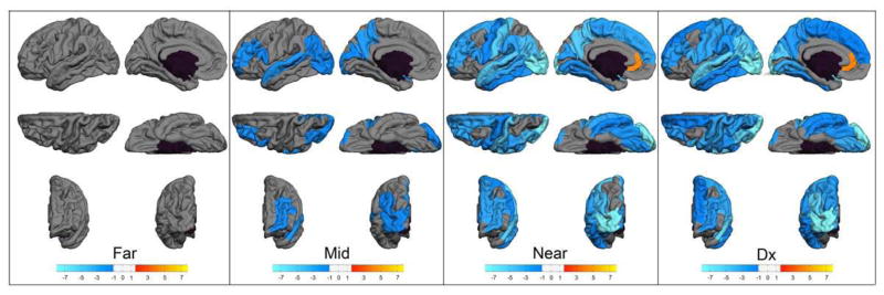
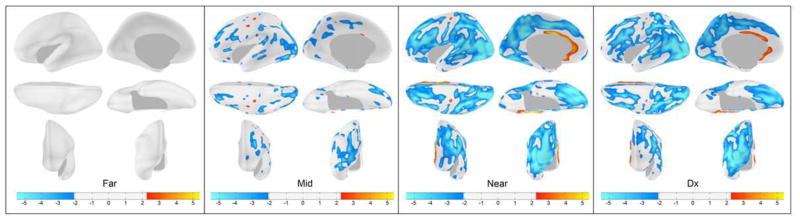
Neuroimaging studies of subjects who are gene-expanded for Huntington Disease, but not yet diagnosed (termed prodromal HD), report that the cortex is "spared," despite the decrement in striatal and cerebral white-matter volume. Measurement of whole-cortex volume can mask more subtle, but potentially clinically relevant regional changes in volume, thinning, or surface area. The current study addressed this limitation by evaluating cortical morphology of 523 prodromal HD subjects. Participants included 693 individuals enrolled in the PREDICT-HD protocol. Of these participants, 523 carried the HD gene mutation (prodromal HD group); the remaining 170 were non gene-expanded and served as the comparison group. Based on age and CAG repeat length, gene-expanded subjects were categorized as "Far from onset," "Midway to onset," "Near onset," and "already diagnosed." MRI scans were processed using FreeSurfer. Cortical volume, thickness, and surface area were not significantly different between the Far from onset group and controls. However, beginning in the Midway to onset group, the cortex showed significant volume decrement, affecting most the posterior and superior cerebral regions. This pattern progressed when evaluating the groups further into the disease process. Areas that remained mostly unaffected included ventral and medial regions of the frontal and temporal cortex. Morphologic changes were mostly in thinning as surface area did not substantially change in most regions. Early in the course of HD, the cortex shows changes that are manifest as cortical thinning and are most robust in the posterior and superior regions of the cerebrum.
Copyright © 2010 Elsevier Inc. All rights reserved.
Figures
References
-
- Abe O, Yamasue H, Aoki S, Suga M, Yamada H, Kasai K, et al. Aging in the CNS: comparison of gray/white matter volume and diffusion tensor data. Neurobiol Aging. 2008;29(1):102–16. - PubMed
-
- Aylward EH, Anderson NB, Bylsma FW, Wagster MV, Barta PE, Sherr M, et al. Frontal lobe volume in patients with Huntington’s disease. Neurology. 1998;50(1):252–8. - PubMed
-
- Bartzokis G, Lu PH, Tishler TA, Fong SM, Oluwadara B, Finn JP, et al. Myelin breakdown and iron changes in Huntington’s disease: pathogenesis and treatment implications. Neurochem Res. 2007;32(10):1655–64. - PubMed
-
- Bartzokis G, Lu PH, Tishler TA, Perlman S. In vivo assessment of iron in Huntington’s disease and other age-related degenerative brain diseases. In: Sigel A, Sigel H, Sigel RKO, editors. Metal Ions in life sciences. Chichester: Wiley; 2006. pp. 151–77.
Publication types
MeSH terms
Grants and funding
LinkOut - more resources
Full Text Sources
Medical
