Review
. 2010 Sep;18(9):388-96.
doi: 10.1016/j.tim.2010.06.010. Epub 2010 Aug 3.

BST-2/tetherin: a new component of the innate immune response to enveloped viruses

Affiliations
Review

BST-2/tetherin: a new component of the innate immune response to enveloped viruses

David T Evans et al. Trends Microbiol. 2010 Sep.

Abstract

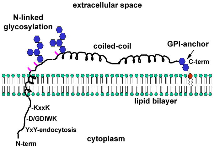
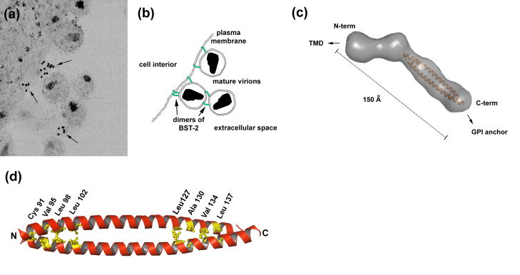
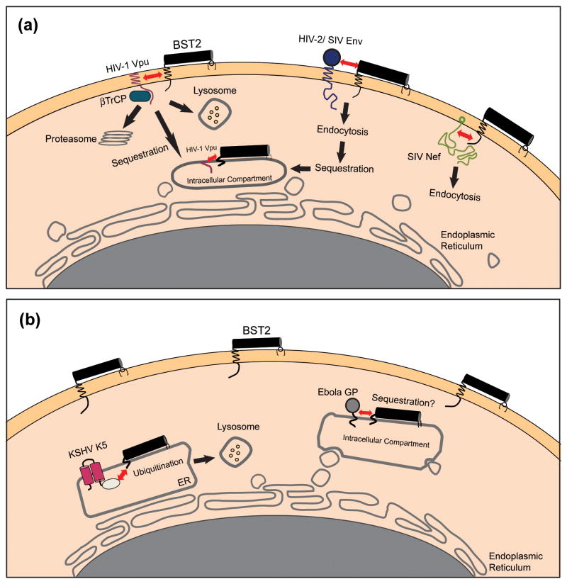
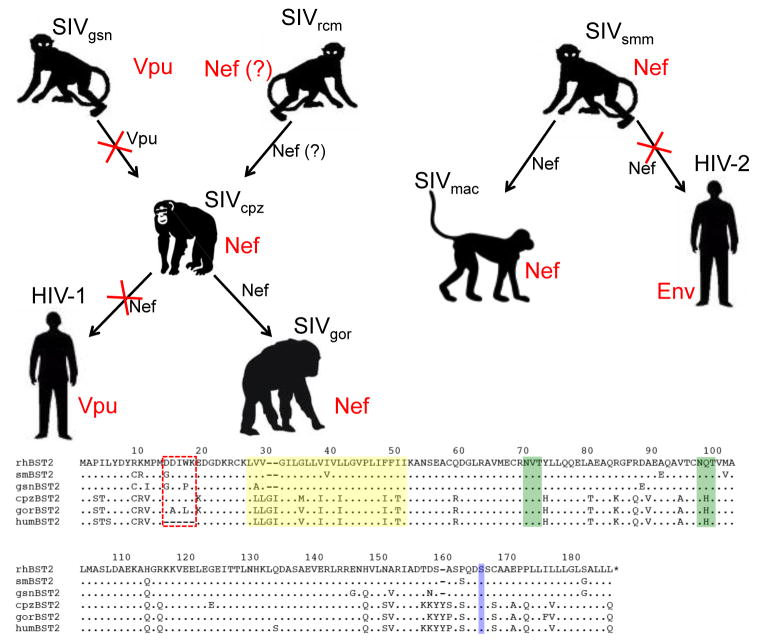
The interferon-inducible, transmembrane protein BST-2 (CD317, tetherin) directly holds fully formed enveloped virus particles to the cells that produce them, inhibiting their spread. BST-2 inhibits members of the retrovirus, filovirus, arenavirus and herpesvirus families. These viruses encode a variety of proteins to degrade BST-2 and/or direct it away from its site of action at the cell surface. Viral antagonism has subjected BST-2 to positive selection, leading to species-specific differences that presented a barrier to the transmission of simian immunodeficiency viruses (SIVs) to humans. This barrier was crossed by HIV-1 when its Vpu protein acquired activity as a BST-2 antagonist. Here, we review this new host-pathogen relationship and discuss its impact on the evolution of primate lentiviruses and the origins of the HIV pandemic.


Figures

Figure 1
Figure 1. Features of the BST-2/tetherin monomer
BST-2 binds the lipid bilayer via an N-terminal transmembrane α-helix and a C-terminal glycosylphophastidyl inositol (GPI) anchor. The cytoplasmic domain contains a YxY sequence that directs endocytosis by interacting with the clathrin adaptor AP-2. Non-human primate BST-2 contains an insertion relative to the human protein (D/GDIWK or a similar sequence), which is targeted by lentiviral Nef proteins. The KxxK sequence is ubiquitinated by the K5 protein of KSHV, leading to degradation. The transmembrane region is required for antagonism by HIV-1 Vpu. The ectodomain of the protein contains three cysteines (locations indicated in pink), which drive homo-dimerization. The ectodomain also contains an extended α-helical region that forms a parallel coiled-coil in BST-2 dimers. The transmembrane and GPI membrane anchors, the cysteines, and the integrity of the coiled-coil region are each required for the restriction of virion release.
Figure 2
Figure 2. Mechanism of restricted virion release by BST-2
(a) Immuno-electron microscopic data indicate that BST-2 is appropriately positioned to directly tether mature virions to the plasma membrane and is incorporated into virions. The cell cytoplasm is at the left; the extracellular space is at the right and includes mature virions. Arrows indicate immuno-label for the BST-2 ectodomain. Reprinted with permission from Ref. [31]. (b) Model in which BST-2 dimers (green) embed one end in the plasma membrane and the other in the virion membrane, retaining virions on the cell surface. BST-2 dimers may also link virions to each other. An alternative model is discussed in the text. (c) Structure of the dimeric BST-2 ectodomain obtained by small angle X-ray scattering (molecular surface in gray) and X-ray crystallography (modified from Ref. [21]). The parallel, dimeric coiled-coil is colored orange. Abbreviations: Å, angstroms. (d) Crystallographic structure of the dimeric coiled-coil indicating residues important for the restriction of virion release (PDB accession code: 2x7a; structure drawn using PyMOL).
Figure 3
Figure 3. Viral antagonists of BST-2 and their potential mechanisms of action
(a) Lentiviral antagonists. The HIV-1 Vpu protein associates with BST-2 through transmembrane domain interactions and recruits βTrCP, leading to the degradation of BST-2 within lysosomes and/or proteasomes. Alternatively, Vpu might simply trap BST-2 within the endosomal system. In either scenario, the concentration of BST-2 is decreased at the plasma membrane, its site of action as a tetherin. The SIV Nef protein interacts with the cytoplasmic domain of BST-2 and mediates downregulation from the cell surface, most likely via endocytosis. The HIV-2 Env protein downregulates and sequesters BST-2 within the trans-Golgi network (TGN) by a mechanism that depends on physical interactions between the two proteins and a conserved GYXXφ motif in the cytoplasmic tail of gp41. (b) Non-lentiviral antagonists. The Ebola GP physically associates with, and possibly sequesters, BST-2 within intracellular compartments. Alternatively, Ebola GP might directly interfere with the restrictive activity of BST-2 or it might direct viral assembly away from membrane domains containing BST-2. The KSHV K5 protein mediates the ubiquitination of BST-2, which induces degradation of the protein within proteasomes and/or lysosomes. The interactions between BST-2 and the lentiviral antagonist proteins Vpu, Nef, and Env are shown here as occurring at the plasma membrane, but they could occur alternatively or additionally within the endoplasmic reticulum (ER), the TGN, or recycling endosomes.
Figure 4
Figure 4. Adaptation of the primate lentiviruses for overcoming restriction by BST-2 in their respective hosts
Chimpanzee SIV (SIVcpz) is thought to be a recombinant derived from Old World monkey SIVs similar to viruses presently found in the greater spot-nosed monkey (SIVgsn) and the red-capped mangabey (SIVrcm). HIV-1 and SIVgor are the result of the cross-species transmission of SIVcpz from chimpanzees to humans and from chimpanzees to gorillas. Likewise, HIV-2 and SIVmac reflect the cross-species transmission of SIVsmm from sooty mangabeys to humans and from sooty mangabeys to macaques. Arrows represent cross-species transmission events and the viral gene products that antagonize BST-2 in each species are indicated in red. The viral gene products that either retained, or lost (indicated by red X’s). Activity against BST-2 are indicated in black next to each arrow. Species-specific differences in BST-2 are depicted in the amino acid sequence alignment. Representative BST-2 sequences are shown for the rhesus macaque (rhBST2), sooty mangabey (smBST2), greater spot-nosed monkey (gsnBST2), chimpanzee (cpzBST2), gorilla (gorBST2) and humans (humBST2). Residues of the cytoplasmic domain that confer susceptibility to Nef are indicated by the red box, the transmembrane domain is highlighted in yellow, potential N-linked glycosylation sites are indicted in green and the site for cleavage and addition of the GPI anchor is indicated in blue. The five-residue deletion in the cytoplasmic domain of human BST-2 renders the protein resistant to antagonism by Nef, leading to the acquisition of BST-2 antagonist activity by HIV-1 Vpu and HIV-2 Env.

Similar articles

Cited by

References

    1. Neil SJ, et al. Tetherin inhibits retrovirus release and is antagonized by HIV-1 Vpu. Nature. 2008;451:425–430. - PubMed
    1. Van Damme N, et al. The interferon-induced protein BST-2 restricts HIV-1 release and is downregulated from the cell surface by the viral Vpu protein. Cell Host Microbe. 2008;3:245–252. - PMC - PubMed
    1. Ishikawa J, et al. Molecular cloning and chromosomal mapping of a bone marrow stromal cell surface gene, BST2, that may be involved in pre-B-cell growth. Genomics. 1995;26:527–534. - PubMed
    1. Sheehy AM, et al. Isolation of a human gene that inhibits HIV-1 infection and is suppressed by the viral Vif protein. Nature. 2002;418:646–650. - PubMed
    1. Stremlau M, et al. The cytoplasmic body component TRIM5alpha restricts HIV-1 infection in Old World monkeys. Nature. 2004;427:848–853. - PubMed

Publication types

MeSH terms

Substances