Review
. 2010 Jul-Sep;9(3):191-205.
doi: 10.14310/horm.2002.1270.

Physiological effects and mechanisms of action of endocrine disrupting chemicals that alter estrogen signaling

Affiliations
Review

Physiological effects and mechanisms of action of endocrine disrupting chemicals that alter estrogen signaling

Derek V Henley et al. Hormones (Athens). 2010 Jul-Sep.
No abstract available


Figures

Figure 1
Figure 1
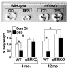
Effect of neonatal DES exposure on seminal vesicle weight in adult WT and αERKO mice. Top, seminal vesicles from 6-month old WT and αERKO mice following neonatal exposure to either vehicle (corn oil, CO) or DES (scale is in centimeters). Bottom, quantitative analysis of seminal vesicle weights from WT and aERKO mice at 4 and 12 months of age following neonatal exposure to CO or DES. *, P<0.05. (Modified from Prins, G.S., Birch, L., Couse, J.F., Choi, I., Katzenellenbogen, B., and Korach, K.S., 2001 Cancer Res., 61: 6089-6097.)
Figure 2
Figure 2
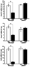
Effect of neonatal DES exposure on uterine expression of Hoxa10, Hoxa11, and Wnt7a in WT and αERKO mice. Top and middle, Quantitative analyses of ribonuclease protection assays (Hoxa10 and Hoxa11) (+/− SEM) showing average percentage of cyclophilin (normalization mRNA) for each treatment group. Bottom, Quantitative analysis of semiquantitative RT-PCR showing the average Wnt7a levels as a percentage of Actb (+/− SEM) for each treatment group. *, P<0.01. (Modified from Couse, J.F., Dixon, D., Yates, M., Moore, A.B., Ma, L., Maas, R., and Korach, K.S., 2001 Dev. Biol., 238: 224-238.).
Figure 3
Figure 3
Role of ERα in modulating genistein-induced uterotropic effects. Ovariectomized WT and αERKO mice (n=4 mice/treatment group) were treated daily with subcutaneous injections of corn oil (vehicle), 10μg/kg estradiol, or 50μg/kg genistein for three days. On the fourth day whole body weights were measured and uteri were collected and weighed. The data represent uterine wet weight with respect to whole body weight. (Unpublished data from Lindzey, J. and Korach, K.S.).
Figure 4
Figure 4
MAA activates the CMV promoter in vitro. A. HeLa cells were transiently transfected with a human ERβ expression vector, the estrogen-responsive 3X-ERE-TATA-Luc reporter plasmid, and the CMV-β-gal reporter plasmid and treated for 18h with either vehicle, increasing concentrations of E2, 5mM MAA, or increasing concentrations of E2 plus 5mM MAA. The histogram represents the average fold over control of duplicate samples from three independent experiments. The error bars represent the standard error of the mean (SEM), *p<0.05 comparing identical treatments +/− 5mM MAA. B. HeLa cells were transiently transfected with a human ERα expression vector, 3X-ERE-TATA-Luc and CMV-β-gal and treated identically to the cells in Figure 1A. The data shown represent the average fold over control of duplicate samples from three independent experiments and the error bars indicated the SEM, *p<0.05 comparing identical treatments +/− 5mM MAA. C. HeLa cells were transiently transfected with a human ERα expression vector and treated with either vehicle or increasing concentrations of MAA for 18h. Total protein was isolated from the cells and ERα protein expression was analyzed by Western blot. The data shown are representative of three independent experiments. D. HeLa cells were transiently transfected with the pRL-CMV reporter plasmid and treated with either vehicle or increasing concentrations of MAA for 18h. The data shown represent the average fold over control of duplicate samples from three independent experiments. The error bars represent the SEM, *p<0.01 comparing treatment with MAA to vehicle control. (Modified from Henley, D.V., Mueller, S., and Korach, K.S. In Press Environ Health Persp.).
Figure 5
Figure 5
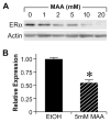
Methoxyacetic Acid (MAA) decreases endogenous ERα expression.. A. MCF-7 cells were treated for 24h with either vehicle or increasing concentrations of MAA. Total protein was isolated from each sample and analyzed for ERα protein expression by Western blot. The data shown are representative of results from three independent experiments. B. MCF-7 cells were treated for 24h with either vehicle or 5mM MAA. Total RNA was isolated from each sample and steady state mRNA levels of ERα were measured by real-time PCR. The data shown represent the average fold over control obtained from duplicate samples in four independent experiments, and the error bars indicate the SEM, *p<0.01 comparing treatment with 5mM MAA to vehicle control. (Modified from Henley, D.V., Mueller, S., and Korach, K.S. In Press Environ Health Persp.).
Figure 6
Figure 6
Methoxyacetic Acid (MAA) disrupts estrogen-mediated endogenous gene expression. MCF-7 cells were pretreated with either vehicle or 5mM MAA for 2h and then treated for 18h with either vehicle or 1nM E2. Total RNA was isolated from the samples and the expression of endogenous estrogen-responsive genes was analyzed by real-time PCR. The data shown represent the average fold over control of duplicate samples from at least three independent experiments. Error bars represent the SEM, *p<0.01 comparing treatment with 1nM E2 + 5mM MAA to treatment with 1nM E2 alone. The error bars represent SEM. (Modified from Henley, D.V., Mueller, S., and Korach, K.S. In Press Environ Health Persp.).
Figure 7
Figure 7
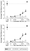
In vitro estrogenic activity of lavender oil and tea tree oil in reporter gene assays. MCF-7 cells were transiently transfected with the estrogen-inducible 3x-ERE-luciferase reporter plasmid and treated with increasing concentrations of either lavender oil or tea tree oil for 18 hours in the presence and absence of fulvestrant, an ER antagonist. Estradiol treatment (1 nM) was included as a positive control. Data are plotted as fold-luciferase activity over vehicle control. (Modified from Henley, D.V., Lipson, N., Korach, K.S., and Bloch, C.A., NEJM, 356, 481, 2007).
Figure 8
Figure 8
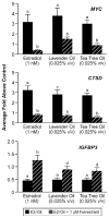
In vitro effects of lavender oil and tea tree oil on the expression of estrogen-regulated endogenous genes. MCF-7 cells were treated with 1 nM estradiol, 0.025% lavender oil, or 0.025% tea tree oil in the presence or absence of fulvestrant and real-time PCR was performed to measure changes in the expression of the estrogen-inducible genes MYC, IGFBP3, and CTSD. Data shown are normalized and relative to a vehicle control. (Modified from Henley, D.V., Lipson, N., Korach, K.S., and Bloch, C.A., NEJM, 356, 481, 2007).
Figure 9
Figure 9
In vitro antiandrogenic activity of lavender oil and tea tree oil in reporter gene assays. MDA-kb2 cells stably transfected with the MMTV-luciferase reporter plasmid were treated for 24 hours with increasing concentrations of lavender oil or tea tree oil in the presence or absence of 0.1 nM dihydrotestosterone (DHT). The androgen receptor antagonist flutamide (1 μM) was included as a control antagonist for the androgen receptor. Luciferase activity was measured and data plotted as the average fold above control. (Modified from Henley, D.V., Lipson, N., Korach, K.S., and Bloch, C.A., NEJM, 356, 483, 2007).
Figure 10
Figure 10
In vitro effects of lavender oil and tea tree oil on androgen-stimulated endogenous gene expression. MDA-kb2 cells were treated with 0.1 nM dihydrotestosterone for 24 hours in the presence or absence of 0.0005% lavender oil, 0.0005% tea tree oil, or 1μM flutamide and real-time PCR was performed to measure changes in the expression of androgen-inducible genes. Data shown are normalized and relative to a vehicle control. (Modified from Henley, D.V., Lipson, N., Korach, K.S., and Bloch, C.A., NEJM, 356, 483, 2007).

Similar articles

Cited by

References

    1. Dodds EC, Lawsaon W, Robinson R. Estrogenic activity of certain synthetic compounds. Nature. 1938:141247–141248.
    1. Smith OW, Smith GBS, Hurwitz D. Increased excretion of pregnanediol in pregnancy from diethylstilbestrol with special reference to the prevention of late pregnancy accidents. Med Rec Ann. 1946;40:1669–1671. - PubMed
    1. Dieckmann WJ, Davis ME, Rynkiewicz LM, Pottinger RE, Gabbe SG. Does the administration of diethylstilbestrol during pregnancy have therapeutic value 1953? Am J Obstet Gynecol. 1999;66:1062–1081. - PubMed
    1. Herbst AL, Ulfelder H, Poskanzer DC. Adenocarcinoma of the vagina. Association of maternal stilbestrol therapy with tumor appearance in young women. N Engl J Med. 1971;284:878–881. - PubMed
    1. Melnick S, Cole P, Anderson D, Herbst A. Rates and risks of diethylstilbestrol-related clear-cell adenocarcinoma of the vagina and cervix. An update. N Engl J Med. 1987;316:514–516. - PubMed

LinkOut - more resources