. 2010 Dec;51(12):6858-67.
doi: 10.1167/iovs.10-5499. Epub 2010 Aug 4.

Intersubject variability of foveal cone photoreceptor density in relation to eye length

Affiliations

Intersubject variability of foveal cone photoreceptor density in relation to eye length

Kaccie Y Li et al. Invest Ophthalmol Vis Sci. 2010 Dec.

Abstract

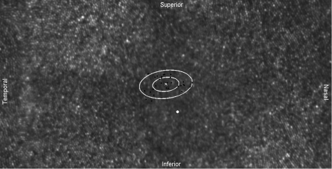
Purpose: Adaptive optics scanning laser ophthalmoscopy (AOSLO) under optimized wavefront correction allows for routine imaging of foveal cone photoreceptors. The intersubject variability of foveal cone density was measured and its relation to eye length evaluated.

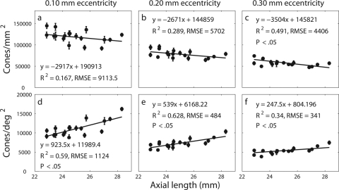
Methods: AOSLO was used to image 18 healthy eyes with axial lengths from 22.86 to 28.31 mm. Ocular biometry and an eye model were used to estimate the retinal magnification factor. Individual cones in the AOSLO images were labeled, and the locations were used to generate topographic maps representing the spatial distribution of density. Representative retinal (cones/mm(2)) and angular (cones/deg(2)) cone densities at specific eccentricities were calculated from these maps.

Results: The entire foveal cone mosaic was resolved in four eyes, whereas the cones within 0.03 mm eccentricity remained unresolved in most eyes. The preferred retinal locus deviated significantly (P < 0.001) from the point of peak cone density for all except one individual. A significant decrease in retinal density (P < 0.05) with increasing axial length was observed at 0.30 mm eccentricity but not closer. Longer, more myopic eyes generally had higher angular density near the foveal center than the shorter eyes, but by 1°, this difference was nullified by retinal expansion, and so angular densities across all eyes were similar.

Conclusions: The AOSLO can resolve the smallest foveal cones in certain eyes. Although myopia causes retinal stretching in the fovea, its effect within the foveola is confounded by factors other than cone density that have high levels of intersubject variability.


Figures

Figure 1.
Figure 1.
(a) A 0.25° × 0.50° (72 × 144 μm) section of a cone mosaic in subject 6 with identified cone locations. The patch is located at approximately 0.75° (216 μm) from the foveal center. (b) Result after taking the distance transform of the (x, y) cone locations. (c) Voronoi tiles generated using the watershed transform.
Figure 2.
Figure 2.
(a) Axial length plotted as a function of the spherical equivalent spectacle refraction with the solid line being a linear regression of the data. (b) Calculated RMF plotted as a function of the spherical equivalent spectacle refraction. Equation 2 was used directly to compute RMF for the uncorrected case, whereas the spectacle corrected RMFs were obtained by multiplying the uncorrected RMFs by the corresponding spectacle magnifications calculated with equation 3. Lines represent linear regressions of the data. The decrease in RMF with less refractive error was significant (P < 0.05) for both the corrected and uncorrected cases.
Figure 3.
Figure 3.
A 1° × 2° (320 × 640 μm) cone mosaic centered about the PRL (white x) for subject 13. Black dots: fixation locations; white ellipses: one and two standard deviations of the fixation points. The PRL is displaced approximately 9.5 arc min (50 μm) from the foveal center. (white dot).
Figure 4.
Figure 4.
A 1° × 1° (336 × 336 μm) cone mosaic centered about the PRL (white x). Small black dots: fixation locations; white dot: location of the anatomic foveal center. The semimajor axis angle for the distribution of fixation is approximately 143°. The PRL is displaced approximately 3.9 arc min (22 μm) from the foveal center.
Figure 5.
Figure 5.
Examples of cone density topographic maps. All maps are oriented as indicated in the top left panel. T, temporal; S, superior; N, nasal; I, inferior. Locations of the foveal center and the PRL are indicated by a white dot and an x, respectively. The size of each map is 0.6 × 0.6 mm, and consecutive contour lines are separated by 5000 cones/mm2. Dark blue areas include both the central foveal region in some eyes where cones could not be resolved and regions outside of the support of the acquired retinal images.
Figure 6.
Figure 6.
Retinal cone density as a function of retinal eccentricity. Representative cone density measurements at particular eccentricities were computed by circular averaging of density estimates around all meridians. The shaded region corresponds to the range of foveal cone density values reported by Curcio et al.
Figure 7.
Figure 7.
Retinal (ac) and angular (df) cone density as a function of axial length at three different retinal eccentricities. Error bars represent one standard deviation in the spread of cone densities at the specified eccentricities. Lines: weighted least squares linear regression of the data.
Figure 8.
Figure 8.
Retinal cone density (ac) and angular cone density (df) as a function of axial length at three different angular eccentricities. Error bars represent one standard deviation in the spread of cone density values at the specified eccentricities. Lines: weighted least squares linear regression of the data.
Figure 9.
Figure 9.
Retinal cone density as a function of axial length at the PRL. Line: linear regression of the data. The regression slope is insignificant (P > 0.05).

References

    1. Chui TYP, Yap MKH, Chan HHL, Thibos LN. Retinal stretching limits peripheral visual acuity in myopia. Vision Res. 2005;45:593–605 - PubMed
    1. Coletta NJ, Watson T. Effect of myopia on visual acuity measured with laser interference fringes. Vision Res. 2006;46:636–651 - PubMed
    1. Chui TYP, Song H, Burns SA. Individual variations in human cone photoreceptor packing density: variations with refractive error. Invest Ophthalmol Vis Sci. 2008;49:4679–4687 - PMC - PubMed
    1. Strang NC, Winn B, Bradley A. The role of neural and optical factors in limiting visual resolution in myopia. Vision Res. 1998;38:1713–1721 - PubMed
    1. Bullimore MA, Gilmartin B, Royston JM. Steady-state accommodation and ocular biometry in late-onset myopia. Doc Ophthalmol. 1992;80:143–155 - PubMed

Publication types