. 2010 Sep 17;285(38):29054-8.
doi: 10.1074/jbc.C110.168641. Epub 2010 Aug 5.

A novel mechanism for ribonuclease regulation: transfer-messenger RNA (tmRNA) and its associated protein SmpB regulate the stability of RNase R

Affiliations

A novel mechanism for ribonuclease regulation: transfer-messenger RNA (tmRNA) and its associated protein SmpB regulate the stability of RNase R

Wenxing Liang et al. J Biol Chem. .

Abstract

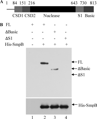
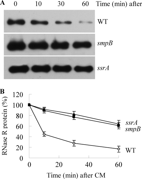
The amount of RNase R, an important degradative exoribonuclease, increases 3-10-fold under a variety of stress conditions. This elevation is due to posttranslational regulation in which the highly unstable RNase R protein becomes stabilized during stress. Here we identify two components of the trans-translation machinery, transfer-messenger RNA (tmRNA) and SmpB, that are responsible for the short half-life of RNase R in exponential phase cells. The absence of either lengthens the half-life of RNase R in vivo >6-fold. SmpB directly interacts with RNase R in vitro and is stimulated by tmRNA. The C-terminal region of RNase R, encompassing its basic region and adjacent S1 domain are required for the interaction; their removal eliminates binding and stabilizes RNase R in vivo. However, the binding of SmpB and tmRNA does not alter RNase R activity. These data define a previously unknown regulatory process in which the stability of an RNase is determined by its interaction with an RNA and an RNA-associated protein.


Figures

FIGURE 1.
FIGURE 1.
Association of RNase R with SmpB. A, chemical cross-linking of soluble proteins in extracts from strain MG1655(Seq)rph+. Samples were treated without (−) or with (+) DMP and analyzed as described under “Experimental Procedures.” The position of RNase R is indicated, and * indicates a band of about 115 kDa. The migration positions of the indicated molecular mass standards are shown on the left. This gel was overexposed to observe the cross-linked bands. B, co-immunoprecipitation of extracts prepared from MG1655(Seq)rph+ (FLAG-SmpB, His-RNase R) cells. Extracts were treated without (−) or with (+) RNase R antibody, and the resulting immunoprecipitates were analyzed by SDS-PAGE by probing with His-probe monoclonal antibody or anti-FLAG monoclonal antibody. C, pulldown of FLAG-SmpB and tmRNA with His-RNase R. Extracts from MG1655(Seq)rph+ (FLAG-SmpB, His-RNase R) and smpB derivative cells were treated with Ni-NTA, and the presence of His-RNase R, FLAG-SmpB, and tmRNA in the eluant was detected by His-probe monoclonal antibody, anti-FLAG monoclonal antibody, or tmRNA-specific probe, respectively. D, pulldown of RNase R with His-SmpB. Purified RNase R, His-SmpB, and tmRNA were prepared and used for a pulldown assay as described under “Experimental Procedures.” The proteins and tmRNA present in each assay are indicated above the lanes. The presence of RNase R protein and recombinant His-SmpB protein was detected as above.
FIGURE 2.
FIGURE 2.
Interaction between SmpB and various forms of RNase R. Full-length RNase R (FL), RNase RΔBasic (ΔBasic), and RNase RΔS1 (ΔS1) proteins (A) were purified and used in a pulldown assay (B) using Ni-NTA resin as described under “Experimental Procedures.” The proteins present are indicated above each lane. The presence of RNase R proteins and recombinant His-SmpB protein was detected as in Fig. 1.
FIGURE 3.
FIGURE 3.
Stability of RNase R in wild type (WT) and smpB and ssrA mutant strains. The indicated cells were grown to exponential phase, treated with chloramphenicol, and assayed for RNase R by immunoblotting as described under “Experimental Procedures.” A, representative Western blot of RNase R from samples at the indicated times after the addition of chloramphenicol. B, quantitation of three independent experiments carried out as in A. RNase R levels at zero time of chloramphenicol (CM) addition were set at 100% for each strain. Error bars indicate S.D.
FIGURE 4.
FIGURE 4.
Stability of full-length and truncated RNase R proteins. Experiments were carried out as in Fig. 3. A, representative Western blot of full-length (FL) and truncated RNase R proteins from exponential phase cells. CM, chloramphenicol. B, quantitation of three independent experiments carried out as shown in Panel A. RNase R levels at zero time of chloramphenicol addition were set at 100% for each RNase R. Error bars indicate S.D.

References

    1. Cheng Z. F., Deutscher M. P. (2002) J. Biol. Chem. 277, 21624–21629 - PubMed
    1. Cheng Z. F., Deutscher M. P. (2005) Mol. Cell 17, 313–318 - PubMed
    1. Richards J., Mehta P., Karzai A. W. (2006) Mol. Microbiol. 62, 1700–1712 - PubMed
    1. Li Z., Reimers S., Pandit S., Deutscher M. P. (2002) EMBO J. 21, 1132–1138 - PMC - PubMed
    1. Cheng Z. F., Deutscher M. P. (2003) Proc. Natl. Acad. Sci. U.S.A. 100, 6388–6393 - PMC - PubMed

Publication types

MeSH terms

LinkOut - more resources